154
Ⅰ. 서 론
일반적인 치조골 소실의 원인으로는 진행된 치주질환, 외 상, 전신질환의 이차적인 치조골소실 및 선천적 기형 등이
있다. 특히 고령의 무치악 환자에서는 심하게 위축된 하악 골의 수직 및 수평적인 치조골의 심한 흡수 양상을 보여 불 충분한 의치의 유지력, 의치에 의한 하치조신경 압박에 의 한 통증, 이부 및 하순의 외모 변화 등 여러 문제점을 가지 염학렬∙전승호∙김윤태∙팽준영∙안강민∙명 훈∙황순정∙서병무∙최진영∙이종호∙정필훈∙김명진
서울대학교 치과대학 구강악안면외과학교실
심하게 위축된 하악골의 완전 무치악 환자에서의 수직 골신장술을 이용한 임플란트 치료
IMPLANT INSTALLATION USING VERTICAL DISTRACTION OSTEOGENESIS AT A SEVERELY ATROPHIED EDENTULOUS MANDIBLE
Hak-Ryol Yeom, Seung-Ho Jeon, Yoon-Tae Kim, Jun-Young Paeng, Kang-Min Ahn, Hoon Myung, Soon-Jung Hwang, Byoung-Moo Seo, Jin-Young Choi, Jong-Ho Lee, Pill-Hoon Choung, Myung-Jin Kim
Department of Oral and Maxillofacial Surgery, College of Dentistry, Seoul National University O
Obbjjeeccttiivveess :: It is difficult to place implants at a severely atrophied edentulous mandible because of ver- tical and horizontal alveolar defect. The autogenous and allogenic bone graft and guided bone regeneration are useful, but there are some limitations such as the resorption and infection of the grafted bone, and insufficiency of soft tissue. Distraction osteogenesis has recently evolved a challenging technique to over- come major draws of conventional augmentation procedures, we, therefore, report the four applications of implant placement after vertical alveolar distraction osteogenesis.
P
Paattiieennttss aanndd MMeetthhooddss :: Four patients(all female, mean age: 60±6 years) with severe alveolar ridge deficiencies at their anterior mandible were treated with vertical alveolar distraction osteogenesis by intrao- ral device(Track 1.5, 15 mm model, KLS Martin, Tuttlingen, Germany) and placement of implants (Branemark MK III, TiUnite). After the latency periods of 5, 7, 8 days, distraction rhythm and rate were 0.5 mm×2 times/day in the case of good bone quality, and 0.25 mm×3 times/day in the case of poor bone quality. After consolidation periods of mean fifteen weeks, five implants for each patients were placed at the interforaminal area.
R
Reessuullttss :: On average, a vertical gain of 11.38±1.38 mm was obtained by distraction. And all distrac- tion zone showed complete ossification by panoramic radiography. There were no postoperative complica- tions other than numbness of lower lip in one case. Total twenty implants in four patients were placed and their outcomes were satisfactory.
C
Coonncclluussiioonn :: It is a useful method to place five implants after vertical distraction osteogenesis of the severely atrophied mandible for the implant-supported fixed prosthesis.
Key words : Vertical Distraction Osteogenesis, Atrophy of Mandible, Dental Implant Abstract
고 있다1).
이러한 무치악 환자의 보철 수복을 위해서 전통적 치료방 법인 완전 연조직 지지 의치 대신 임플란트를 이용한 성공 적인 보철 치료를 위해서 임플란트 식립을 위한 적절한 치 조골의 높이와 폭이 필수적이다. 치조골 결손을 해결하기 위해 자가골을 이용한 onlay graft 또는 수산화인회석같은 합성골이식술 및 골유도재생술 등이 이용될 수 있으나 이는 이식골의 흡수 및 노출, 그리고 이식부위의 감염 및 연조직 의 부족 등 많은 한계점을 가진다2-4).
이러한 한계점을 해결하기 위해 치조골신장술이 최근 많 이 이용되고 있으며 이런 골신장술 개념은 1989년 러시아 정형외과의사인 Ilizarov5,6)에 의해 처음 소개된 후 1996년 Block이 처음으로 개의 하악골에 수직 치조골신장술 실험 을 시행하였으며7), 동년 Chin 등이 무치악 환자의 하악 치 조제에 처음으로 적용하여 양호한 임상결과를 얻었다8).
본 교실에서는 하악골의 정중부 수직 높이가 평균 9(±
3.06) mm인 위축된 완전 무치악 환자 4명에서 통상적인 골이식술 대신 구내 수직 골신장술을 시행한 후 임플란트 지지 고정성 의치 치료를 위해 이공간에 다섯 개의 임플란 트 식립술을 시행하여 양호한 결과를 얻었기에 이를 문헌고 찰과 함께 보고하고자 하였다.
Ⅱ. 환자 및 방법 1. 환 자
심하게 위축된 하악골의 완전 무치악과 기존 총의치 대신 임플란트 보철 치료를 주소로 서울대학교 치과병원 구강악 안면외과에 내원 또는 의뢰된 4명의 여성환자(평균 연령 60세)를 대상으로 하였다. 모두 전신마취하에 구내 수직 치 조골신장 장치(Track 1.5, 15 mm model, KLS Martin, Tuttlingen, Germany)를 이용한 하악골 수직 골신장술 및 골신장술 후 임플란트 식립술을 시행 받았다. 이중 한 환 자는 상악골의 심한 수평적 및 수직적 골흡수가 동반되어 있어 하악골 수직 골신장술과 동시에 LeFort I형 골절단술 을 이용한 전진술 및 장골이식을 이용, 양측 상악동 이식술 및 interpositional bone graft와 동시 임플란트 식립술(총 10개, Branemark MK III, TiUnite, Length : 15 mm) 을 시행 받았다. 환자의 의치 사용 기간은 평균 15년 이상 이었으며 이공간(interforaminal distance)의 하악골의 정 중부 수직 높이는 평균 9(±3.06) mm이었다(Table 1).
입원기간은 장골이식을 동반한 상하악 수술 환자의 경우 10일이었고, 하악 골신장술만 시술 받은 환자의 경우에는 3-5일이었다. 모든 환자에서 골다공증을 포함한 전신질환 은 없었다.
2. 수술방법
(1) 치조골신장 장치
4명의 환자 모두 구내 수직 골신장술 장치인 Track 1.5, 15mm model(KLS Martin, Tuttlingen, Germany)을 사용하였다. Track 1.5 시스템은 골신장 길이에 따라 10 mm와 15 mm의 두 가지 모델이 있으며 하나의 수직암인 permucosal pin과 하악골과의 고정을 위한 두 개의 수평암 으로 구성된 단순한 형태의 장치이며, 쉽게 하악골에 적합 할 수 있다. 한번의 완전한 회전으로 0.5 mm의 골신장이 얻어지며 보통 하루에 2번 회전시켜 하루에 총 1 mm의 골 신장량을 얻도록 고안되어 있다.
(2) 수술 방법(Fig. 1)
4명의 환자 모두 전신마취하에 수술이 이루어졌다. 모든 증례에서 수술 전 임상사진, lateral cephalography, PA cephalography, panoramic radiography 및 3D CT를 촬 영하였으며 3D CT를 이용하여 RP 모형을 미리 제작하였 다. 제작된 RP 모델상에 연필을 이용, 이공부 및 수직, 수 평 골절단부를 디자인한 후 장치를 2-3 mm 정도 신장시킨 후 시적하여 수평암을 미리 구부려 놓는다. 이때 passive adaptation을 유지하도록 주의하여야 한다. 몇 개의 금속 나사를 이용, 부착될 모형상의 위치에 고정한 후 다시 제거 한다. Reciprocating saw 및 chisel을 이용, RP 모형을 디 자인한대로 골절단한 후 장치를 다시 적합시키고, interfer- ence 유무 및 골신장 정도 및 방향 등을 확인한 후 장치 멸 균을 위해 소독하였다.
절개는 점막 치은 경계부 하방 5 mm에 수평전정절개를 하였으며 수직이완절개는 하지 않았다. 신장될 Transport disc의 혈류공급을 유지하기 위해 상방 및 설측면의 박리는 최소로 하며 점막골막피판을 주의 깊게 박리해 나간다. 이 때 하치조신경의 손상을 주의하여야 한다. 골절단 전에 미 리 구부려 놓은 장치를 시적하여 몇 개의 금속나사를 사용, 부착할 위치에 고정하고, 골신장방향 등을 확인한다. 먼저 No.701 tapered fissure bur를 사용하여 생리식염수 주수 하에 골 절단부를 indentation한 후 장치를 제거하고, rec- iprocating saw 및 chisel을 사용하여 골절단을 완료한다.
수직 골절단을 시행할 때 골신장시 측벽과의 interference 가 없도록 치조능측의 길이가 기저측의 길이보다 약간 길게 형성하여야 한다. 골절단을 완료한 후 장치를 다시 시적, 신 장시켜 interference 유무, 골신장량 및 방향을 최종 확인한 후 원래 위치로 골편을 위치시키고 금속나사를 사용하여 장 치를 최종 고정한다. 이때 드릴로 인한 나사의 느슨해짐을 예방하기 위해 self-drilling screw로 고정하는 것도 고려해 볼 수 있다. 봉합은 세심한 지혈과 생리식염수 세척 후 흡수
156
성 봉합사인 Vicryl을 사용하였다. 술 후 부종을 예방하기 위해 봉합 후 수술부위의 압박 드레싱을 시행하였으며, 감 염 예방을 위해 술 후 5일에서 7일간의 항생제를 투여하 였다.
3. 골신장술 Protocol
수술 후 5-8일정도의 잠복기(latency period)가 지난 후 에 골신장을 시작하였다. 골신장율 및 리듬은 2명의 환자에 서는 1회 0.5 mm씩 하루에 2번, 총 1 mm의 골신장을 시 행하였으며 수술 중 해면골의 상태가 비교적 양호하지 않았 던 2명의 환자에서는 1회 0.25 mm씩 하루에 3번 총 0.75 mm의 골신장을 시행하였다. 각각 10.5, 10, 12, 13 mm 의 평균 11.38(±1.38) mm의 골신장량이 얻어졌으며, 1 명의 환자에서만 절개에 의한 이신경 손상에 의해 술 후 2- 3주까지 턱이 조이는 듯한 통증과 하순의 감각저하증상을 호소하였으나, 약 6개월 후에 회복되었다. 다른 환자에서 특별한 술 후 합병증은 없었다. 모든 환자에서 양호한 골신 장이 이루어 졌으며, 골신장완료 후 평균 15(±3)주 후에 국소마취하에 장치의 제거 및 임플란트를 식립하였다 (Table 2).
4. 임플란트 식립
먼저 골신장 장치를 노출시킨 후 임플란트 스텐트를 이용 하여 통상적인 순서대로 충분한 생리식염수 주수 하에 드릴 링을 시행하였다. 이때 임플란트 식립시 발생할 수 있는 골
편의 변위를 최소화하기 위해서 장치를 제거하지 않고 유지 하여 미리 tapping을 시행해야 하며 permucosal pin의 방 향과 평행을 유지하면서 식립해야 한다. 식립된 임플란트는 모두 직경 4 mm, 길이 18 mm의 Branemark 시스템, MKIII, TiUnite이었고 모두 초기 고정이 양호하였다.
5. 치료 결과
모든 증례에서 평균 11.38(±1.38) mm의 양호한 골신 장량 및 골신장방향을 보였다. 또한 신장부위의 열개나 골 편의 괴사, 술 후 감염 및 장치의 파절 등은 발생하지 않았 다. 단, 한 환자에서 술 후 하순의 감각저하를 호소하였으나 이는 골절단 및 골신장에 의한 하치조신경의 손상이 아닌 절개 시 치조정 상방에 위치한 이신경의 부분적 손상에 의 해 발생하였다. 각 환자당 5개씩 총 20개의 임플란트가 식립되었으며 초기 고정 및 술 후 경과가 모두 양호하였다.
Ⅲ. 고 찰
지금까지 심하게 위축된 하악골의 보철치료를 위한 여러 방법이 소개되어 왔으며 근래 골유착 임플란트의 도입으로 전통적인 의치가 지닌 여러 한계점을 많이 극복할 수 있게 되었다. 그러나 여전히 수직적 및 수평적인 치조골의 부족 으로 인해 임플란트의 식립에 많은 어려움이 있어, 이를 해 결 하고자 자가골이식 및 골유도 재생술 등이 이용 되어져
왔다2-4,9-12). 하지만 이 역시 골 흡수 및 감염, 연조직의 부족,
예측하기 힘든 골 증대량 등 한계점이 있다.
Table 1.Patients who Underwent Alveolar Distraction Osteogenesis of the Atrophied Anterior Mandible Patients Age/Sex Denture Period Intraoral State Initial Bone Distraction
Height length
PMD 68/F 40 years Mx,Mn: CD 6.5 mm 10.5 mm
POR 57/F 15 years Mn : CD 9.0 mm 10.0 mm
HOH 54/F 15 years Mx:RPD,Mn:CD 6.5 mm 12.0 mm
KJJ 61/F 20 years Mx,Mn: CD 14.0 mm 13.0 mm
* CD : Complete denture, RPD : Removable partial denture
Table 2.Distraction Protocol
Patients Latency Period Rhythm & Rate Distraction Length Consolidation Period
PMD 5 days 0.25 mm*3/day 10.5 mm 18 weeks
POR 7 days 0.25 mm*3/day 10.0 mm 17 weeks
HOH 8 days 0.5 mm*2/day 12.0 mm 11 weeks
KJJ 5 days 0.5 mm*2/day 13.0 mm 14 weeks
통상적으로 심하게 흡수된 하악골에서 임플란트 식립을 이용한 보철치료를 요약하면 다음과 같다13).
1) 짧은 골내 임플란트을 이용한 고정성 및 가철성 보철 2) Transosseous implant 시스템을 이용한 가철성
보철14,15)
3) 골이식술 또는 골신장술을 이용한 하악골의 치조제 증 대술 후 긴 골내 임플란트를 이용한 고정성 및 가철성 보철
비록 무치악의 하악골에 식립된 임플란트에 대해 많은 연 구가 이루어졌지만, 어떤 치료 방법이 최선의 치료방법인지 에 대해서는 아직 많은 논란의 여지가 있다. 먼저 이공간에 5개 내지 6개의 짧은 골내 임플란트를 이용한 고정성 보철 물의 제작이 Branemark 그룹에 의해 처음으로 소개되었 고16) 이 방법의 임플란트 생존율에 대해 여러 문헌에서 90 에서 98%라고 보고하였다17-19). 그러나 항상 5개 내지 6개 의 임플란트 식립이 가능한 것은 아니어서 2개 내지 4개의 짧은 임플란트 식립을 이용한 볼 attachments, clip-bar attachments, magnet attachments, 및 milled bar with precision attachments 등의 가철성 피개의치 방법 이 이어서 소개되었다20,21). 비교적 술식이 간단하고 합병증 도 적은 방법이나 짧은 임플란트의 사용은 임플란트 길이와 교합면사이의 거리의 비율이 생역학적으로 불리하고 이부 의 비심미성 등의 문제점이 있으며, 하악골 골절 등 합병증 의 유발 가능성도 보고되고 있다22).
Transmandibular implant(TMI) 시스템은 1983년 Bosker 등이 처음으로 도입하여 심하게 흡수된 하악골에서 가장 적절한 치료방법이라고 보고 하였지만, 골내 임플란트 와 성공율을 비교하지는 않았다23). 1996년 Geertman 등은 심하게 흡수된 하악골에 식립된 두 시스템의 결과를 비교하 였는데 첫 1년은 두 군간에 유의한 차이가 없었으나 골유착 의 실패, 감염 및 상부 구조물의 부적절한 적합 등의 합병증 은 TMI 시스템에서 더 많이 발생하였다고 보고하였다24). 2001년 Meijer 등은 6년 후 두 시스템간의 생존율을 비교 하여 짧은 골내 임플란트의 생존율이 97%로 TMI 시스템 의 72%에 비해 유의적으로 양호함을 보고하였다25). 또한 Stellingsma 등은 하악골 정중부의 길이가 12 cm 미만인 환자 60명을 대상으로 TMI 시스템, 자가골이식 후 4개의 골내 임플란트를 식립한 증례와 4개의 골내 임플란트만을 식립한 증례를 비교하여 골내 임플란트만을 식립한 증례에 서 제일 양호한 결과를 보였다고 보고 하였다26).
이런 치료방법과 더불어 하악골의 수직 길이 증진을 위해 장골 이식 등의 자가골 이식과 동종골 이식을 이용한 onlay graft, 골유도재생술 등 다양한 방법이 이용 되어 왔지만, 술 후 감염, 골증대량의 한계 및 골 흡수, 공여부 합병증 같 은 여러 문제점이 있다. 최근 이런 한계점을 극복하기 위해
치조골신장술의 이용이 증가되고 있다.
치조골신장술의 성공은 골형성조직(osteogenic tissue) 의 보존, 신장될 치조골편 장치의 안정성(stability), 적절 한 잠복기(latency period) 및 골신장율과 리듬 그리고 적 절한 경화기(consolidation period) 등에 달려있다27).
골형성조직을 보존하기 위해서는 반드시 구강내 전정절개 를 해야 하며, 신장될 치조골편에 대한 혈류공급을 위해 최 대한 치조골정 및 설측의 골막 박리를 피해야 한다. 이때 심 하게 퇴축된 하악골일수록 이신경이 치조정에 위치하므로 절개 및 박리 시 신경손상을 주의하여야 한다.
장치의 안정은 술 후 감염 및 치조골편의 흡수 및 미세동 요에 의한 부정 유합 등을 방지하기 위해 필수적이다. 이를 위해 술 전에 모든 환자에서 3D CT를 촬영하여 RP 모형을 제작하고, RP 모델 상에 미리 모의 수술을 시행함으로써 수 술 시 골신장장치의 보다 안정된 적합을 얻을 수 있었다. 또 한 이상적인 골절단 부위의 디자인과 골신장 방향 등을 미 리 확인함으로써 수술 시간의 단축 및 수술의 정확성 등을 얻을 수 있었다. 이때 상악도 무치악인 경우 반드시 상악골 과 하악골의 전후방 및 수직적 위치를 환자 및 교합기상에 서 평가하여 상악골 후퇴가 동반된 경우에는 LeFort I형 전 진술을, 수직적 흡수가 심한 경우에는 장골이식을 이용한 양측 상악동 이식술 및 interpositional bone graft를 고려 해야 한다(Fig. 2).
잠복기는 골 절단 후 골신장을 시작하기까지의 기간으로 신장될 치조골편과 기저골편 사이의 골절단부에 섬유혈관 가교(fibrovascular bridge)의 형성을 허용한다. 여러 문헌 에 의하면 5일에서 10일의 잠복기가 적정하다고 하였다. 그 러나 수술 시의 치조골편의 접촉상태, 신장될 길이, 환자의 연령 등에 따라 변경될 수 있다1,27-29).
골신장율 및 리듬은 악안면영역의 골에서는 하루 0.5에서 1 mm씩 신장시키는 것이 추천되고 있으며1,27,30-32), 하루 0.5 mm이하일 때 골화의 부족이 야기되고, 1 mm이상에 서는 과도한 힘이 가해져서 골 흡수 및 부정 유합을 일으킨 다고 보고되고 있다33). Li 등은 하루 0.7 mm의 골신장량이 세포증식을 위해 적정하며, 하루 1.3 mm이상의 골신장의 경우 조직 손상이 야기된다고 보고하였다34). 본 교실에서는 술 후 5-8일 정도의 잠복기가 지난 후에 골신장을 시작하였 으며, 해면골의 상태가 양호하지 않은 경우에는 1회 0.25 mm씩 하루에 3번 즉, 하루에 총 0.75 mm의 골신장을 시 행하였고, 양호한 경우에는 1회 0.5 mm씩 하루에 2번 즉, 하루에 총 1 mm의 골신장을 시행하였다.
경화기는 신장 완료 후 신장된 골편을 그대로 유지시킴으 로써 신장된 공간이 성숙화(maturation), 개조(remodel- ing)되는 기간으로 Ilizarov는 최소한 골신장시기 만큼의 경화기가 필요하다고 하였다5,6). 그리고 Urbani35)와 Block
158
등36)은 회귀현상 예방 및 임플란트 매식을 위해서는 최소 60일정도, 약 8내지 12주의 경화기가 필요하다고 보고하였 다. 본 교실에서는 양호한 골신장이 이루어짐을 파노라마 방사선사진으로 확인하고 골신장완료 후 평균 15(±3)주 후에 국소마취하에 장치 제거 및 임플란트 식립을 시행하였 다. 임플란트를 식립하면서 임플란트 나사선에 의한 신장된 골편의 변위를 방지하기 위해 미리 tapping을 시행해야 하 며, 또한 같은 목적으로 임플란트 식립 이전에 보강을 위해 골신장장치를 미리 제거하지 않고 유지하는 것이 좋다. 임 플란트 식립은 수직암에 평행하게 하여 이공 사이에 5개의 임플란트를 식립하였다.
치조골신장술시 발생 가능한 합병증은 잘못된 신장 방향, 저신장(underdistraction), 신장부위의 열개, transport disc 흡수, 하악골 골절, 이신경 손상, 감염 및 신장시 동통 및 불편감 등이 있다37). 잘못된 신장방향을 예방하기 위해 미리 RP 모형상에서 장치를 적합, 모의수술을 시행하여 수 직암의 방향이 반대 악골의 순측 전정부를 향하도록 하였 다. 하악의 퇴축이 심해 잔존골이 9 mm이하인 경우에 치조 골신장술 시행 시 하악골 골절이 발생할 수 있으므로 주의 해야 하고, 이를 예방하기 위해 reconstruction plate를 기 저골에 장착할 수 있다. 또한 신장골편의 흡수를 방지하기 위해 Robiony는 segment의 수직높이가 최소 4 mm 이상 이어야 한다고 하였다38).
수직골신장술은 통상적인 골이식에 비해 합병증도 적으며 그 치료기간도 비슷하거나 짧다. 즉, 약 1주의 잠복기, 약 1-2주의 골신장기, 약 8-12주의 경화기 및 임플란트 식립 후 3-6개월의 골유착기, 총 5-9개월을 필요로 한다. 이에 비해 onlay graft 또는 골유도재생술을 이용한 임플란트 동 시 식립의 경우에는 일반적으로 6-9개월 정도가 걸리며, 먼 저 onlay graft 및 골유도재생술 후 임플란트를 식립하는 경우에는 총 9-15개월이 걸린다. 또한 골흡수 정도도 통상 적인 골이식술에 비해 적다고 알려져 있다. Rachmiel 등은 신장술 후 6개월에서 20개월 안에 어떠한 골 흡수도 없었다 고 하였고39), Jensen 등은 골신장술 후 5년 동안 약 1.6 mm의 골흡수량을 보고하였다40). 신장된 골에 식립된 임플 란트 역시 Chiapasco 등은 1년 후 100% 생존율41), Jensen 등은 5년 후 90%의 생존율을 보여 통상적인 임플 란트의 성공율과 유사하였다고 보고하였다40). 이와 같이 통 상적인 골이식술 등에 비해 골신장술은 술 후 적은 골 흡수 량, 골 채취의 불필요성, 낮은 감염율 및 임플란트의 조기 식립 및 환자의 이부의 심미적 개선 등 여러 이점이 있고, 무엇보다도 가장 큰 장점은 결과에 대한 예측가능성(pre- dictability)이다31). 하지만 잔존골이 부족한 상하악 구치부 에서의 한계점과 수평적인 골결손의 해결 문제와 경화기의 단축노력 등은 향후 연구해야 할 부분이라고 생각된다.
Ⅳ. 결 론
1. 하악 정중부 수직 높이가 평균 9(±3.06) mm인 4명의 환자에서 구내 수직 골신장술을 이용하여 모든 환자에서 특별한 합병증 없이 평균 11.38(±1.38) mm의 양호한 골신장량을 얻었다.
2. 골신장 완료 후 평균 15(±3)주 후에 골신장 장치의 수 직암에 평행하게 이공간에 5개의 18 mm 골내 임플란트 를 성공적으로 식립하였다.
3. 임플란트의 골유착 후 상부 고정성 보철치료를 이용하여 환자의 심미 및 기능적 만족을 얻을 수 있었다.
구내 수직 골신장술은 골결손을 완벽하게 해결할 수는 없 지만 그 결과가 어느 정도 예측 가능하며 특히 심하게 흡수 된 하악골의 무치악 환자에서 임플란트 보철 치료를 위한 치조골증식술의 한 방법으로 아주 유용한 방법이라고 생각 된다.
참고문헌
1. Uckan S, Haydar SG, Dolanmaz D : Alveolar distraction : analysis of 10 cases. Oral Surg Oral Med Oral Pathol Oral Radiol Endod 94 : 561, 2002.
2. Bell RB, Blakey GH, White RP et al : Staged reconstruc- tion of the severely atrophic mandible with autogenous bone graft and endosteal implants. J Oral Maxillofac Surg 60 : 1135, 2002.
3. Stellingsma C, Raghoebar GM, Meijer HJ et al : Reconstruction of the extremely resorbed mandible with interposed bone grafts and placement of endosseous implants. Br J Oral Maxillofac Surg 36 : 290, 1998.
4. Williamson RA : Rehabilitation of the resorbed maxilla and mandible using autogenous bone grafts and osseointe- grated implants. Int J Oral Maxillofac Implants 11 : 476, 1996.
5. Ilizarov GA : The tension-stress effect on the genesis and growth of tissues. Part I. The influence of stability of fixa- tion and soft-tissue preservation. Clin Orthop Relat Res 238 : 249, 1989.
6. Ilizarov GA : The tension-stress effect on the genesis and growth of tissues. Part II. The influence of the rate and frequency of distraction. Clin Orthop Relat Res 239 : 263, 1989.
7. Block MS, Chang A, Crawford C : Mandibular alveolar ridge augmentation in the dog using distraction osteogene- sis. J Oral Maxillofac Surg 54 : 309, 1996.
8. Chin M, Toth BA : Distraction osteogenesis in maxillofa- cial surgery using internal devices : review of five cases. J Oral Maxillofac Surg 54 : 45, 1996.
9. Ersanli S, Olgac V, Leblebicioglu B : Histologic analysis of alveolar bone following guided bone regeneration. J Periodontol 75 : 750, 2004.
10. Zitzmann NU, Scharer P, Marinello CP : Factors influenc- ing the success of GBR. J Clin Periodontol 26 : 673, 1999.
11. Capelli M : Autogenous bone graft from the mandibular ramus : a technique for bone augmentation. Int J Periodontics Restorative Dent 23 : 277, 2003.
12. Fugazzotto PA : Immediate implant placement and GBR in humans : a case report and histologic evaluation. Int J Periodontics Restorative Dent 19 : 457, 1999.
13. Stellingsma C, Vissink A, Meijer HJ et al : Implantology and the severely resorbed edentulous mandible. Crit Rev Oral Biol Med 15 : 240, 2004.
14. Nomura T, Gold E, Powers MP et al : Interface analysis of a successful well-functioning transmandibular implant from a cadaver mandible. J Biomed Mater Res B Appl Biomater, 2005.
15. Powers MP, Bosker H : The transmandibular implant : from progressive bone loss to controlled bone growth. J Oral Maxillofac Surg 52 : 904, 1994.
16. Branemark PI, Hansson BO, Adell R et al : Intraosseous implants. Odontostomatol Implantoprotesi 1 : 24, 1980.
17. Albrektsson T, Jansson T, Lekholm U : Osseointegrated dental implants. Dent Clin North Am 30 : 151, 1986.
18. Naert I, Quirynen M, van Steenberghe D et al : A study of 589 consecutive implants supporting complete fixed pros- theses. J Prosthet Dent 68 : 949, 1992.
19. Lindquist LW, Carlsson GE, Jemt T : A prospective 15- year follow-up study of mandibular fixed prostheses sup- ported by osseointegrated implants. Clin Oral Implants Res 7 : 329, 1996.
20. Davis DM, Packer ME : Mandibular overdentures stabi- lized by Astra Tech implants with either ball attachments or magnets. Int J Prosthodont 12 : 222, 1999.
21. Fujimoto T, Niimi A, MurakamiI et al : Use of new mag- netic attachments for implant-supported overdentures. J Oral Implantol 24 : 147, 1998.
22. Mason ME, Triplett RG, Van Sickels JE et al : Mandibular fractures through endosseous cylinder implants. J Oral Maxillofac Surg 48 : 311, 1990.
23. Bosker H, van Dijk L : The transmandibular implant. Ned Tijdschr Tandheelkd 90 : 381,1983.
24. Geertman ME, Boerrigter EM, Van Waas et al : Clinical aspects of a multicenter clinical trial of implant-retained mandibular overdentures in patients with severely resorbed mandibles. J Prosthet Dent 75 : 194, 1996.
25. Meijer HJ, Geertman ME, Raghoebar GM et al : Implant- retained mandibular overdentures: 6-year results of a multicenter clinical trial on 3 different implant systems. J Oral Maxillofac Surg 59 : 1260, 2001.
26. Stellingsma K, Raghoebar GM, Meijer HJ et al : The extremely resorbed mandible : a comparative prospective study of 2-year results with 3 treatment strategies. Int J Oral Maxillofac Implants 19 : 563, 2004.
27. Ley J, Cranin AN : Distraction osteogenesis for augment- ing the deficient alveolar ridge in preparation for dental
implant placement. J Oral Implantol 30 : 14, 2004.
28. Gaggl A, Schultes G, Karcher H : Vertical alveolar ridge distraction with prosthetic treatable distractors. Int J Oral Maxillofac Implants 15 : 701, 2000.
29. Ole T. Jensen D, MS : Alveolar distraction osteogenesis, Quintessence Publishing, 2002.
30. Aronson J : Experimental and clinical experience with dis- traction osteogenesis. Cleft Palate Craniofac J 31 : 473, 1994.
31. Mazzonetto R, Serra ES, Ribeiro To : Clinical assessment of 40 patients subjected to alveolar distraction osteogene- sis. Implant Dent 14 : 149, 2005.
32. Urbani G, Lombardo G, Santi E et al : Distraction osteo- genesis to achieve mandibular vertical bone regeneration.
Int J Periodontics Restorative Dent 19 : 321, 1999.
33. Block MS, Almerico B, Crawford C et al : Bone response to functioning implants in dog mandibular alveolar ridges augmented with distraction osteogenesis. Int J Oral Maxillofac Implants 13 : 342, 1998.
34. Li G, Simpson AH, Kenwright J : Assessment of cell pro- liferation in regenerating bone during distraction osteogen- esis at different distraction rates. J Orthop Res 15 : 765, 1997.
35. Urbani G : Alveolar distraction before implantation. Int J Periodontics Restorative Dent 21 : 569, 2001.
36. Block MS, Gardiner D, Almerico B et al : Loaded hydroxy- lapatite-coated implants and uncoated titanium-threaded implants in distracted dog alveolar ridges. Oral Surg Oral Med Oral Pathol Oral Radiol Endod 89 : 676, 2000.
37. Enislidis G, Fock N, Millesi-Schobel G : Analysis of com- plications following alveolar distraction osteogenesis and implant placement in the partially edentulous mandible.
Oral Surg Oral Med Oral Pathol Oral Radiol Endod 100 : 25, 2005.
38. Robiony M, Polini F, Costa F et al : Osteogenesis distrac- tion and platelet-rich plasma for bone restoration of the severely atrophic mandible. J Oral Maxillofac Surg 60 : 630, 2002.
39. Rachmiel A, Srouji S, Peled M : Alveolar ridge augmenta- tion by distraction osteogenesis. Int J Oral Maxillofac Surg 30 : 510, 2001.
40. Jensen OT, Cockrell R, Kuhike L : Anterior maxillary alveolar distraction osteogenesis. Int J Oral Maxillofac Implants 17 : 52, 2002.
41. Chiapasco M, Romeo E, Vogel G : Vertical distraction osteogenesis of edentulous ridges for improvement of oral implant positioning. Int J Oral Maxillofac Implants 16 : 43, 2001.
저자 연락처
우편번호 110-749
서울특별시 종로구 연건동 275-1 서울대학교 치과대학 구강악안면외과학교실 김 명 진
원고 접수일 2005년 12월 21일 게재 확정일 2006년 2월 28일
Reprint Requests Myung-Jin Kim
Dept. of OMFS, School of Dentistry, Seoul National University
#275-1 Yeongun-dong. Jongno-Gu, Seoul, 110-749, South Korea Tel: 82-2-2072-3813 Fax: 82-2-766-4948
E-mail: [email protected] Paper received 21 December 2005 Paper accepted 28 February 2006
사진부도 설명
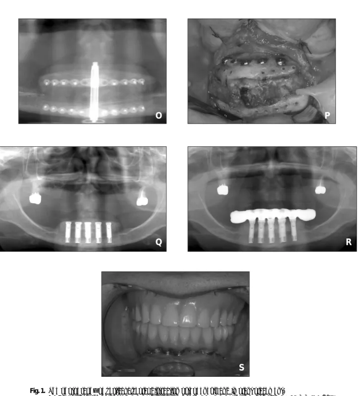
Fig. 1. The photography of vertical alveolar distraction osteogenesis and implant placement.
- F/68, initial bone height : 6.5 mm, distraction length : 10.5 mm, rhythm and rate : 0.25 mm ×3/day A) The appearance of the distraction device that was previously adapted to RP model.
B) After the activation of distraction device as a plan, at this time, ideal vector direction should be checked.
C) Design of incision line at anterior vestibule about 5 mm apart from mucogingival junction.
D) Mucoperiosteal flap dissection, superior and lingual aspect of the segment should endure only mini- mal reflection.
E) Temporary adaptation of pre-formed device at mandible after the activation of 2-3 mm.
F) Indentation using a No. 701 tapered fissure bur under copious saline irrigation.
G,H) After removal of device, a reciprocating saw was used for the osteotomy.
I,J) Osteotomy by small osteotome.
K,L,M) Confirmation of activation degree and vector direction again.
N) Photography after suture with 4-0 vicryl.
O) Radiography after activation and consolidation.
P) Photography of distracted zone.
Q) Five implants are placed in the distracted zone between interforaminal area.
R,S) Panoramic radiography and intraoral photography after fixed prostheses.
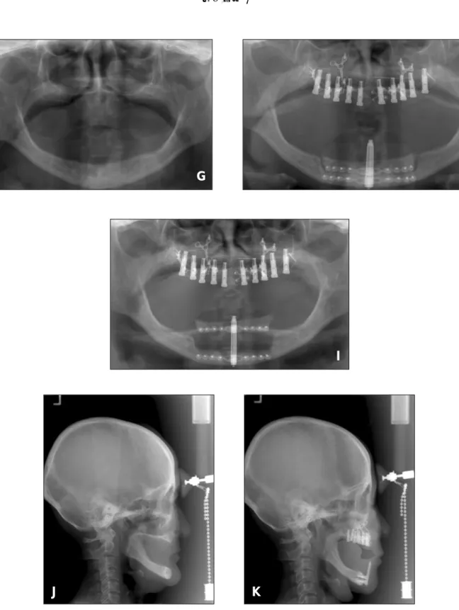
Fig. 2. The photography of LeFort I osteotomy maxillary advancement, both sinus lifting and sandwich bone graft with iliac bone graft and simultaneous implant installation(×10) and vertical alveolar DO of mandible.
- F/61, initial bone height : 14 mm, distraction length : 13 mm, rhythm and rate : 0.50 mm × 2/day A) Incision line designing.
B,C) Photography after both sinus lifting.
D) Photography after downfracture and advancement of maxilla.
E) Photography after sandwich bone graft with iliac bone graft.
F) Guide pin insertion after drilling.
G,J) Preoperative panoramic radiography and lateral cephalogram.
H,K) Postoperative panoramic radiography and lateral cephalogram.
I) Panoramic radiography after 13 mm activation.
사진부도 ①
A B
C D
E F
G H
162
사진부도 ②
I J
K L
M N
사진부도 ③
O P
Q R
S
Fig. 1. The photography of vertical alveolar distraction osteogenesis and implant placement.
- F/68, initial bone height : 6.5 mm, distraction length : 10.5 mm, rhythm and rate : 0.25 mm ×3/day
164
사진부도 ④
A B
C D
E F
사진부도 ⑤
G
I
J K
H
Fig. 2. The photography of LeFort I osteotomy maxillary advancement, both sinus lifting and sandwich bone graft with iliac bone graft and simultaneous implant installation(×10) and vertical alveolar DO of mandible.
F/61, initial bone height : 14 mm, distraction length : 13 mm, rhythm and rate : 0.50 mm × 2/day