This is an Open Access ar ticle distributed under the terms of the Creative Commons Attribution Non-Commercial License (http:
//creativecommons.org/licenses/by-nc/4.0/) which permits unrestricted non-commercial use, distribution, and reproduction in any medium, provided the original work is properly cited.
© 2021 THE KOREAN SOCIETY OF MYCOLOGY.
Accepted: June 24, 2021 Revised: June 23, 2021 Received: April 1, 2021
https://doi.org/10.4489/KJM.20210018 Kor. J. Mycol. 2021 March , 49(2): 183-197
OPEN ACCESS pISSN : 0253-651X eISSN : 2383-5249
RESEARCH ARTICLE
Seven Previously Unrecorded Fungal Species Isolated from Freshwater Ecosystems in
Korea
Jaeduk Goh
*, Hye Yeon Mun, and Yoosun Oh
Fungi Research Team, Nakdonggang National Institute of Biological Resources, Sangju 37242, Korea
*
Corresponding author: [email protected]
ABSTRACT
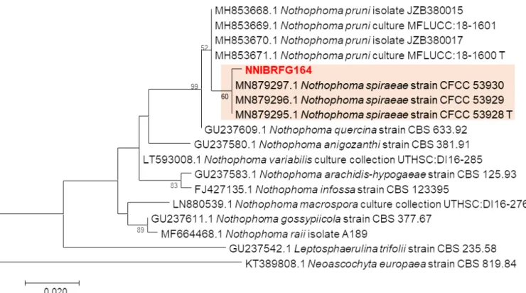
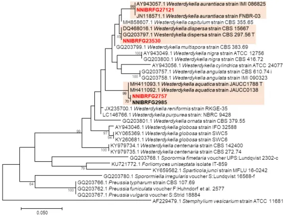
Various freshwater ecosystems, such as streams, lakes and rivers, provide a dynamic habitat for fungi. In this study, we isolated several fungal strains from freshwater sediment and plant litter. The strains were identified using molecular phylogenetic analyses of rDNA and beta tubulin (TUB) sequences. Morphological characteristics of the fungi were also investigated using microscopy and culture characteristics of the fungi grown on several media. We identified seven species previously unrecorded in Korea, Nothophoma spiraeae, Westerdykella aquatica, W. aurantiaca, W. dispersa, Chrysosporium sanyaense, C.pseudomerdarium and Taeniolella phialosperma.
Keywords: Freshwater, Plant litter, Sediment, Unreported species
INTRODUCTION
Freshwater fungi are taxonomically diverse and a polyphylogenetic group that is found in aquatic or semi-aquatic environments [1]. Well-known habitats of freshwater fungi include soil, plant litter such as submerged wood and leaves, and plants and animals of which the exist as endophytes [2]. The main ecological role of freshwater fungi is the degradation of organic materials in water, including plant litter and/
or dead animal tissues. Some of these fungi are pathogens that can cause plant diseases or endophytes living symbiotically on plant tissues [3]. However, many of the biological roles of fungi in aquatic ecosystems, and their physiological and biochemical characteristics remain undetermined. Four fungal genera relative to the current study are Nothophoma, Westerdykella, Chrysosporium, and Taeniolella.
The genus Nothophoma belongs to Didymellaceae, one of the largest families within the order Pleosporales of the phylum Ascomycota. To date, 21 species have been classified under genus [4]. Many species of this genus are reported to be plant pathogens [5]. Members of this genus are characterized by anamorphic stage and conidia morphology [6].
The genus Westerdykella belongs to Sporormiaceae, one of the families in the order Pleosporales. To date,
18 species belonging to this genus have been identified with many being reported to be saprobic isolates
from dung [7,8]. Westerdykella species are known to be a potential bioresource capable of producing
various bioactive substances [7]. Westerdykella species are characterized by their black globose astomatous ascomata, multi-spored asci, and brown ascospores [9].
The genus Chrysosporium belongs to Onygenaceae, one of the largest families of the Ascomycota order Onygenales. Currently, more than 60 species belonging to this genus have been reported [10]. Some species of this genus are isolated from human and animal skin, and exhibited keratinophilic activity [11]. In addition, this genus is commonly found in soils. This genus is classified according to characteristics of an anamorphic stage and its conidia morphology [12].
The genus Taeniolella belongs to Mytilinidiaceae, which is the largest family of Ascomycota order Mytilinidiales. More than 20 species belonging to this genus have been reported to date [13]. Fungal species belonging to this genus come from saprobes lichenicolous taxa and demonstrate a wide range of habits and ecological preferences [14]. Taeniolella is classified based on its morphology of asexual stage - conidiophores, holoblastic conidiogenous cells, and aseptate to pluriseptate conidia formed in acropetal
chains [15].
For the first time, we isolated seven subphylum Pezizomycotina species from environmental samples, Nothophoma spiraeae, Westerdykella aquatica, W. aurantiaca, W. dispersa, Chrysosporium sanyaense, C. pseudomerdarium and Taeniolella phialosperma. The environmental samples included submerged plant litter, and sediment from freshwater environments including rivers, wetland, and streams. Molecular phylogenetic and morphological characteristics of the isolates were investigated.
MATERIALS AND METHODS
Isolation of fungal strains and culture conditions
Fungal strains were collected from plant litter and sediment sampled from freshwater environment. The collection information of all strains identified in this study is listed in Table 1. To isolate fungal strains, plant litter samples were washed with distilled water at least twice, and incubated in a pretreatment liquid medium (0.05% 3-morpholinopropane-1-sulfonic acid [weight/volume (w/v)], 0.05% KNO
3[w/v], 0.025%
KH
2PO
4[w/v], and 0.025% K
2HPO
4[w/v]) at 20℃ for three days. Then, 100 µL of the pretreatment
medium was spread on a 1% water agar plate and incubated at 20 ℃ for two days. Hyphal tips and
germinated conidia were isolated under a microscope and transferred onto a 24-well plate containing V8
agar (V8A; 8% V8 juice [v/v] and 1.5% agar [w/v], adjusted to pH 6.0 using 10 N NaOH) and incubated at
25℃ in the dark. To isolate fungal strains from sediments, a dilution method was used. Diluted suspension
(200 μL) of freshwater sediment and distilled water (1:200 and 1:2,000) were spread on potato dextrose
agar (PDA; 3.9% potato dextrose agar powder [w/v]; Difco, Sparks, MD, USA) containing 50 ppm
streptomycin, and fungal strains were isolated in the pure form after incubation for 4-5 days at 25 ℃ by
repeating this step. All strains identified in this study were grown on malt extract agar (MEA; 2% malt
extract [w/v] and 2% agar [w/v]), oatmeal agar (OA; 7.25% oatmeal agar powder [w/v]; Difco, Sparks, MD,
USA), potato carrot agar (PCA; 2.4% potato carrot agar powder [w/v]; HiMedia, Mumbai, India), corn
meal agar (CMA; 3.9% corn meal agar powder [w/v]; Difco, Sparks, MD, USA), Czapek-dox solution agar (CDA; Difco Sparks, MD, USA), dichloran glycerol chloramphenicol agor (DG18A; Merck Millipore, Billerica, MA, USA), and corn meal dextrose agar (CMDA; 2% cornmeal [w/v], 2% glucose [w/v], and 2% agar), and yeast extract peptone dextrose agar (YPDA; Duchefa Biochemie, Haarlem, the Netherlands).
All strains were preserved at Nakdonggang National Institute of Biological Resources (NNIBR), Sangju, Korea
Morphological analysis
Microstructures of fungal species were observed under an Eclipse Ni light microscope (Nikon, Tokyo, Japan) equipped with a Ds-Ri2 digital camera (Nikon, Tokyo, Japan). At least 50 individuals were examined for observation and measurement of each structure.
DNA extraction, polymerase chain reaction (PCR), and DNA sequencing
Fungal genomic DNA was isolated using a NucleoSpin
®Plant II DNA extraction kit (Macherey-Nagal, Düren, Germany). For molecular identification of the fungi, PCR amplifications were performed for the internal transcribed spacer (ITS) rDNA regions using primers ITS1 (5′-TCCGTAGGTGAACCTGCGG-3′) and ITS4 (5 ′-TCCTCCGCTTATTGATATGC-3′) [16], for the large subunit of rDNA (LSU) using primers LROR (5′-ACCCGCTGAACTTAAGC-3’) and LR7 (5′-TACTACCACCAAGATCT-3′) [17], for the Table 1. Information of strains used in this study.
Species Strain No. Source Collection date Locations (GPS) GenBank acc. no.
Nothophoma spiraeae NNIBRFG164 Plant litter 23-Oct-2015 Bukcheon, Sangju-si, Naeseomyeon, Gyeongsangbul-do
(36°24'2 6.7"N, 128°4'17.8"E) MW841132
aWesterdykella aquatica NNIBRFG2757 Sediment 5-Jun-2016
Seomjingang river, Sina-li, Jinwol-myeon, Gwangyang-si, Junlanam-do
(34°58'5 6.4"N, 127°46'5.1"E)
MW830126
bWesterdykella aurantiaca NNIBRFG27121 Sediment 18-Sep-2019 Maehwamareum Habitat, Choji-li, Gilsang-myeon, Ganghwa-gun, Incheon
(37°38'6"N, 126°31'47"E) MW830128
bWesterdykella dispersa NNIBRFG23530 Sediment 21-Jun-2019
Younggang river, Hwasan-li, Nongam-myeon, Mungyeoung-si, Gyeongsangbuk-do
(36°34'2 9"N, 127°59'33"E)
MW830129
bChrysosporium sanyaense NNIBRFG4460 Sediment 19-Jul-2017 Danjang-cheon, Danjang-myeon, Milyang-si, Gyeongsangnam-do
(35°29'15"N, 128°55'53"E) MW830132
bChrysosporium pseudomerdarium NNIBRFG23908 Sediment 9-May-2019 Sangokcheon, Sangok-li, Taean-gun,
Chungcheongnam-do
(36°46'44"N, 126°19'27"E) MW830130
bTaeniolella phialosperma NNIBRFG5365 Sediment 24-Mar-2018 Wicheon, Yiyeon-li , Danbuk-myeon,
Uiseong-gun, Gyeongsangbuk-do
(36°22'43"N, 128°23'27"E) MW821354
Ca