Coronary Vessel Segmentation by Coarse-to-Fine Strategy using Otsu Algorithm and Decimation-Free
Directional Filter Bank
Tan Dat Trinh*, Thieu Bao Tran*, Le Nhi Lam Thuy*, Ikuko Shimizu**, Jin Young Kim***, Pham The Bao*★
Abstract
In this study, a novel hierarchical approach is investigated to extract coronary vessel from X-ray angiogram.
First, we propose to combine Decimation-free Directional Filter Bank (DDFB) and Homographic Filtering (HF) in order to enhance X-ray coronary angiographic image for segmentation purposes. Because the blood vessel ensures that blood flows in only one direction on vessel branch, the DDFB filter is suitable to be used to enhance the vessels at different orientations and radius. In the combination with HF filter, our method can simultaneously normalize the brightness across the image and increases contrast. Next, a coarse-to-fine strategy for iterative segmentation based on Otsu algorithm is applied to extract the main coronary vessels in different sizes. Furthermore, we also propose a new approach to segment very small vessels. Specifically, based on information of the main extracted vessels, we introduce a new method to extract junctions on the vascular tree and level of nodes on the tree. Then, the window based segmentation is applied to locate and extract the small vessels. Experimental results on our coronary X-ray angiography dataset demonstrate that the proposed approach can outperform standard method and attain the accuracy of 71.34%.
Key words:Coronary vessel segmentation, DDFB, Coarse-to-fine strategy, Otsu algorithm, Hierarchical segmentation approach
* Computer Science Department, Sai Gon University, Vietnam
** Computer Science Department, Sai Gon University, Vietnam
** Tokyo University of Agriculture and Technology, Japan
*** Chonnam National University, Korea
★ Corresponding author [email protected]
※ Acknowledgment
Manuscript received Apr. 16, 2019; revised Jun. 6, 2019; accepted Jun. 18. 2019.
This is an Open-Access article distributed under the terms of the Creative Commons Attribution Non-Commercial License (http://creativecommons.org/licenses/by-nc/3.0) which permits unrestricted non-commercial use, distribution, and reproduction in any medium, provided the original work is properly cited.
Ⅰ. Introduction
Blood vessels in the human body are highly organized and complex structure. Extraction and visualization of blood vessels from X-ray angiogram plays an important role in different medical diagnostic tasks [1]. Specifically, it is necessary and important to measure the blood
vessel width, reflectivity, and abnormal branching so that we can detect vessel disease signs and symptoms such as stenosis, vascular malformation, and atherosclerosis, so on, to determine the treatment therapy. Based on the X-ray angiogram, medical experts can manually detect and delineate the blood vessels. However, this is very slow or even impossible when the number of vessels in 208
an X-ray angiogram is huge or when a large of number of images is required. Therefore, the development of automatic and accurate blood vessels detection and segmentation methods from angiograms is highly necessary.
Coronary vessel segmentation is also one of the most successful applications of medical image analysis and understanding. It is a complex image processing problem in real world applications and has been an active research area during the last few decades. It has proven to be a difficult and challenging task due to weak contrast between the coronary arteries and the background, illumination problem in X-ray angiogram, complex and unknown deformable shape of the vessel tree, and strong overlapping shadows of the bones [1].
There have been a great variety of proposed coronary vessel enhancement and segmentation algorithms. The algorithms and techniques used in coronary angiography can be divided into seven main categories: pattern recognition approaches, model-based approaches, tracking-based approaches, artificial intelligence-based approaches, neural network-based approaches and miscellaneous tube-like object detection approaches [2].
Sarwal and Dhawan [3] proposed a method for obtaining a 3-D description of the arterial tree based on the matching of coronary arteries from multiple views. The simplex based linear programming and relaxation based consistent labeling are applied to skeletons from three views in order to obtain a matching of branches between views. At the lower resolution scale, the stronger vessel tree branches are extracted.
Otherwise the weaker branches are extracted at higher scale. R. Liao et. al. [4] presented a robust method for 3-D reconstruction of the coronary artery tree from multiple ECG-gated views of an X-ray angiography. An enhanced multi-scale approach was applied to extract 2-D coronary artery centerlines from X-ray projection images.
Authors proposed the 3-D symbolic reconstruction
based on an energy minimization problem incorporating a soft epipolar line constraint and a smoothness term evaluated in 3-D. O’Brien and Ezquerra [5] used the temporal, spatial, and structural constraints to segment the coronary vessels in angiograms. First, a low pass filtering is applied to the image as preprocessing. Then, a predefined seed point is initialized. The region-growing approach is applied to find the boundary of the coronary vessels. To reconstruct 3D catheter paths from biplane angiograms, Molina et. al. [6] proposed to applied 3D snakes.
The correction of geometric distortions in both images introduced by the X-ray projections of the vessels is obtained by finding and matching markers affixed to the input screens of both image intensifiers. Then, authors used a ridge detector to extract the catheter in both images.
Petrocelli et. al. [7] introduced the deformable template matcher that is a combination of a priori knowledge of the arterial tree in the form of mathematical templates and a stochastic deformation process described by a Hidden Markov model (HMM). The arterial tree is extracted by deforming the structure model and calculating the likelihood estimate of the deformation. Lu and Eiho [8] proposed a three-step approach for tracing the coronary arterial boundaries with sub-branches from XRAs. First, edged detector is applied by fixed by employing a smoothing differential operator on the scan line perpendicular to the direction of the vessel. Second, branch positions and branches are detected automatically by checking the gray profile on the scan line. Finally, sequential contour tracing achieved by incorporating the features, such as the central point, the searching direction, and the search range, detected from previous step into the next step.
Bombardier et. al. [9] proposed an automatic identification of artery boundaries from angiogram images by two fuzzy segmentation operators to extract different anatomical structures in their
knowledge-based approach using fuzzy set theory. First, authors identified the region of interest (ROI) which is the renal artery. Then, they detect the boundaries of the ROI regions.
Besides, a number of vessel enhancement approaches has been proposed to improve the performance of segmentation results. Poli and Valli [10] proposed to apply a set of linear filters that obtained as linear combinations of properly shifted Gaussian kernels, sensitive to vessels of different orientation and radius. Morphological operators such as erosion, dilation, and top-hat are used to enhance the shape of the artery and remove outliers [11, 12]. Non-linear anisotropic filtering [13, 14] was also applied for enhancing the blood vessels. This method performs anisotropic smoothing without blurring its edge on the local orientation of a vessel. Hessian-based multi-scale filtering [15-18] has been proposed for the vessel enhancement. One advantage of this approach, in this category, is that vessels in a wide range of diameters can be captured due to the multi-scale analysis. In this technique, an input image is filtered by the derivatives of a Gaussian at multiple scales. Then, the Hessian matrix is analyzed at each pixel in the filtered image, to determine the local shape of the structures at that pixel. However, the Hessian-based approaches are highly sensitive to noise, due to the second-order derivatives. Furthermore, they lead to suppress junctions, as junctions are characterized similar to the blob-like structures.
To overcome that problem, Agam et al. [17]
introduced a filter model to avoid the need for second-order derivatives. This model is based on the correlation matrix of the regularized gradient vector. However, when dealing with angiography images, which are noisier and suffer from non-uniform illumination, it shares the same limitations of Hessian-based filters in finding small and low-contrast vessels. In [18] author proposed a new framework for the vessel enhancement by applying the directional
information present in an image. Specifically, the input image is first decomposed by a decimation-free directional filter bank (DDFB) into a set of directional images, each of which contains line-like features in a narrow directional range. The directional decomposition leads to two advantages.
The first is, noise in each directional image can be suppressed compared to that in the original one due to its omni-directional nature. The second is, because one-directional image contains only vessels with similar directions, the Hessian eigenvalue calculation in it is facilitated [18]. Next, distinct appropriate enhancement filters are used to enhance vessels in the respective directional images. Finally, the enhanced directional images are recombined to generate the output image with enhanced vessels. The enhanced experimental results show that this approach is less noise-sensitive, can reveal the small vessel network, and avoid junction suppression.
In this study, a hierarchical approach is proposed to extract coronary vessels in X-ray coronary angiographic image. We apply the vessel enhancement by combining the decimation-free directional filter bank (DDFB) and homographic filtering (HF) to for segmentation purposes. The proposed method can be lower sensitive to noise and enhance significantly the small vessels, and avoid the junction suppression. Furthermore, it can also simultaneously normalize the brightness across the image and increases contrast. Next, the main coronary vessels are segmented in different sizes by using a coarse-to-fine strategy for iterative segmentation based on Otsu algorithm.
Beside, by using information of the main extracted vessels, we propose a new method to extract very small vessels based junctions on the vascular tree and level of nodes on the tree. We use a window analysis approach to detect the small vessel regions. After that, we reduce effect of the main vessels on these regions and segment small vessels based on the DDFB and Otsu algorithm.
Ⅱ. Proposed Method
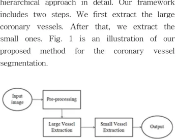
In this section, we describe the proposed hierarchical approach in detail. Our framework includes two steps. We first extract the large coronary vessels. After that, we extract the small ones. Fig. 1 is an illustration of our proposed method for the coronary vessel segmentation.
Fig. 1. An illustration of the proposed method for the coronary vessel segmentation.
The pre-processing procedure is applied to remove small noise and unnecessary areas that do not include coronary vessels areas and also enhance the quality of X-ray coronary angiographic image. Next, we apply a coarse-to-fine strategy for iterative segmentation. First, we segment regions that include the main coronary vessels based on their high contrast. The main coronary vessels include features as vascular tree, junctions and level of vascular tree. After that, we use coarse information that extracted in the previous step to detect the small vessels that often have low contrast and are affected by noise. We describe each step of the proposed technique in detail in the following sub-sections.
2.1. Pre-processing Process
The pre-processing is necessary before the vessel extraction method to be performed. We first apply Gaussian filter to smooth the vessel image and remove small noises. Then, a histogram equalization method [19] is applied to adjust the contrast of the image.
The vessel images are often affected by illumination problem. The boundary between the black background and the foreground causes a
sudden change which can be considered as a vessel when we apply segmentation process in the vessel image. Fig. 2 shows our pre-processing process to remove the background.
Fig. 2. Our pre-processing process.
First, we apply Hough transform [20] to detect the circle boundary between the black background and the foreground. Then, we extract the mask that includes the foreground.
To reduce the effect of a sudden change in brightness intensity from foreground to background, we use foreground diffusion base on the heat equation [21]. The foreground information on the boundary can be diffused through the background.
Fig. 3 shows an example of diffusion on our database.
Fig. 3. An example of diffusion on our atabase.
a) Input image.
b) Boundary between background and foreground based edge detector.
c) Circle mask based on Hough Transform d) Diffused result from input.
2.2. Large Vessel Extraction based on Decimation- free Directional Filter Bank and Coarse-to- fine Segmentation Strategy
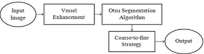
In this section, we proposed a new method to extract large vessels by using decimation-free directional filter bank (DDFB) and coarse-to-fine segmentation strategy. Fig. 4 shows a block diagram of the large vessel extraction.
Fig. 4. Large vessel extraction based coarse-to-fine segmentation strategy.
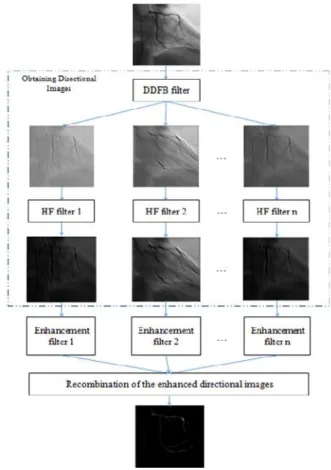
We apply the DDFB and homographic filtering (HF) [18, 19] to X-ray coronary angiographic image for the vessel enhancement. The input image first is decomposed into different directions to obtain directional images by using the DDFB [18]. This leads to some advantages. Because the blood vessel ensures that blood flows in only one direction on vessel branch, the DDFB filter is suitable to be used to enhance the vessels at different orientations and radius. The oriented noise in each directional image will be significantly eliminated compared to that in the input image. Furthermore, this method can reveal the small vessel network and helps to preserve junctions because a one-directional image contains only vessel with similar directions [18]. Besides, to reduce the effect of non-uniform illumination, we apply the homographic filtering on the directional images. This helps to suppress the low-frequency component (the illumination component) while enhance the high-frequency one (the reflectance component) and simultaneously normalize the brightness across the image and increases contrast. It also provides us a better control on the parameters of individual homomorphic filter for each directional image. Next, each directional image is enhanced in its directional range and is called the enhanced directional image. Finally, the enhanced directional images are by simply summing all directions to yield the final filtered image. Fig. 5 shows a block diagram of the vessel enhancement based on the DDFB and HF filter. There are three steps:
obtaining directional images, vessel enhancement, and recombination of the enhanced directional images.
For vessel enhancement, a vessel model is represented as a tube with a Gaussian profile across its axis, which is identical to the x-axis [18].
πσ
σ
(1)
where C is structureness measurement, σ is a Gaussian of standard deviation. Assume that the directional image … corresponds to the orientations ranging from m in to m ax (followed counterclockwise angle). We can transform the coordinate of each to new coordinate ′′ by rotating an amount as large as the mean value .
min max
(2)
Therefore, the Hessian matrix of in the new coordinates ′′ is described as ′
′
∂′∂∂′′
∂
∂′′
∂
∂′
∂
(3)where
∂′
∂
∂
∂
θ ∂∂
∂
sinθ
∂
∂
θ (4)
∂′
∂
∂
∂
θ ∂∂
∂
sinθ
∂
∂
θ (5)
∂′∂′
∂
∂
∂
θ
∂∂
∂
θ
∂
∂
θ (6)
The Hessian eigenvalues are then defined by the diagonal values of ′. These values described
through the following equations
and
σ σ
′ σ σ
′′ , (7)
where σ is the standard deviation of the Gaussian kernel used in the multi-scale analysis. Because the vessel axis is not, in general, identical to the x′- axis and so . Inside the vessel, ′
σ σ, and.
≪ . To distinguish background pixels from others, we define a structureness measurement
(8)
Because the background has no structure and small derivative magnitude, so the value of C should be small. The vessel filter output can be defined as [15]:
ϕσ ηexp
β
exp
γ
(9)where ′ ′,
, β and γ are constants, and
i f ≥ i f (10) The vessel filter is analyzed at various scales σ . When the scale matches the size of the vessel, the filter response will be maximum.Therefore, the enhanced vessel filter response is
maxϕ (11)
σϵ
The enhanced vessel filter in Eq. 11 is applied to one-directional image to enhance vessel structures in it. Finally, the enhanced directional images are by simply summing all directions to yield the final filtered image.
∑ Φ (12)
After the vessel enhancement, we apply the Otsu algorithm [22] to segment the main coronary vessels. Furthermore, to extract the main vessels at different sizes and lengths, we propose to coarse-to-fine segmentation strategy by applying iterative Otsu algorithm segmentation.
In our experiments, we use three iterations for coarse-to-fine segmentation strategy.
Fig. 5. An block diagram of the vessel enhancement based on the DDGFB and HF filter.
Fig. 6 shows an example of coarse-to-fine segmentation strategy based on Otsu algorithm to extract the large vessels. From Fig. 6, we realize that the large vessels can be extracted.
However, there exists some noise and false alarm in Fig .6. To overcome this problem, we use a post-processing to reduce the false alarm to obtain the clearly large vessels by removing small dots based their sizes. Furthermore, we also apply the neighborhood relationship to remove noise. Specifically, we first choose the largest connected component in the output binary
image. Then, we use neighborhood relationship of the largest object to find neighborhood components based distance between them.
Fig. 6. An example of coarse-to-fine segmentation strategy based on Otsu algorithm to extract the large vessels.
a) Input image, b) Segmentation result.
2.3. Small Vessel Extraction based on Level of the Vessel Tree and Local Segmentation Approach.
In previous section, we represent the approach to extract the main vessels. However, it cannot reveal well small vessels due to their blurring and low contrast compared with the background.
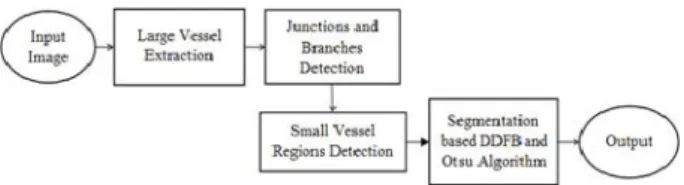
To solve this problem, we use information of the main extracted vessels and propose a new method to extract junctions on the vascular tree and level of nodes on the tree. Then, we apply a window analysis approach to detect the small vessel regions. After that, we reduce effect of the main vessels on these regions and segment small vessels based on the DDFB and Otsu algorithm. Fig. 7 shows a block diagram of our method for small vessel extraction.
Fig. 7. A block diagram of our method for small vessel extraction.
The branching geometry and junction of the blood vessel tree pose a challenge in applying coarse-to-fine Otsu segmentation framework to
vessel segmentation. We introduce a new method to construct the vessel tree based on the branch and junction detection. The approach is summary in Algorithm 1.
Algorithm 1. Branch and junction detection Input:
Binary image after applying large vessel extraction
Output:
Branch and junction information of the vessel tree Level of the vessel tree and degree of nodes
Step 1. Find the center-line of vessels. For each point in the centerline, construct a circle S of radius R.
Step 2. Connect the nearest the center-lines.
Step 3. Cluster the center-lines into the same vessel branch.
Step 4. Determine level of the vessel tree and degree of nodes
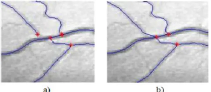
Each individual vessel can be modeled by a center-line and a radius function defined at each point of this center-line [23]. The centerline of the blood vessel includes several small segmented lines … . Each small line includes set of points … . All the points in … will generate seed point, end point and branch points of each center-line. Based on the segmentation results, in some case the branch points are miss-classified as the seed point or the end point. To overcome this problem, we use neighborhood relationship to correct the branch point by connecting the nearest the center-lines. Fig. 8 illustrates an example of connecting the nearest the center-lines.
In X-ray angiogram, there are a lot of vessel branches. Our aim is to distinguish each vessel branch. We need to cluster the center-lines into the same vessel branch. For each center-line, we first choose a seed point that has highest radius value. From the seed point, we trace on the center-line of the vessel to find the next candidate point. At the current point, if there are one or two neighborhood points, these points are
Fig. 10. An example of eliminating the invalid branches.
In this case, the branches corresponding to directions and are eliminated.
considered as next candidate points that lie on the same vessel branch with the seed point, and the point that have two neighborhood points is considered as the joint point or junction. Fig. 9 shows an example of tracing the vessel branch and junction.
Fig. 8. An example of connecting the nearest the center-lines.
a) Input image,
b) Image after connecting the nearest the center-lines
Fig. 9. An example of tracing the vessel branch and junction.
a) The seed point,
b) Tracking candidate points and the junction
Otherwise, if the current point has more than two neighborhood points, we will eliminate invalid points based on angle information. Specifically, we assume that the current point A has three neighborhood points. At point A, we determine its direction as a vector and three branches at A have directions as , and , respectively.
We assume that α is angle between and and β is angle between and as shown in Fig. 10.
Because the blood vessel ensures that blood flows in only one direction on vessel branch, we can eliminate the direction because the value α is large. Furthermore, because the value of b
is smaller than α, we can eliminate the direction
(the direction and may be belongs to the same vessel branch). Finally, we can select
as a suitable branch.
After determining exactly the information of the vessel branches, we need to level of the vessel tree and degree of nodes. Using the center-line information of vessel branch, we first select a seed point on each branch. The seed point has level 0 and lies on the segment line 1.
Next, we trace to the end point of line 1. If the end point of line 1 has no next neighborhood point, it is consider as leaf node and has level 1.
Otherwise, if the end point of the line 1 has next neighborhood point, it is set to level 1 and is a start point of the segment line 2. This process is repeated until the final point in vessel branch is reach. Fig. 11 shows an example of the level of the vessel branch and degree of nodes. Fig. 12 represents the level of the vessel tree on our dataset.
Fig. 11. An example of the level of the vessel branch and degree of nodes.
Fig. 12. An example of the level of the vessel tree and degree of nodes on our dataset
In this study, we propose a method to extract small vessels based on window analysis. Usually, the small vessels from X-ray angiogram are blurring and have low contrast. It is not easy to extract large and small vessels simultaneously.
So, a local segmentation approach is possible to be used to extract the small ones. The proposed approach is summary in Algorithm 2.
Algorithm 2. Small vessel detection and segmentation based on window analysis.
Input:
Binary image after applying large vessel extraction Level of the vessel tree and degree of nodes Output:
Small vessel segmentation result While node i Î set of nodes
Step 1. Create a window that include node i and father of node i
Step 2. Enhance the contrast of small vessels in the window.
Step 3. Apply DDFB-based enhancement and Otsu algorithm to obtain the results
Based on idea from local thresholding, we apply a local window analysis to segment the small vessels. This helps reduce the effect of changing in gray-scale value between the vessels and the background compared to global approach.
For each node in the vessel tree, we will construct a window between node i and its father node, fi.
The width (w) and height (h) of the window are described by
, (13)
. (14)
In our experiment, we chose bias of 20 pixels so that we can obtain small vessels near to node i. Fig. 13 describes an example of a local window that includes small vessels. In the local window, a part of large vessel that has high intensity and high contrast to the background compared to small vessels can be included. Thus, we remove the effect of the large vessel in the window, and then apply contrast adjustment to areas that include the small vessels. Fig. 14 shows a contrast enhancement of in the window. After that, we apply the DDFB-based approach and Otsu method to segment the small vessels. Fig.
15 presents a segmentation result of small vessels. Finally, combining the large and small vessels extraction approaches, we can obtain the final results.
Fig. 13. An example of window constructed between two nodes.
Fig. 14. An example of contrast enhancement in the window analysis.
a) Input image,
b) Result from contrast enhancement
Fig. 15. Small vessel extraction.
a) Part of input image,
b) Result from large vessel extraction approach, c) Result from small vessel extraction approach
Ⅲ. Experiment Results
3.1. Dataset
The performance of the proposed method is evaluated using the X-ray angiogram dataset which was collected and supported from a hospital at HCM city, Vietnam. The database contains 48 different vessel images corresponding to two categories: Ds1 and Ds2. The size of each image is 512 x 512 pixels, with 256 grey levels per pixel. The Ds1 dataset includes 20 images that obtain a direct front view of the coronary vessels. The Ds2 dataset includes 28 images that are taken from four different angles of the coronary vessels. Fig. 16 shows an image in our dataset.
Fig. 16. An image IM-0001-000130 in our dataset.
In our experiments, we use software MIPAR of John Sosa [24] to create ground truth in order to evaluate the performance of the proposed algorithm.
Furthermore, to evaluate the efficiency of our technique; we calculate the dice similarity coefficient [25] between binary segmentation result and the
ground truth. Fig. 17 presents a ground truth of the image IM-0001-000130 in our dataset. Our experiments are implemented on an Intel Core i7-3520M, CPU @ 2.9GHz with 4.0 GB. The average run-time of the proposed algorithm to apply to each image is 17 minutes. We use a 16-band Decimation-Free Directional Filter Bank (DDFB) was for the enhancement purpose. Based on the experiment, we analysis and evaluate the effect of various parameters on DDFB. Table 1 shows optimal parameters for the DDFB in our experiments.
Fig. 17. Ground truth of the IM-0001-000130 image.
Table 1. The optimal parameters for the DDFB in our experiments.
Parameters Values Description
σ Standard deviation of Gaussian kernel
β Adjusting constants
15 Structureness
measurement
γ 1 Parameter to normalize
derivatives of the image.
3.2. Results
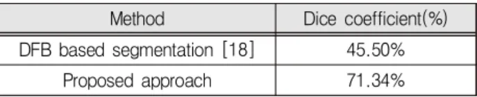
In our experiments, we investigate performance of the coronary vessel segmentation based on the coarse-to-fine strategy for iterative segmentation and comprising with baseline case and another approach. Fig. 18 shows a segmentation result of the proposed approach on our dataset. We found through experimental analysis that our proposed method can extract significantly the large coronary vessels. The performance of large coronary vessel segmentation attains 75.53%. Furthermore, the proposed approach can reveal a large number of small
vessels. However, there are certain disadvantages in small vessels segmentation case. Some vessels are very small and their intensities are similarity to the background, so the proposed approach cannot detect them. Besides, the approach can obtain some false alarms due to the chosen of parameters in the DDFB and number of iterations in coarse-to-fine strategy. Finally, the performance of coronary vessel segmentation based on our method obtains 71.34%. Fig. 19 shows the false alarms case from the proposed approach on our dataset. We also compare the performance of the proposed approach with the well-known technique in [18] on our database. Fig. 20 shows a comparison of segmentation results on our dataset. Table 2 presents a summary of comparison performance of the coronary vessel segmentation in terms of dice coefficient.
Fig. 18. Segmentation result of the proposed approach on our dataset. a) Input image, b) Segmented vessels, c) Ground truth
Fig. 19. Some false alarms of the proposed approach on our dataset. a) Input image
b) False alarms from segmented result, c) Ground truth
Fig. 20. Comparison of the segmentation results on our dataset a) Input image, b) Our result, c) Result in [18]
Table 2. Comparison performance of the coronary vessel segmentation on our dataset.
Method Dice coefficient(%) DFB based segmentation [18] 45.50%
Proposed approach 71.34%
From Fig. 20 and Table 2, it may be observed that our method using the hierarchical approach and coarse-to-fine strategy obtains better segmentation results and outperforms the DFB based approach.
Looking at the left side in the images in Fig. 20, we can realize that the DFB based segmentation [18] leads to more artifacts and fails to correctly enhance small vessels compared to our approach.
Also, it cannot detect the small vessels that have low intensity and miss parts of large vessels.
This method detects the large and small vessels at the same time, so when there is a large difference intensity between the large vessels (that are high contrast objects) and the small vessels (that are low contrast objects), the DFB based method cannot significantly to extract small vessels.
Our method is proposed to overcome that problem by separately detecting the large vessels and small vessels based hierarchical technique.
Because we observe that the large vessels always have high contrast to the background than the small vessels and the blood vessel ensures that blood flows in only one direction on vessel branch, the DDFB filter is suitable to be used to enhance the vessels at different orientations and radius. The coarse-to-fine strategy based segmentation guarantees that the method can correctly enhance and extract the large vessels.
In the small vessel extraction stage, we first reduce the effect of large vessels and make a contrast enhancement on the window that includes small vessels regions. This deals to low contrast problem on small vessels regions. When the small vessels regions are increased the contrast, they were easily detected and segmented. This is significantly to extract small vessels.
The experimental results show that our method overcomes the limitations of the DFB based method such as small vessels intensity and noise sensitivity. It also performs better on real angiography images. So, the proposed method is one of the promising methods for enhancing the performances of the coronary vessel segmentation.
Ⅳ. Conclusion
In this study, we have proposed an improved coronary vessel segmentation technique by combining the decimation-free directional filter bank and homographic filtering in order to enhance X-ray coronary angiographic image for segmentation purposes. Further, we have also represented a coarse-to-fine strategy for iterative segmentation.
Through experiments results, it has been confirmed that our proposed method can enhance the performance of the vessel segmentation. In the future, we intend to improve the computation cost of the proposed method and explore an extended method to deal with 3D images.
References
[1] M. T. Dehkordi, S. Sadri, and A. Doosthoseini,
“A review of coronary vessel segmentation algorithms,” Journal of Medical Signals and Sensors, vol.1, no.1, pp.49-54, 2011.
[2] C. Kirbas and F. Quek, “A review of vessel extraction techniques and algorithms,” ACM Computing Surveys, vol.36, no.2, pp.81-121, 2004.
DOI: 10.1145/1031120.1031121
[3] A. Sarwal and A. Dhawan, “3-D reconstruction of coronary arteries,” Annual International Conference of the IEEE Engineering in Medicine and Biology Society, vol.1, pp.504-505, 1994.
DOI: 10.1109/IEMBS.1994.411932
[4] R. Liao, D. Luc, Y. Sun, and K. Kirchberg, “3-D reconstruction of the coronary artery tree from multiple views of a rotational X-ray angiography,”
The International Journal of Cardiovascular Imaging, vol.26, no.7, pp.733-749, 2010.
DOI: 10.1007/s10554-009-9528-0
[5] J. O’Brien and N. Ezquerra, “Automated segmentation of coronary vessels in angiographic image sequences utilizing temporal, spatial and structural constraints,” Visualization in Biomedical Computing, Rochester, 1994.
DOI: 10.1117/12.185183
[6] C. Molina, G. Prause, P. Radeva, and M. Sonka,
“3-D catheter path reconstruction from biplane angiograms,” in SPIE, vol.3338, pp.504-512, 1998.
DOI: 10.1117/12.310929
[7] R. R. Petrocelli, J. Elion, and K. M. Manbeck,
“A new method for structure recognition in unsubtracted digital angiograms,” in IEEE Computers in Cardiology, pp.207-210, 1992.
DOI: 10.1109/CIC.1992.269410
[8] S. Lu and S. Eiho, “Automatic detection of the coronary arterial contours with sub-branches from an x-ray angiogram,” IEEE Computers in Cardiology, pp. 575-578, 1993.
DOI: 10.1109/CIC.1993.378337
[9] V. Bombardier, M. C. Jaluent, A. Bubel, and J.
Bremont, “Cooperation of two fuzzy segmentation operators for digital subtracted angiograms analysis,”
IEEE Conference on Fuzzy Systems, vol.2, pp.
1057-1062, 1997. DOI: 10.1109/FUZZY.1997.622856 [10] R. Poli and G. Vall, “An algorithm for real-time vessel enhancement and detection,”
Comput Meth Prog Biomed, vol.52, no.1, pp.1-22, 1997. DOI: 10.1016/S0169-2607(96)01773-7
[11] S. Eiho and Y. Qian, “Detection of coronary artery tree using morphological operator,” Proc.
IEEE Comput. Cardiol, pp.525-528, 1997.
DOI: 10.1109/CIC.1997.647950
[12] F. Zana and J. C. Klein, “Segmentation of vessel-like patterns using mathematical morphology and curvature evaluation,” IEEE Trans Image Process, vol.10, no.7, pp.1010-1019, 2001.
DOI: 10.1109/83.931095
[13] C. Yan, S. Hirano, and Y. Hata, “Extraction of blood vessel in CT angiography image aided
by fuzzy logic,” Proc. IEEE Int. Conf. Signal Processing, pp.926-929, 2000.
DOI: 10.1109/ICOSP.2000.891673
[14] M. Orkisz, C. Bresson, I. Magnin, O. Champin, and P. Douek, “Improved vessel visualization in MR angiography by nonlinear anisotropic filtering,”
MagnReson Med, vol.37, no.6, pp.914-919, 1997.
DOI: 10.1002/mrm.1910370617|
[15] A. Frangi, W. Niessen, K. Vincken, and M.
Viergever, “Multiscale vessel enhancement filtering,”
Medical Image Computing Computer-Assisted Intervention. Lect Notes ComputSci, vol.1496, pp.130-137, 1998. DOI: 10.1007/BFb0056195 [16] C. Lorenz, I. C. Carlsen, T. Buzug, C.
Fassnacht, and J. Weese, “A multiscale line filter with automatic scale selection based on the Hessian matrix for medical image segmentation,”
Proc. Scale-Space Theories in Computer Vision, LNCS, vol.1252, pp.152-163, 1997.
DOI: 10.1007/3-540-63167-4_47
[17] G. Agam, S. G. Armato, C. Wu, “Vessel tree reconstruction in thoracic CT scans with application to nodule detection,” IEEE Trans Med Imaging, vol.24, no.4, pp.486-499, 2005.
DOI: 10.1109/TMI.2005.844167
[18] P. T. H. Truc, M. A. Khan, Y. K. Lee, S.
Lee and T. S. Kim, “Vessel enhancement filter using directional filter bank,” Computer Vision and Image Understanding, vol.113, no.1, pp.101-112, 2009. DOI: 10.1016/j.cviu.2008.07.009
[19] R. Gonzalez, R. Woods, Digital Image Processing, Prentice Hall, New Jersey, USA, 2002.
[20] H. K. Yuen, J. Princen, J. Illingworth, and J.
Kittler, “Comparative study of Hough Transform methods for circle finding,” Image and Vision Computing, vol.8, no.1, pp.71-77, 1990.
DOI: 10.1016/0262-8856(90)90059-E
[21] N. Huynh, “A filter bank approach to automate vessel extraction with applications,” Master Thesis, University of California, 2013.
[22] N. Otsu, “A threshold selection method from gray-level histograms,” IEEE Transactions on Systems, Man, and Cybernetics, vol.9, no.1, pp.
62-66, 1979. DOI: 10.1109/TSMC.1979.4310076 [23] V. Mohan, el. al., “Vessel segmentation with automatic centerline extraction using tubular tree segmentation,” Workshop on Cardiovascular Interventional Imaging and Biophysical Modeling, pp.1-8, 2009.
[24] J. Sosa, “MIPAR - Premier Image Analysis
& Image Segmentation Software,” [Online]. Available:
https://www.mipar.us/.
[25] K. H. Zou, el. al., “Statistical validation of image segmentation quality based on a spatial overlap index,” Academic Radiology, vol.11, no.2, pp.178-189, 2004.
DOI: 10.1016/S1076-6332(03)00671-8
BIOGRAPHY
Tan Dat Trinh (Member)
2010:BS degree in Mathematics and Computer Sciences fm
HCMC University of Natural Sciences.
2013: MS degree, in Dept. of Electronics Eng, Chonnam National University
2017:Ph.D degree, in Dept. of Electronics Eng, Chonnam National University
2018~now:Lecturer at Computer Science Department, Sai Gon University, Vietnam
Research interests:speaker recognition, computer vision, machine learning and pattern recognition.
Thieu Bao Tran (Member)
2012~2015 and 2016-2017:BS. Honours Programme, Faculty of Mathematics & Computer Science, University of Science, Ho Chi Minh City, Vietnam
2015~2016:Short-term Exchange Program, in Tokyo University of Agriculture and Technology, Japan 2017 to 2018:Teaching Assistant, in Ho Chi Minh City University of Science, Vietnam
10/2018~now:Research Engineer in Trustring Social, Vietnam
Research interests: Image processing, artificial intelligence and data science
Le Nhi Lam Thuy (Member)
2004:Bachelor of Science, degrees in University of Natural Science 2009:Master of Science, degrees in VietNam National University 2005~2/2018: Lecturer of the Department Computer Science, Ho Chi Minh City College of Economics, Vietnam.
2/2018~now:Lecturer of the Department Computer Science, Faculty of Information Science, Sai Gon University, Hochiminh city, Vietnam.
Ikuko Shimizu (Member)
Ikuko Shimizu received the B.S., M.S., and Ph.D degrees in Mathmatical Engineering and Information Phisycs from the University of Tokyo. She is an associate professor of Tokyo University of Agriculture and Technology.
Her research interests include basic algorithms and applications of computer vision.
Jin Young Kim (Member)
1986:BS degree, in Dept. of Electronics Eng, Seoul National University
1988:MS degree, in Dept. of Electronics Eng, Seoul National University
1994:Ph.D degree, in Dept. of Electronics Eng, Seoul National University
1995~now:Professor in Department of Electronics and Computer Engineering, Chonnam National University Research interests: Digital Signal Processing, Image Processing, Speech Signal Processing, ML, DL
Pham The Bao (Member)
1995:BS., 2000:MSc., 2009: Ph.D.
degrees in University of Science 1995 to 11/2018: Lecturer(1995~2013) and Professor(2013~2018) of the Department Computer Science, Faculty of Mathematics & Computer Science, University of Science, Hochiminh city, Vietnam.
11/2018~now:Professor of the Department Computer Science, Faculty of Information Science, Sai Gon University, Hochiminh city, Vietnam.
Research interests:Image processing & pattern recognition, intelligent computing