Inhibition of Cell Growth by Anoikis in Various Human Cancer Cell Lines Treated with an Extract of Smilax china L.
Min-Jae Kim1, Hyeon-Ji Kim1, Moo-Gyeong Kim1, Sung-Ho Lee2 and Byeong-Gyun Jeon1,3*
1Department of Biology Education, Gyeongsang National University, Jinju 52828, Korea
2Division of Life Science, Gyeongsang National University, Jinju 52828, Korea
3Institute of Education, Gyeongsang National University, Jinju 52828, Korea
Received September 21, 2020 /Revised November 18, 2021 /Accepted November 30, 2020
The present study examined the cytotoxic effects of a Smilax china L. extract (SCLE) in human cancer (A-549, MCF-7, MDA-MB-231, U87-MG, AGS, MKN-74, and SNU-601) and normal MRC-5 fibroblasts, as well as in mesenchymal stem cells derived from dental tissue (DSC). The 50% inhibitory concen- tration (IC50) values for SCLE were significantly (p<0.05) lower in the cancer cell lines (A-549, MCF-7, MDA-MB-231, U87-MG, AGS, MKN-74 and SNU-601) than in the MRC-5 and DSC cells. Cell growth was significantly (p<0.05) more inhibited in the cancer cell lines treated with 200 μg/ml SCLE than in the normal MRC-5 and DSC, and anoikis-like floating cell morphology was observed in the SCLE-treated cancer cells. The cells detached by SCLE treatment were retrieved daily and assayed for viability and telomerase activity. Cells retrieved at 4 days showed significantly decreased viability and telomerase activity (p<0.05), as well as apoptosis-like abnormal morphology, when compared to cells retrieved in the previous 3 days. The ratio of apoptosis and cells in the G1 phase was significantly (p<0.05) increased in the A-549, AGS, and MCF-7 cancer cells treated with SCLE for 4 days compared to untreated controls. However, after SCLE treatment, cell adhesion was not increased by application of an inhibitor of the associated protein kinase (ROCK) that mainly contributes to the increase in cell attachment. This suggests that the cellular detachment by SCLE is probably controlled by a Rho-in- dependent mechanism(s). These observations indicate that SCLE readily induces anoikis in cancer cells and could serve as a potent agent for cancer chemotherapy.
Key words : Anoikis, cancer cells, human, Smilax china L. extract
*Corresponding author
*Tel : +82-55-772-2236, Fax : +82-55-772-2239
*E-mail : [email protected]
This is an Open-Access article distributed under the terms of the Creative Commons Attribution Non-Commercial License (http://creativecommons.org/licenses/by-nc/3.0) which permits unrestricted non-commercial use, distribution, and reproduction in any medium, provided the original work is properly cited.
Introduction
The benign tumor is a cell mass that is caused by abnor- mal cell growth, and can be easily removed by surgical treatment. However, the tumor cells are converted to cancer cells by the more accumulation of the cellular alteration, such as genetic modification with carcinogen. Thus, cancer cells are generally characterized by an un-controlled cell growth and cell division, and the overgrowing cancer cells can form a huge cell mass in the tissue. Further, the cancer cells can invade into neighboring tissues or spread into oth- er new secondary tissues of the host body, as per blood- stream or lymph system, as called to metastasis, and the metastasized and invaded cancer cells are survived in the
secondary tissue, unlike normal cells and tissues [16]. The high mortality rates by various types of cancer diseases are generally associated with tissue invasion by the metastasis of cancer cells [17]. The cellular and molecular mechanisms converting into the cancer cells with metastasis capacity from primary tumor cells are still unclear, however, the processes of metastasis are included in loss of cell-to-cell interaction and adhesion, increased cell motility and in- vasion into surrounding tissues and circulation system, and high survival and proliferation of the moved cancer cells in the new tissue sites [43].
The adhesion of cells to neighboring cells and tissues, basement membranes or extracellular matrix (ECM) is in- evitably necessary for normal cell growth of most of the animal cells, including human. The cell adhesion is mainly mediated with integrin proteins on the cell surface and actin filament via Rho signal pathway and high expression of Rho activity is induced to rounded cell shape by loss of cell adhesion [12]. However, the inappropriate cell adhesion by loss of cell contact mediated by integrin and actin fila- ment leads to cellular apoptosis, as called anoikis [12, 13].
The cell death by anoikis in the other tissues is important to remove the cells which are located in incorrect position.
While the tumor cells are gradually changing into the malig- nant cancer cells, the cells acquire the capacity to resist anoi- kis cell death and survive at circulation system and metasta- sized to others sites without undergoing anoikis cell death after detachment from their primary site [41]. And the cell death by anoikis pathway is also induced by intrinsic and extrinsic pathway, and BCL-2 family and cytochrome C in mitochondria are key proteins [36]. It has been demon- strated in comparison to normal somatic cells, cancer cells are resistant to anoikis induced cell death [23, 37]. However, the attachment of malignant cancer cells with resisting anoi- kis is also necessary for their growth and survival in the metastasized new site or tissue, and the detached and float- ed cancer cells results in the cell death by anoikis during cancer treatment. However, chemicals and drugs induced anoikis cell death is one of the example among various can- cer chemotherapies [23, 37].
Since thousands of years, plants and herbs derived nu- merous compounds and drugs have been used to treat vari- ous human diseases, including cancer. It has been widely observed that glycoside compounds e.g. amygdalin (cyano- genic glycoside) and saponins are useful chemicals for che- motherapy treatment and prevention of different kinds of cancer. Moreover, natural plants derived various types of polyphenols or taxols have also been used as anticancer agents for treating cancer [4, 26, 34]. Smilax china L. is a climbing plant species that grows in some parts of east Asia, including Korea, China, Taiwan and Japan. Its root extracts have traditionally been prescribed as an alternative drug for treatment of rheumatic arthritic and inflammatory dis- eases [10]. Recently, Smilax china L. extract (SCLE) have shown anti-tumor effects by inhibiting nuclear factor-κB in A2780 ovarian cancer cells [20]. Further, it was suggested that inhibition of cancer metastasis is probably induced by modulation of ECM degradation in MDA-MB-231 breast cancer cells [35]. However, the anti-tumor and/or anti-meta- stasis effects by treatment of SCLE in various human cancer and normal cells are still not fully investigated.
To further evaluate the anti-tumor effects of SCLE, the human cancer and normal cell lines derived from various origins were exposed to Smilax china L. extract, and the con- centration of treatment was determined with the half max- imal inhibitory concentration (IC50) values. Under estab- lished concentration of treatment, the effect of the extracts
on the fundamental cell properties, including cell pro- liferation, cell morphology status and telomerase activity was investigated in the human normal and cancer cell lines of various origins. Further, the frequency of cellular apopto- sis and cell cycle phase was investigated by flow cytometry after exposure of SCLE. And, the effects of Rho signal path- way that most importantly regulates cell adhesion, was also examined in the cancer cells after administration of SCLE.
Materials and Methods
Culture and treatment of cells
All media and chemicals for cell culture were purchased from Thermo Fisher Scientific (USA) and Sigma Chemical Company (USA), unless otherwise specified. The cancer cell lines, including A-549 lung adenocarcinoma, MCF-7 breast adenocarcinoma, MDA-MB-231 breast adenocarcinoma, U87- MG brain glioblastoma astrocytoma, AGS stomach ad- enocarcinoma, MKN-74 stomach adenocarcinoma, SNU-601 stomach carcinoma cancer cell lines and normal MRC-5 fetal lung fibroblasts were purchased from the American Type Culture Collection (USA). Additionally, mesenchymal stem cells derived from papilla and pulp tissues of the third mo- lar tooth (DSC) at passage 5 were also used as per pre- viously described studies [21, 22, 30]. All types of cells were cultured in advanced-Dulbecco's modified eagle medium (A-DMEM) supplemented with 3% fetal bovine serum (FBS) and 1.0% penicillin (10,000 IU/ ml)-streptomycin (10,000 μg/
ml). The SCLE used in this research was obtained from Korea Plant Extract Bank at the Korea Research Institute of Bioscience and Biotechnology (Daejeon, Korea) and the extract was dissolved at 1 mg/ml in dimethyl sulfoxide (DMSO) and each treatment concentration was freshly pre- pared by adding extract stock in basic culture medium. The cell culture for each treatment was accomplished in a hu- midified atmosphere of 5% CO2, incubator at 37.5℃.
Determination of IC50 by MTT assay
The values of half maximal inhibitory concentration (IC50) against SCLE were estimated by 3-(4,5-dimethyl-2-thia- zolyl)-2,5-diphenyl-2H-tetrazolium bromide (MTT) assay in the cancer and normal cell lines of various origins. In Brief, each of normal and cancer cells were implanted into a 6-well plate at the density of 5×10⁴ cells per well using com- plete A-DMEM media containing 0 (control), 25, 50, 100, 200 and 300 μg/ml SCLE and cultured at 37.5℃ in a hu-
midified atmosphere of 5% CO2 in air for 7 days. The treat- ed cells were rinsed with Dulbecco's phosphate buffered saline (D-PBS) for 3 times, and 1 ml of 5 mg/ml MTT stock solution was added into each well for formation of for- mazan by mitochondrial reductase. Following incubation at 37℃ for 4 hr, MTT stock solution was removed and each well was again washed thrice with D-PBS, the crystalized formazan was resolved with 300 μl DMSO for 15 min at room temperature. After retrieval of resolved formazan into a new read plate, intensity of formazan was measured with an ELISA micro plate reader (Perkin Elmer, USA) at 570 nm wavelength. The survival rate of untreated controls was calculated as 100% for comparison with the each treatment group of at least three replications, and the IC50 value was then calculated in each of cell lines.
Analysis of growth inhibition by PDT
The effect of SCLE on cell growth and proliferation was analyzed with the evaluation of population doubling time (PDT) assay using cell counting in the cancer and normal cell lines of various origins. Following determination of IC50
value, the treatment concentration of the SCLE was de- termined as 200 ug/ml in treatment groups, compared to those of untreated controls. The each of normal and cancer cells were transplanted at the cell density of 1×10⁴ cells/well into a 6-well plate containing basic A-DMEM cell culture media supplemented with 0 μg/ml (untreated control) and 200 μg/ml of SCLE, respectively, and cultured at 37.5℃
in a 5% CO2 incubator for 7 days with changing each cell culture media every 3 days. After treatment of SCLE in the each media, cells were harvested with 0.25% trypsin EDTA solution treatment and the cell number was measured with a hemocytometer. The PDT was calculated following for- mula: PDT = duration × log(2)/log (final concentration)−
log(initial concentration).
Analysis of cell morphology and viability in detached cells
Following administration of SCLE, the cells were exam- ined under an inverted microscope (Nikon, Japan) equipped with CCD image system. Most of the cells were not attached to the culturing plate and were floating. And the viability of the detached cells was important for the evaluation of treatment day(s) along with determination of IC50 value and analysis of growth inhibition. The viability of detached and floated cells was investigated in the A-549, AGS and MCF-7
cancer cell lines. After treatment with SCLE, the detached and floated cells were daily harvested up to 6 days, and the cells were cultured in the fresh medium. And the cell viability along with exposed day(s) was then carried out with MTT assay, as described above.
Analysis of TERT expression and telomerase activity The expression level of telomerase reverse transcriptase (TERT) related to telomerase activity was analyzed by re- verse transcription polymerase chain reaction (RT–PCR) assay in both the untreated control and SCLE-treated A-549, AGS and MCF-7 cancer cells. Briefly, the total RNA was extracted with Ribospin extraction kit (GeneAll, Korea), as per the manufacturer’s instructions. 1 μg of total RNA was converted to cDNA using RTase kit (Qiagen, USA) and the expression level of transcripts was analyzed using Rotor Gene Q (Qiagen, USA), real-time PCR machine. Each PCR tube in 20 μl of reaction containing 2 μl of cDNA sample was amplified with each primer. The primer sequences for TERT and glyceraldehyde 3-phosphate dehydrogenase (GAPDH) was described in the early study [21]. The ampli- fication protocol of PCR was consisted of denaturation for 15 s at 95℃, annealing for 10 s at 55℃ and extension for 16 s at 72℃ in 35 cycles and expression level was relatively quantified by analysis of threshold value (Ct value) using Rotor Gene Q software (Qiagen, USA), based on the ex- pression level of a reference gene, GAPDH.
Analysis of cell cycle and apoptosis by flow cy- tometry
Following treatment of 200 ug/ml SCLE in the A-549, AGS and MCF-7 cancer cell lines, the rate of cellular apopto- sis by anoikis and cell cycle was analyzed by flow cytometry (BD Bioscience, USA) according to previously published protocols [20, 22]. Briefly, the cells were harvested with trypsinization and centrifugation. The rate of apoptotic cells was examined with flow cytometry using a fluorescein iso- thiocyanate (FITC) Annexin V Apoptosis Detection Kit (BD Bioscience, USA) following the manufacturer’s instructions.
Briefly, the cells were collected, washed with cold D-PBS and suspended with 1× binding buffer. The phosphati- dylserine on the cell membrane and nuclei were respectively stained with FITC-conjugated Annexin V for 15 min at 25
℃ in darkness. After being supplemented with additional 1× binding buffer, the cells were immediately sorted with flow cytometry using 488 nm laser excitation and fluo-
rescence emission at 530 nm (FL1) and >575 nm (FL3). At least 50,000 cells per sample were carried out in triplicate and the ratio of apoptosis was analyzed with cell Quest Pro software (BD Bioscience, USA). For cell cycle, the cells were also harvested, washed with D-PBS and fixed with 70% ethanol for 30 min at 4℃. The nucleic DNA of cells was subsequently stained with 50 μg/ml and propidium iodide (PI) stock solution for 30 min at 25℃. The phase of cell cycle was classified into G1, S and G2/M as per DNA content of each cell.
Analysis of Rho-associated protein kinase inhibitor effect on cell adhesion
The Rho-associated protein kinase (ROCK) is a main reg- ulator for cell adhesion and Y-27632 is widely used for in- crease of cell adhesion by ROCK inhibition. The rounded and floated cells were simultaneously and respectively treat- ed with SCLE and Y-27632, and the effect of Rho signal pathway on cell detachment was examined in the SCLE- treated A-549, AGS and MCF-7 cancer cell lines. The A-549, AGS and MCF-7 cancer cells were cultured in the fresh me- dium, SCLE-added medium, Y-27632-added medium, and SCLE-added medium with Y-27632, respectively. The cells were also observed under an inverted microscope (Nikon, Japan) equipped with CCD image system. Further, ex- pression level of cell adhesion-related transcripts, RhoA and RhoC was investigated in the A-549, AGS and MCF-7 cancer cells treated with SCLE. The PCR protocol was mentioned above. The primer sequences for RhoA and RhoC was de- scribed in the early study [28]. The expression level was relatively quantified by analysis of threshold value (Ct val- ue) using Rotor Gene Q software (Qiagen, USA), based on the expression level of a reference gene GAPDH.
Statistical analysis
Each treatment group was independently carried out for at least three replicates, and one representative figure was shown among repetitive treatments. All data are expressed as mean ± standard error of the mean (mean ± SEM). The significance of the statistical differences in the acquired data was computed by one-way analysis of variance (ANOVA) using SPSS statistics software (version 15.0, IBM, USA). The results were considered to be significant when p<0.05.
Results
Determination of IC50 value by MTT assay
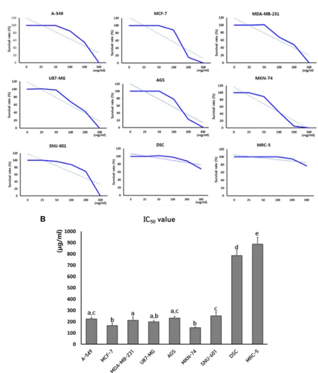
The cytotoxicity and IC50 values by treatment of SCLE was determined with MTT assay in the normal and cancer cell lines, including A-549, MCF-7, MDA-MB-231, U87-MG, AGS, MKN-74, SNU-601, MRC-5 fibroblasts and dental tis- sue-derived mesenchymal stem cells (DSCs), as shown in Fig. 1. The mean IC50 value (mean ± SEM) in three replicates was 222±13.5, 165±22.9, 211±31.1, 198±10.9, 231±15.8, 147±
6.8, 251±35.6, 784±45.8 and 886±61.3 μg/ml in the A-549, MCF-7, MDA-MB-231, U87-MG, AGS, MKN-74, SNU-601, DSCs and MRC-5 fibroblasts, respectively. The IC50 values in the cancer cell lines such as A-549, MCF-7, MDA-MB-231, U-87 MG, AGS, MKN-74 and SNU-601 was found to be at the concentration of approximately 200 μg/ml extract however, the IC50 values for normal cell lines, such as MRC- 5 fibroblasts and DSCs was found to be four times higher i.e. 800 μg/ml extract, than cancel cell lines. Therefore, the IC50 values in the normal cell lines were significantly (p<
0.05) higher, compared to those of cancer cell lines.
Analysis of population doubling time (PDT) After determination of the IC50 values, the cancer and normal cell lines were cultured in the media containing 200 μg/ml of SCLE. During treatment of SCLE, a lots of cells were not attached and floated in the cell culture flask, and the number of detached cells dramatically increased in the cancer cell lines, as shown in Fig. 2. Further, the cell number was investigated and PDT was calculated in each of the cell lines treated with 200 μg/ml of SCLE for 7 days, and PDT was compared to those of untreated controls. The re- sults was shown in the Fig. 3. The mean PDT in the un- treated control A-549, MCF-7, MDA-MB-231, U87-MG, AGS, MKN-74 and SNU-601 cancer cells was 26.9±3.33, 52.4±2.56, 45.2±3.67, 55.3±4.45, 35.1±3.22, 65.5±5.56 and 59.2±3.23, re- spectively. However, the mean PDT in the SCLE-treated A-549, MCF-7, MDA-MB-231, U-87 MG, AGS, MKN-74 and SNU-601 cancer cells was 46.2±3.67, 82.9±4.89m 63.4±4.15, 80.3±5.22, 60.1±2.45, 110,0±4.89 and 79.5±4.11, respectively.
In the cancer cell lines, PDT was significantly (p<0.05) in- creased by decrease in cell proliferation rate after treatment of SCLE. Further, the mean PDT in untreated normal DSCs and MRC-5 fibroblasts was 32.7±2.24 and 43.3±1.34, re- spectively, whereas the mean PDT in SCLE -treated normal DSCs and MRC-5 fibroblasts was found to be 34.4±3.47 and
A
B
Fig. 1. Analysis of cell cytotoxicity by MTT assay and determination of IC50 values in cancer cell lines (A-549, MCF-7, MDA-MB-231, U87-MG, AGS, MKN-74 and SNU-601) and normal cell lines (DSC and MRC-5) treated with SCLE. A: Inhibition curves of cell growth by MTT assay in each cell line. Figures shown are representatives of three independent experiments. B:
Mean IC50 values of each cell line in triplicate. a, b, c, d and e indicate significant (p<0.05) difference among each cell lines.
45.2±4.67, respectively. The PDT after treatment of SCLE in the normal cell lines tended to be slightly increased be- tween untreated control and treated cell lines, there was no significant (p<0.05) difference.
Analysis of viability of detached cells
After treatment of SCLE, the viability of detached cells
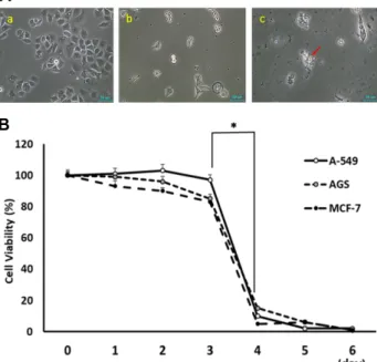
was analyzed with MTT assay in the A-549, AGS and MCF- 7 cancer cell lines. All cell lines were exposed to the medium containing SCLE, and the detached or floated cells were daily retrieved for up to 6 days. The retrieved cells were transferred and cultured in the fresh medium (Fig. 4A, Fig.
4B). The detached cells retrieved at 1 day, 2 days and 3 days after SCLE treatment were mostly survived in the fresh
A
B
Fig. 2. Changes of cell morphology in cancer cell lines (A, A-549, MCF-7, MDA-MB-231, U87-MG, AGS, MKN-74 and SNU-601) and normal cell lines (B, DSC and MRC-5) at 1 day after treatment of SCLE. The high number of detached and rounded cells were specially observed in SCLE-treated cancer cell lines. Scale bars; 50 μm.
medium. However, the cell viability of detached cells re- trieved at 4 days was significantly (p<0.05) decreased in all used cancer cells with apoptosis-like shrunk cell shape.
Therefore, SCLE induced detached cells were investigated for their survival rate for at least 3 days.
Analysis of telomerase activity
After treatment with 200 ug/ml of SCLE for 4 days, the quantitative expression of TERT transcript related with telo- merase activity in untreated control and SCLE-treated A- 549, AGS and MCF-7 cancer cell lines, as shown in Fig.
5A. GAPDH transcript was used as control reference genes.
The expression of TERT was highly decreased in the cells exposed to SCLE, as compared with their untreated control.
Further, the relative telomerase activity was measured in the A-549, AGS and MCF-7 cancer cell lines (Fig. 5B). The mean level of telomerase activity analyzed by RQ-TRAP as- say was 786±24.1%, 760±27.7% and 858±60.4% in the un- treated control A-549, AGS and MCF-7 cancer cell lines, re- spectively, as compared to normal MRC-5 fibroblasts.
Whereas, the level of telomerase activity was 277±16.5%, 354.0±10.5% and 315.7±6.0% in the extract-treated A-549, AGS and MCF-7 cancer cell lines, respectively. The relative telomerase activity was significantly (p<0.05) down-regu-
Fig. 3. Analysis of PDT in cancer cell lines (A- 549, MCF-7, MDA-MB-231, U87-MG, AGS, MKN-74 and SNU-601) and nor- mal cell lines (DSC and MRC-5) at 7 days after treatment of SCLE. The PDT values indicated mean±SEM of three replicates in each cell lines. Bars with an asterisk indicate significant (p<0.05) differences between untreated control (■) and SCLE-treated cells (■).
A
B
Fig. 4. The viability analysis of detached cells in A-549, AGS and MCF-7 cancer cells treated with SCLE. A: A repre- sentative example in untreated normal AGS cancer cells (a). The detached AGS cancer cells at 2 day after treat- ment of SCLE (b) and the apoptosis-like shrunken AGS cancer cells (arrow) at 4 days after treatment of SCLE (c). B: Viability rate after daily retrieval of detached cells. The cell viability was significantly (p<0.05) de- creased in the cells at retrieved 4 days after SCLE treat- ment. Bars with an asterisk indicate significant (p<0.05) differences between cells retrieved at 3 days and 4 days.
lated by treatment of SCLE.
Analysis of apoptosis rate
After treatment with 200 ug/ml of SCLE for 4 days, the
apoptosis rate was analyzed by flow cytometry in the A-549, AGS and MCF-7 cancer cells, as exhibited in Fig. 6. The mean percentages of live and apoptotic cells was 89.3±5.1 and 10.6±3.67; 92.3±4.56 and 6.25±1.5; 84.9±3.33 and 15.1±
5.45% in untreated control A-549, AGS and MCF-7 cancer cells, respectively. However, The mean percentages of live and apoptotic cells was 44.2±4.44 and 55.8±3.58; 48.2±3.33 and 51.8±2.52; 84.9±4.46 and 51.1±5.17% in the extract-treated A-549, AGS and MCF-7 cancer cell lines, respectively. The ratio of cells at apoptotic status was significantly (p<0.05) increased after exposure of SCLE for 5 days.
Analysis of cell cycle phase
After treatment with 200 ug/ml of SCLE for 4 days, the cell cycle was also examined in the A-549, AGS and MCF-7 cancer cells (Fig. 7). In the untreated control A-549 cancer cells, the proportions of cells in G1, S and G2/M phase were 66±4.2, 19±3.3 and 15±2.5%, respectively. In the un- treated control AGS cancer cells, the proportions of cells in G1, S and G2/M phase were 60±4.4, 13±2.6 and 27±2.8%, respectively. In the untreated control MCF-7 cancer cells, the proportions of cells in G1, S and G2/M phase were 42±6.6, 21±4.6 and 37±2.9%, respectively. Whereas, In the SCLE treated A-549 cancer cells, the proportions of cells in G1, S and G2/M phase were 84±2.6, 9.1±3.3 and 7.5±
2.0%, respectively. In the SCLE treated AGS cancer cells, the proportions of cells in G1, S and G2/M phase were 75.4±2.4, 6.0±2.3 and 18.6±2.4%, respectively. In the SCLE treated MCF-7 cancer cells, the proportions of cells in G1, S and G2/M phase were 76±8.7, 8±4.3 and 16±2.5%, respectively. Under SCLE induction, G1 phase of cell cycle
A B
Fig. 5. Telomerase activity in A-549, AGS and MCF-7 cancer cells treated with SCLE for 4 days. A: Expression of telomerase reverse transcriptase (TERT) transcript by RT-PCR in untreated control and SCLE-treated cell lines. B: Mean telomerase activity by RQ-TRAP in untreated control and SCLE-treated cell lines. Bars with an asterisk indicate significant (p<0.05) differences between untreated control (■) and SCLE-treated cells (■).
A
B
Fig. 6. Analysis of apoptosis rate in A-549, AGS and MCF-7 cancer cells treated with SCLE for 4 days. A: A representative images by cytometric analysis of apoptotic cells in untreated control and SCLE-treated cell lines. B: Mean percentage (%) of live, early apoptosis, late apoptosis and necrosis cells. Bars with an asterisk indicate significant (p<0.05) differences between untreated control (■) and extract-treated cells (■).
was significantly (p<0.05) increased in the A-549, AGS and MCF-7 cancer cells.
Analysis of Rho-associated protein kinase inhibitor on cell adhesion
The detached and floated cells by treatment of SCLE were simultaneously and respectively treated with SCLE and Y- 27632, an inhibitor of Rho-associated protein kinase (ROCK) which is tightly regulated in cell morphology and move- ment by acting on the cytoskeleton. Star-shape cell morphol-
A
B
Fig. 7. Analysis of cell cycle phase in A-549, AGS and MCF-7 cancer cells treated with SCLE for 4 days. A: A representative images by cytometric analysis of cell cycle in untreated control and SCLE-treated cell lines. B: Mean percentage (%) of live, early apoptosis, late apoptosis and necrosis cells. Bars with an asterisk indicate significant (p<0.05) differences between untreated control (■) and SCLE-treated cells (■).
ogy were frequently observed after only Y-27632 treatment in specially MCF-7 cancer cells. However, detached A-549, AGS and MCF-7 cancer cell lines by SCLE treatment did not show any morphological alternations under Y-276532 treatment, as shown in Fig. 8A. And SCLE induced cell de- tachment and anoikis might have resulted due to Rho signal pathway-independent of others mechanisms. Therefore, the expression level of transcripts related to cell adhesion was measured using real time RT-PCR, as shown in Fig. 8B and C. The mean expression level of RhoA and RhoC transcripts were 55±6.3 and 25±4.5; 63±6.7 and 30±2.3; and 48±3.4 and 26±3.3 in the untreated control (0D) A-549, AGS and MCF-7 cancer cells, respectively, comparing to expression level of GAPDH. The mean expression level of RhoA transcript at 1 day (1D), 2 days (2D), 3 days (3D) and 4 days (4D) after SCLE treatment was 53±5.3, 49±4.3, 23±2.1 and 12±2.6;
64±5.9, 60±3.3, 41±1.7 and 12±1.3; and 38±4.3, 26±2.9, 27±2.6 and 9±1.3% in the A-549, AGS and MCF-7 cancer cells. The mean expression level of RhoC transcript at 1 day (1D), 2 days (2D), 3 days (3D) and 4 days (4D) after SCLE treat- ment was 23±3.3, 19±4.3, 13±2.1 and 6±2.6; 29±6.0, 26±3.0, 18±1.7 and 8±1.2; and 19±2.2, 15±2.0, 11±2.3 and 2±0.9 in the A-549, AGS and MCF-7 cancer cells, respectively. The expression level of RhoA and RhoC was significantly (p<
0.05) decreased in A-549, AGS and MCF-7 cancer cells at 3 days after SCLE treatment, compared to those of cells at untreated control, 1 day and 2 days. In the present study, the inhibition of Rho activity did not result into the increased cell adhesion. Further, SCLE treatment resulted into floated status within 1 day after SCLE treatment, as mentioned above, however, expression of RhoA and RhoC was still at a high level up to 1 day and 2 days after SCLE treatment.
A
B
Fig. 8. Cell morphology in the A-549, AGS and MCF-7 cancer cells treated with Y-27632, a Rho-associated protein kinase (ROCK) inhibitor, and SCLE. A: The detached and spheroid cells were observed in the A-549, AGS and MCF-7 cancer cells at 2 days after simultaneous treatment of SCLE and Y-27632, and SCLE alone, respectively. Scale bars; 50 μm. B: Expression of RhoA and RhoC transcripts by RT-PCR in A-549, AGS and MCF-7 cancer cells treated with SCLE for 0 day (0D, untreated control), 1 day (1D), 2 days (2D), 3 days (3D) and 4 days (4D). C: Mean expression level (%) of RhoA (■) and RhoC (■) transcript. A, B and C indicate significant (p<0.05) differences among treatment day(s) in the expression of RhoA transcript. a, b, c and d indicate significant (p<0.05) differences among treatment day(s) in the expression of RhoC transcript.
Discussion
In the present study, the various types of cancer cells (A-549, MCF-7, MDA-MB-231, U87-MG, AGS, MKN-74 and SNU-601), normal MRC-5 fibroblasts and mesenchymal stem cells derived from dental tissues (DSCs) were treated with Smilax china L. extract (SCLE). All of these different cell lines were investigated for their fundamental cellular characterizations, including cell morphology, cell growth, phase of cell cycle, apoptosis and telomerase activity. The present study have demonstrated that SCLE treatment in- duces the inhibition of cell growth and cellular cytotoxicity by cellular anoikis in the human cancer and normal cell lines. However, the induction of anoikis and cytotoxic ef- fects by SCLE treatment were prominently observed in the cancer cell lines with extremely lower IC50 values, compared to those of normal cell lines, such as MRC-5 fibroblasts and
DSCs.
The natural compounds and drugs derived from herbs or plants have unceasingly suggested and applied for the treatment of a number of diseases including cancer [4]. The SCLE has been used as a conventional and alternative drug for inflammatory, rheumatic arthritis, diuretic and detox- ification treatments in traditional oriental medicine which usually comprises a number of different medical herbs, as per the symptoms of individual patient [10, 38, 40]. Previous studies have also demonstrated that SCLE treatment also induces anti-cancer effects in several types of cancer cells [20, 31, 35]. These studies strengthen the use of SCLE as a meaningful alternative chemotherapeutic drug for tumor treatment. In the present study, the anti-cancer effects were induced by anoikis pathway with the decreased cell attach- ment in the cancer cells treated with SCLE. Similar ob- servations have also been made by other studies in which
SCLE treatment induces anti-metastatic effect in human MDA-MB-231 breast cancer cells, possibly due to modu- lation of extracellular matrix (ECM) degradation [35]. How- ever, anti-cancer effects by SCLE treatment in various types of human cells have not been fully elucidated and are still under investigation.
Most of the animal cells in in vivo tissues and organs are attached to mesenchymal cells, such as fibroblasts or ECM matrix [29]. However, the loss of cell attachment or inappropriate ECM environment is induced to cellular apoptosis, known as anoikis [14]. Whereas, the progression to cancer cells from tumor cells first gradually acquire meta- stasis potential and the transformed cancer cells are sur- vived in inappropriate tissue environments of different tar- get organs. The resistance against anoikis cell death can pos- sess capacity to survive in the other tissues from the pri- mary sites [37]. Even though, the cancer cells display high anoikis resistance, and it has consistently been demon- strated that the loss of detachment to cell-to-cell or ECM during long time can be induced to apoptotic cascade proce- dure [27, 36, 37]. In the present study, the anoikis cell death was also observed to be induced in the specially cancer cell lines than normal cell lines. The morphological alter- ation of the cell to shrinkage status by the loss of cell vol- ume was considered as one of main fundamental character- ization for defining signaling based cellular apoptosis [7, 11]. The present study have also shown that the shrinkage and contracted cell morphology is also observed in the de- tached cancer cells after treatment of SCLE. During early apoptotic events, the flipped phosphatidyl serine strongly combined with fluorophore-conjugated Annexin V and has been widely and easily used for detecting early apoptosis using flow cytometry [8]. The present results have shown that the rate of early cellular apoptosis is increased in the cancer cells treated with SCLE by analysis of Annexin V protein using flow cytometry. Besides, one of the main cel- lular characterizations in the cells displaying cellular apop- tosis is arresting at the G0/G1 phase of cell the cycle [33].
In the previous studies, ovarian cancer cells at the spheroid or floated state by loss of cell adhesion were exhibited to be arrested in the G0/G1 phase of the cell cycle [9, 25].
The present study has also shown that the rate of cell ar- rested at G0/G1 phase of the cell cycle is increased in the cancer cells treated with SCLE. Further, accordingly to flow cytometry analysis, the ratio of cell population at sub-G1 phase was highly increased in the cancer cells treated with
SCLE (data not shown). Previous studies have suggested that the high peak of cell at sub-G1 phase is tightly related with increased DNA fragmentation during cellular apopto- sis [32]. Our results have clearly shown that SCLE treatment resulted in the increase of G0/G1 phase and sub-G1 of cell cycle by cellular anoikis.
In the present study, SCLE treatment clearly induced loss of cell adhesion and subsequent anoikis in the cancer cells, however, the detached cancer cells were more resistant to the cell death by cellular anoikis than normal cells, such as MRC-5 fibroblasts and mesenchymal stem cells. Whereas, the detached normal cell lines by SCLE treatment were easi- ly and quickly induced to anoikis cell death than the de- tached cancer cells (data not shown). It have well demon- strated that the cancer cells are displayed to more anoikis resistance after detachment in primary tissues, or metastasis to others tissues and circulatory systems [27, 41]. As shown in the present results, the detached cancer cells were sur- vived for 3 days, and induced to cell death after 4 days after cell detachment. However, the detached normal cells were quickly induced to cell death within 1 day after cell attachment. The previous study has also shown that over 90% of human intestinal epithelial cells are induced to anoi- kis cell death by caspase-2 and 9 apoptosis pathways within 3 hr after loss of cell anchorage [15].
Cell attachment arise normally through interactions be- tween integrin molecules on the cell membrane and ECM fibronectins, and many cellular events and functions, includ- ing cell growth and motility as well as gene expression can be regulated by the appropriate interaction and attachment of cell-to-cell or cell-to-ECM [12, 13]. Thus, the cellular and tissue morphology, maintenance, distribution, junction and differentiation can be also induced by the interaction of cells and tissues through various signal pathways [1, 6]. The cell adhesion molecules, such as integrin and cadherin proteins on the outer cell membrane are generally known for cell interactions combined with other cells or ECM [1, 6]. The integrin and cadherin proteins are tightly linked to intra- cellular actin filament and a number of interacting adapter proteins. The appropriate expression and position of outer integrins and cadherins as well as intracellular actin fila- ment can lead to normal cell adhesion. Moreover, the small GTP binding Rho proteins play an essential part in cell ad- hesion and motility through integrin by regulating actin cy- toskeleton [1, 5, 19]. During mitosis, the cells are formed to rounded and floated cell shape by decreased actin fila-
ment and integrin with high activity of Rho protein. And it has been previously showed that inhibitor of Rho signal pathway limit invasive progress of cancer cells [42]. And Rho signal pathway is highly up-regulated in the cell re- programming procedure to induced pluripotent stem cells (iPSCs) from differentiated normal cells. Thus, iPSCs are easily floated on the cell culture flask, and entered to anoikis cell death. It has been demonstrated that the cell viability and adhesion capacity of iPSCs can be increased by regu- lation of integrin and actin filaments as per inhibition of Rho signal pathway [39]. In the present study, the cancer cells floated with SCLE treatment were also administrated with Y-27632 Rho-associated protein kinase inhibitor, how- ever, the adhesion capacity of cell detached by SCLE treat- ment was not increased with inhibition of Rho signal path- way using Y-27632. Besides, the expression of RhoA and RhoC transcripts by real time PCR analysis was continually remained for 2 days in the A-549 cancer cells during floated status by SCLE treatment. However, the expression level of their transcripts was detected at a very low level and reached to apoptotic cell death after 4 days of treatment.
The lower expression of RhoA and RhoC at 4 days might be due to the apoptotic cell death rather than cell adhe- sion-related signal pathway induced by SCLE. And the most of the cancer cell were immediately floated after admin- istration of SCLE under high expression of cell adhesion-re- lated transcripts. Thus, we assumed that influences on the cell adhesion in the cancer cells treated with SCLE were not related with Rho signal pathway regulating activity of integrin and actin filaments, and loss of cell adhesion and subsequent anoikis cell death by SCLE treatment is thought to be a consequence of others mechanisms rather than in- trinsic Rho signal pathway. Further, the telomerase activity was significantly decreased in the cancer cells after SCLE treatment. The high level of telomerase activity is generally expressed in the immortal cells, such as embryonic stem cells and cancer cells, and the decreased level of telomerase activity was displayed in the cancer cells reached at nearly cellular apoptosis levels [3, 24]. Thus, SCLE treatment seems to be induced to decreased telomerase activity in the cancer cells and also reached to cellular apoptosis levels, as men- tioned above.
The chemical components of SCLE are not well-known in detail, however, polyphenols, flavonoids and triterpenes are commonly suggested as major components of SCLE. It has been previously reported that the cytotoxic effects result
from the various types of polyphenols [44], flavonoid glyco- side [32] and steroidal saponins [38]. However, the available investigations on the SCLE-derived chemical components which can exhibit the anti-cancer effects as per induction of anoikis are not still reported in the various types of hu- man cancer cell lines. Previous another study has been sug- gested that anti-metastatic effect by SCLE treatment is ex- hibited in the human breast cancer cells, probably caused by modulation of ECM degradation [28]. We presumed that induction of anoikis by SCLE might be caused by physical disturbance of cell-to-cell contact, rather than modulation of intracellular integrin and Rho signal pathways by chem- ical components derived from SCLE. Any further additional analysis and studies associated with cell death by anoikis will be needed for investigation of chemical components derived from SCLE.
The present study has clearly demonstrated that SCLE outstandingly exhibits the inhibition of cell growth and in- crease of anoikis cell death along with reduced (lost) cell adhesion in in vitro cultures various types of human cancer cells than normal cells. Therefore, the SCLE alone or in com- bination with other treatments might be a potential chemo- therapy agent or an alternative treatment against cancer cells. However, it has suggested that first step for metastasis of the tumor cells is to detach and escape from the primary tissue, and metastasize to secondary tissue with long last resistance against anoikis cell death [2]. The clinical applica- tion of SCLE treatment should be carefully examined in the more cancer and normal somatic cell lines with intrinsic cellular mechanisms and component analysis of SCLE.
The Conflict of Interest Statement
The authors declare that they have no conflicts of interest with the contents of this article.
References
1. Albelda, S. M. and Buck, C. A. 1990. Integrins and other cell adhesion molecules. FASEB. J. 4, 2868-2880.
2. Alizadeh, A. M., Shiri, S. and Farsinejad, S. 2014. Metastasis review: from bench to bedside. Tumour. Biol. 35, 8483-8523.
3. Artandi, S. E. and DePinho, R. A. 2010. Telomeres and telo- merase in cancer. Carcinogenesis 31, 9-18.
4. Atanasov, A. G., Waltenberger, B., Pferschy-Wenzig, E. M., Linder, T., Wawrosch, C., Uhrin, P., Temml, V., Wang, L., Schwaiger, S., Heiss, E. H., Rollinger, J. M., Schuster, D., Breuss, J. M., Bochkov, V., Mihovilovic, M. D., Kopp, B.,
Bauer, R., Dirsch, V. M. and Stuppner, H. 2015. Discovery and resupply of pharmacologically active plant-derived nat- ural products: A review. Biotechnol. Adv. 33, 1582-1614.
5. Barry, S. T., Flinn, H. M., Humphries, M. J., Critchley, D.
R. and Ridley, A. J. 1997. Requirement for Rho in integrin signaling. Cell Adhes. Commun. 4, 387-398.
6. Borghi, N., Lowndes, M., Maruthamuthu, V., Gardel, M.
L. and Nelson, W. J. 2010. Regulation of cell motile behavior by crosstalk between cadherin- and integrin-mediated ad- hesions. Proc. Natl. Acad. Sci. USA. 107, 13324-13329.
7. Bortner, C. D. and Cidlowski, J. A. 1998. A necessary role for cell shrinkage in apoptosis. Biochem. Pharmacol. 56, 1549- 1559.
8. Brumatti, G., Sheridan, C. and Martin, S. J. 2008. Expression and purification of recombinant annexin V for the detection of membrane alterations on apoptotic cells. Methods 44, 235- 240
9. Carduner, L., Picot, C. R., Leroy-Dudal, J., Blay, L., Kellouche, S. and Carreiras, F. 2014. Cell cycle arrest or survival signal- ing through αv integrins, activation of PKC and ERK1/2 lead to anoikis resistance of ovarian cancer spheroids. Exp.
Cell Res. 320, 329-242.
10. Chen, L., Yin, H., Lan, Z., Ma, S., Zhang, C., Yang, Z., Li, P. and Lin, B. 2011. Anti-hyperuricemic and nephropro- tective effects of Smilax china L. J. Ethnopharmacol. 135, 399- 405.
11. Friis, M. B., Friborg, C. R., Schneider, L., Nielsen, M. B., Lambert, I. H., Christensen, S. T. and Hoffmann, E. K. 2005.
Cell shrinkage as a signal to apoptosis in NIH 3T3 fibro- blasts. J. Physiol. 567, 427-443.
12. Frisch, S. M. and Ruoslahti, E. 1997. Integrins and anoikis.
Curr. Opin. Cell Biol. 9, 701-706.
13. Frisch, S. M. and Screaton, R. A. 2001. Anoikis mechanisms.
Curr. Opin. Cell Biol. 13, 555-562.
14. Gilmore, A. P. 2005. Anoikis. Cell Death Differ. 12, 1473-1477.
15. Grossmann, J., Walther, K., Artinger, M., Kiessling, S. and Schölmerich, J. 2001. Apoptotic signaling during initiation of detachment-induced apoptosis ("anoikis") of primary hu- man intestinal epithelial cells. Cell Growth Differ. 12, 147-155.
16. Gupta, G. P. and Massagué, J. 2006. Cancer metastasis:
building a framework. Cell 127, 679-695.
17. Hanahan, D. and Weinberg, R. A. 2000. The hallmarks of cancer. Cell 100, 57-70.
18. Heng, Y. W. and Koh, C. G. 2010. Actin cytoskeleton dy- namics and the cell division cycle. Int. J. Biochem. Cell. Biol.
42, 1622-1633.
19. Hotchin, N. A. and Hall, A. 1996. Regulation of the actin cytoskeleton, integrins and cell growth by the Rho family of small GTPases. Cancer Surv. 27, 311-322.
20. Hu, L. L., Chen, D. S., Wang, Y. Y., Qin, Y., Huang, P., Yu, L. X., Liao, J. and Hua, X. L. 2015. Smilax china L.
rhizome extract inhibits nuclear factor-κB and induces apoptosis in ovarian cancer cells. Chin. J. Integr. Med. 21, 907-915.
21. Jeon, B. G., Jang, S. J., Park, J. S., Subbara, R. B., Jeong, G. J., Park, B. W. and Rho, G. J. 2015. Differentiation poten-
tial of mesenchymal stem cells isolated from human dental tissues into non-mesodermal lineage. Anim. Cell. Syst. 19, 321-331.
22. Jeon, B. G., Kumar, B. M., Kang, E. J., Ock, S. A., Lee, S. L., Kwack, D. O., Byun, J. H., Park, B. W. and Rho, G. J. 2019. Characterization and comparison of telomere length, telomerase and reverse transcriptase activity and gene expression in human mesenchymal stem cells and can- cer cells of various origins. Cell Tissue Res. 345, 149-161.
23. Keledjian, K. and Kyprianou, N. 2003. Anoikis induction by quinazoline based alpha 1-adrenoceptor antagonists in prostate cancer cells: antagonistic effect of bcl-2. J. Urol. 169, 1150-1156.
24. Kim, H. I., Moon, S. H., Lee, W. C., Lee, H. J., Shivakumar, S. B., Lee, S. H., Park, B. W., Rho, G. J. and Jeon, B. G.
2018. Inhibition of cell growth by cellular differentiation into adipocyte-like cells in dexamethasone sensitive cancer cell lines. Anim. Cell. Syst. 22, 178-188.
25. Kim, K., Pang, K. M., Evans, M. and Hay, E. D. 2000.
Overexpression of beta-catenin induces apoptosis in- dependent of its transactivation function with LEF-1 or the involvement of major G1 cell cycle regulators. Mol. Biol.
Cell. 11, 3509-3523.
26. Kim, Y. D., Jang, S. J., Lim, E. J., Ha, J. S., Shivakumar, S. B., Jeong, G. J., Rho, G. J. and Jeon, B. G. 2017. Induction of telomere shortening and cellular apoptosis by sodium meta-arsenite in human cancer cell lines. Anim. Cell. Syst.
21, 241-254.
27. Kim, Y. N., Koo, K. H., Sung, J. Y., Yun, U. J. and Kim, H. 2012. Anoikis resistance: an essential prerequisite for tu- mor metastasis. Int. J. Cell. Biol. 2012, 306879.
28. Lang, J. Y., Chen, H., Zhou, J., Zhang, Y. X., Zhang, X. W., Li, M. H., Lin, L. P., Zhang, J. S., Waalkes, M. P. and Ding, J. 2005. Antimetastatic effect of salvicine on human breast cancer MDA-MB-435 orthotopic xenograft is closely related to Rho-dependent pathway. Clin. Cancer Res. 11, 3455-3464.
29. Langhans, S. A. 2018. Three-dimensional in vitro cell culture models in drug discovery and drug repositioning. Front.
Pharmacol. 9, 6.
30. Lee, W. C., Kim, D. Y., Kim, M. J., Lee, H. J., Bharti, D., Lee, S. H., Kang, Y. H., Rho, G. J. and Jeon, B. G. 2019.
Delay of cell growth and loss of stemness by inhibition of reverse transcription in human mesenchymal stem cells derived from dental tissue. Anim. Cell. Syst. 23, 335-345.
31. Li, Y. L., Gan, G. P., Zhang, H. Z., Wu, H. Z., Li, C. L., Huang, Y. P., Liu, Y. W. and Liu, J. W. 2007. A flavonoid glycoside isolated from Smilax china L. rhizome in vitro anticancer effects on human cancer cell lines. J. Ethnophar- macol. 113, 115-124.
32. Liu, Y., Zhao, J., Zhang, W., Gan, J., Hu, C., Huang, G.
and Zhang, Y. 2015. lncRNA GAS5 enhances G1 cell cycle arrest via binding to YBX1 to regulate p21 expression in stomach cancer. Sci. Rep. 5, 10159.
33. Mao, Z., Ke, Z., Gorbunova, V. and Seluanov, A. 2012.
Replicatively senescent cells are arrested in G1 and G2 phases. Aging 4, 431-435.
초록:토복령 추출물이 처리된 여러 종류의 사람 암세포주에서 아노이키스 세포 사멸에 의한 세포 성장의 억제
김민재1․김현지1․김무경1․이성호2․전병균1,3*
(1경상대학교 사범대학 생물교육과, 2경상대학교 생명과학부, 3경상대학교 교육연구원)
본 연구에서는 다양한 사람의 암세포주(A-549, MCF-7, MDA-MB-231, U87-MG, AGS, MKN-74 및 SNU-601 세 포)와 정상세포주(MRC-5 섬유아세포 및 사랑니 유래 중간엽성 줄기세포에 토복령 추출물(Smilax china L. extract, SCLE)을 처리하여 세포 사멸 효과를 조사하였다. SCLE 처리 후, MTT 분석에서 여러 암세포주는 정상세포주보다 유의적으로 휠씬 낮은 반억제농도값을 나타내었고, 세포는 세포부착력의 소실로 인한 세포사멸(anoikis)이 관찰되 었다. 또한, SCLE를 처리한 A-549, AGS 및 MCF-7 암세포주에서 세포의 생존성과 말단소립 복원효소의 활성도를 조사하였을 때, SCLE 처리 후 4일째에 세포의 생존성과 말단소립 복원효소의 활성도가 현저히 줄어드는 것을 관 찰하였다. 또한, SCLE를 처리한 A-549, AGS 및 MCF-7 암세포주에서 세포 주기의 G1기에서 세포 성장이 정지되 었고,세포 사멸이 유의적으로 증가하는 것을 알 수 있었다. 그러나, SCLE 처리는 rho 단백질의 활성과 관련 없는 세포부착력의 소실과 세포 사멸이 유도되는 것을 관찰하였다. 이 연구의 결과를 바탕으로 토복령 추출물은 정상 세포보다는 암세포에 특이적으로 세포부착력의 소실과 세포 사멸을 유도하여, 이 추출물에 포함된 물질을 이용한 항암 연구에 응용될 수 있을 것으로 판단된다.
34. Moon, J. Y., Kim, S. W., Yun, G. M., Lee, H. S., Kim, Y.
D., Jeong, G. J., Ullah, I., Rho, G. J. and Jeon, B. G. 2015.
Inhibition of cell growth and downregulation of telomerase activity by amygdalin in human cancer cell lines. Anim.
Cell. Syst. 19, 295-304.
35. Nho, K. J., Chun, J. M. and Kim, H. K. 2015. Anti-metastatic effect of Smilax china L. extract on MDA-MB-231 cells. Mol.
Med. Rep. 11, 499-502.
36. Paoli, P., Giannoni, E. and Chiarugi, P. 2013. Anoikis molec- ular pathways and its role in cancer progression. Biochim.
Biophys. Acta. 1833, 3481-3498.
37. Sakamoto, S. and Kyprianou, N. 2010. Targeting anoikis resistance in prostate cancer metastasis. Mol. Aspects Med.
31, 205-214.
38. Shao, B., Guo, H., Cui, Y., Ye, M., Han, J. and Guo, D.
2007. Steroidal saponins from Smilax china and their anti- inflammatory activities. Phytochemistry 68, 623-630.
39. Shim, Y. J., Kang, Y. H., Kim, H. J., Kim, M. J., Lee, H.
J., Son, Y. B., Lee, S. H. and Jeon, B. G. 2019. Improvement
of cell viability using a Rho-associated protein kinase (ROCK) inhibitor in human dental papilla derived sin- gle-induced pluripotent stem cells. J. Life Sci. 29, 895-903.
40. Shu, X. S., Gao, Z. H. and Yang, X. L. 2006. Anti-inflamma- tory and anti-nociceptive activities of Smilax china L. aque- ous extract. J. Ethnopharmacol. 103, 327-332.
41. Simpson, C. D., Anyiwe, K. and Schimmer, A. D. 2008.
Anoikis resistance and tumor metastasis. Cancer Lett. 272, 177-185.
42. Somlyo, A. V., Bradshaw, D., Ramos, S., Murphy, C., Myers, C. E. and Somlyo, A. P. 2000. Rho-kinase inhibitor retards migration and in vivo dissemination of human prostate can- cer cells. Biochem. Biophys. Res. Commun. 269, 652-659.
43. van Zijl, F., Krupitza, G. and Mikulits, W. 2011. Initial steps of metastasis: cell invasion and endothelial transmigration.
Mutat. Res. 728, 23-34.
44. Wu, L. S., Wang, X. J., Wang, H., Yang, H. W., Jia, A.
Q. and Ding, Q. 2010. Cytotoxic polyphenols against breast tumor cell in Smilax china L. J. Ethnopharmacol. 130, 460-464.