cause of death in high-income countries, accounting for 3%–
8% of total deaths, and it was the sixth leading cause of death in nations of low and middle income, accounting for 4%–9%
of total deaths
3. In this same report, COPD was also estimated to be the seventh and 10th leading cause of disability-adjusted life years in countries of high income and in those of low or middle income, respectively
3. COPD tissues are characterized by chronic inflammation, mucus metaplasia, alveolar destruc- tion, and structural cell apoptosis
2. It is important to point out;
however, that the underlying mechanisms of the pathogenesis of COPD have not yet been adequately elucidated, hindering the successful development of disease-modifying therapeu- tics.
Recently, mitochondria/mitochondrial dysfunction has been highlighted in a variety of disorders and human health
4. Mitochondria take important parts not only in cellular respira- tion but also in other fundamental cellular functions including metabolism, innate and adaptive immune signaling, calcium homeostasis, senescence, and cell death. Accordingly, recent studies have revealed unprecedented roles of mitochondrial molecules which play in the context of COPD pathogenesis.
These recent advancements on mitochondrial biology have enabled us to envisage that COPD pathogenesis could be understood better if it is focused on from the mitochondrial
Introduction
Chronic obstructive pulmonary disease (COPD) encom- passes several clinical syndromes, most notably emphysema and chronic bronchitis
1,2. It is a major unmet medical burden worldwide and, in western society, is strongly associated with cigarette smoke (CS) exposure. Estimates from World Health Organization (WHO)’s Global Burden of Disease and Risk Factors project show that in 2001, COPD was the fifth leading
A Mitochondrial Perspective of Chronic
Obstructive Pulmonary Disease Pathogenesis
Min-Jong Kang, M.D., M.P.H., Ph.D.
1and Gerald S. Shadel, Ph.D.
2,31
Section of Pulmonary, Critical Care and Sleep Medicine, Department of Internal Medicine, Departments of
2Pathology and
3
Genetics, Yale University School of Medicine, New Haven, CT, USA
Chronic obstructive pulmonary disease (COPD) encompasses several clinical syndromes, most notably emphysema and chronic bronchitis. Most of the current treatments fail to attenuate severity and progression of the disease, thereby requiring better mechanistic understandings of pathogenesis to develop disease-modifying therapeutics. A number of theories on COPD pathogenesis have been promulgated wherein an increase in protease burden from chronic inflammation, exaggerated production of reactive oxygen species and the resulting oxidant injury, or superfluous cell death responses caused by enhanced cellular injury/damage were proposed as the culprit. These hypotheses are not mutually exclusive and together likely represent the multifaceted biological processes involved in COPD pathogenesis.
Recent studies demonstrate that mitochondria are involved in innate immune signaling that plays important roles in cigarette smoke-induced inflammasome activation, pulmonary inflammation and tissue remodeling responses. These responses are reviewed herein and synthesized into a view of COPD pathogenesis whereby mitochondria play a central role.
Keywords: Pulmonary Disease, Chronic Obstructive; Mitochondria; NLRX1 Protein, Human
Copyright © 2016
The Korean Academy of Tuberculosis and Respiratory Diseases.
All rights reserved.
Address for correspondence: Min-Jong Kang, M.D., M.P.H., Ph.D.
Department of Internal Medicine, Yale University School of Medicine, 300 Cedar Street, TAC Building #S425B, New Haven, CT 06520-8057, USA Phone: 1-203-737-6343, Fax: 1-203-785-3826
E-mail: min-jong.kang@yale.edu Received: Apr. 5, 2016
Revised: Apr. 15, 2016 Accepted: May. 26, 2016
cc It is identical to the Creative Commons Attribution Non-Commercial License (http://creativecommons.org/licenses/by-nc/4.0/).
perspective. In this review, current state-of-art understanding of mitochondrial biology and mitochondrial molecules in the context of COPD pathogenesis is discussed.
Current Theories on COPD Pathogenesis
A number of major theories of COPD pathogenesis have been promulgated. Initially, since the 1960s, the protease/anti- protease hypothesis dominated thinking in this area. And, the concept has built up that the increase in protease burden is thought to derive from inflammatory cells (hence the “Inflam- mation Hypothesis” of pathogenesis). In addition, the “Apop- tosis Hypothesis,” which proposes that apoptosis/cell death response caused by cellular injury/damage is a primary event in the pathogenesis of pulmonary emphysema, has been highlighted in the field of COPD research. And for a long time, exaggerated production of reactive oxygen species (ROS) and resulting oxidant injury have been postulated to be a major event in the pathogenesis of COPD (Oxidant Injury Hypoth- esis). These concepts and others that are widely discussed to explain COPD pathogenesis are briefly summarized below.
1. Protease-antiprotease imbalance
In 1964, researchers reported that a deficiency of α1- antitrypsin was associated with emphysema
5. A few years later, neutrophil elastase was reported to be the target of α1- antitrypsin. These findings, together with the observation of increased numbers of neutrophils and macrophages in the lungs of smokers, link various proteases from these inflam- matory cells as the primary effectors of lung destruction in COPD
6-8. In this concept, the normal lung is believed to be protected by an antiprotease “shield” that negates the function of proteolytic enzymes that are released into the airway or parenchyma, and emphysema is believed to be caused by an increase in proteases and or a reduction in antiproteases.
2. Inflammation
As noted in the current definition, COPD is characterized by airflow limitation that is usually progressive and associated with an enhanced chronic inflammatory response in the air- ways and the lung to noxious particles or gases
9. Inflammation with infiltrating macrophages, neutrophils, lymphocytes and occasionally eosinophils, is seen throughout the bronchial tree and parenchyma of lungs from patients with COPD. In addi- tion, airway inflammation is believed to start at an early stage, many years prior to the onset of clinical symptoms, in patients with COPD
10. It is important to note that substantial heteroge- neity is observed in lungs from patients with COPD. Although exaggerated type 1 inflammation plays an important role in the pathogenesis of emphysema
11,12, recent reports have also
highlighted type 2- and type 17-cytokine productions in this disorder (reviewed in Brusselle et al.
13). In this regard, it is in- teresting that a study demonstrated that a pleiotrophic proin- flammatory cytokine, interleukin (IL)-18, can simultaneously induce the signature cytokines associated with type 1, type 2 and type 17 responses and that each of these plays a specific role in the pathogenesis of the IL-18 effector repertoire
14. This study demonstrated also that IL-18 mediated signaling in- duces chronic inflammation, emphysema, mucus metaplasia, airway fibrosis and vascular remodeling with intimal hyper- plasia, which mimic, in many ways, the pathologies in human COPD
14. As known, the increase of IL-1β and IL-18, target cytokines of inflammasome activation, in COPD patients has been demonstrated in multiple studies including ours
15,16. Overall, the inflammation in COPD lung tissues is believed to be causally related to emphysema development and other pathologic alterations in the lungs that worsen with disease progression.
3. Oxidative stress/oxidant injury
An increased burden of oxidative stress is an important feature of the pathogenesis of COPD and, for a long time, oxi- dative stress resulting from an imbalance between oxidants and antioxidants has been proposed as the basis for COPD.
Patients with COPD experience an increased burden of oxi- dative stress due to the combined effects of excess ROS and reactive nitrogen species generation, antioxidant depletion and reduced antioxidant enzyme activity. In addition, CS in- creases the burden of oxidants in the respiratory tract, either directly included in the CS or generated by inflammatory cells, depleting antioxidant defenses and injuring lung cells
17. Fur- thermore, the increase of oxidative stress is compounded by alterations in the antioxidant defenses in patients with COPD
8. Current knowledge on this theme has been extensively re- viewed in other publications.
4. Apoptosis/cell death hypothesis
The lung is unique in that alveolar cells are constantly exposed to external physical and chemical stresses. As physi- ologic responses upon these cellular injury/damage, apop- totic cell removal and cellular replacement are believed to be highly regulated to maintain physiologic homeostasis of the entire alveolar unit
18. Therefore, overinduction of apoptosis and/or blockade of cellular replenishment would be expected to disrupt alveolar septal homeostasis and the “Apoptosis Hy- pothesis” highlights that pulmonary structural cell apoptosis/
death response due to ongoing cellular injury/damage as a
critical element in the COPD pathogenesis (reviewed in Tuder
et al.
18and Yoshida and Tuder
19). Indeed, alveolar septal cell
apoptosis was confirmed in lungs from patients with COPD
20.
Interestingly, this study also suggested that CS-induced injury
might activate mitochondrial intrinsic pathway of apoptosis, which was demonstrated by immunohistochemistry in COPD lungs
20.
5. Accelerated aging/cellular senescence
Aging is thought to be a degenerative process caused by ac- cumulated damage that leads to cellular dysfunction, tissue failure, and death
21,22. Similar to other organ systems, biological aging of the pulmonary system is associated with structural changes leading to a progressive decline in function, and the term “aging lung” is usually applied to designate the organ
23,24. Interestingly, the hallmarks of ageing, which include telomere shortening, genomic instability, epigenetic alterations, loss of proteostasis, mitochondrial dysfunction, deregulated nutrient- sensing and stem cell exhaustion, are all observed in COPD lungs
25. In this sense, studies have long been sought to under- stand COPD pathogenesis in the perspective of accelerated aging.
6. Immunologic perspective
In this theory, a series of innate and adaptive immunologic responses are proposed as major events which contribute to COPD pathogenesis; CS activates innate immune cells such as epithelial cells and macrophages by triggering pattern recognition receptors (PRRs) or via the release of danger- associated molecular patterns (DAMPs) from stressed or dying cells; the signals from various innate immune stimula- tion lead to the activation of dendritic cells and subsequently, induce adaptive immune responses encompassing T helper (Th1 and Th17) CD4
+T cells, CD8
+cytotoxicity, and B-cell re- sponses; in addition, viral and bacterial infections contribute to not only the pathogenesis of acute exacerbations of COPD but also the amplification and perpetuation of chronic inflam- mation in stable COPD via dysregulation of a variety of innate and adaptive immune signaling pathways. Furthermore, the potential role of autoimmunity including autoantibodies in the development of COPD has been suggested also. These re- cent understandings have been comprehensively reviewed in other publications
12,13,26.
Multi-Faceted Mitochondria
Historically, the major role of mitochondria has been thought to be the powerhouse of the cell because these organ- elles catalyze oxidation of metabolites to drive ATP production via the process of oxidative phosphorylation (OXPHOS)
27. OX- PHOS involves transfer of high-energy electrons derived from NADH and FADH2 generated by the TCA cycle through the electron transfer chain (ETC) complexes that ultimately do- nate electrons to molecular oxygen. During this process, pro-
tons are pumped across the inner mitochondrial membrane resulting in a proton-motive force that is utilized to produce ATP by the F0F1 ATP synthase
27. While this is clearly a major function of mitochondria, studies over the past two or three decades have revealed additional critical functions of mito- chondria, including regulation of cell proliferation, death and differentiation, redox and calcium homeostasis and others.
One exciting new area is how mitochondria dictate immune responses via regulation of a broad range of innate immune pathways and the determination of immune cell phenotype and their functions
28,29. Hence, considering that mitochondrial function and behavior are fundamental to the physiology of cellular and organismal health, it is not surprising “mitochon- drial dysfunction” has been implicated in a wide range of diseases that encompass all aspects of medicine (reviewed in Nunnari and Suomalainen
4). For example, the pathogenic role of mitochondrial dysfunction has been increasingly identi- fied in a large number of human diseases including diabetes, Alzheimer’s, Parkinson’s, cardiac dysfunction, tumorigenesis, pulmonary disorders as well as aging
30,31. Below we describe the role of mitochondria in COPD pathogenesis based on re- cent studies in this area.
1. Mitochondrial regulation of inflammasome activation Inflammation is a fundamental response of the innate im- mune system to noxious stimuli. The concept of inflamma- somes was introduced nearly a decade ago, and since then, they have been increasingly recognized as central players in innate immune and inflammatory responses in settings of infection and sterile inflammation
32. Active caspase-1, formed by the activation of inflammasomes, is used for the activation of pro-inflammatory cytokines IL-1β and IL-18, and release of these cytokines results in the recruitment of effector cell populations important in the immune response and tissue repair. During infection or injury, inflammasomes are directly or indirectly activated by a wide array of pathogen-associated molecular patterns (PAMPs) or DAMPs and, under normal circumstances, activation of inflammasomes culminates in the resolution of infection or inflammation and contributes to homeostatic processes. However, in a pathologic context, perpetuation of inflammasome activation can lead to a variety of chronic inflammatory conditions such as metabolic disor- ders, tumorigenesis and autoimmunity
32-34. The roles of mito- chondria on inflammasome regulation have been explored in multiple circumstances and we have recently addressed this in the context of COPD pathogenesis
35.
2. Mitochondrial regulation of cellular redox state and redox signaling
Mitochondria are an important source of ROS within most
mammalian cells
36. Mitochondrial ROS have a crucial roles in
a variety of physiological and pathological circumstances
30,37. In addition, mitochondria are themselves prone to oxidative damage, which results in further perturbation of mitochon- drial function. While ROS have long been appreciated for their damage-promoting, detrimental effects, there is now a greater understanding of their roles as signaling molecule and mi- tochondrial ROS-mediated signaling pathways are involved in various basal and adaptive physiological responses
38,39. It should be noted that our understanding of mitochondrial redox signaling pathways is still in its infancy, but unraveling these holds therapeutic potential for many diseases including COPD.
3. Mitochondrial regulation of cell death
Mitochondria represent a physical point of convergence for many cell death-inducing signals in mammalian cells
40,41. Mi- tochondria not only adjudicate the decision of whether or not to commit to cell death, but also release pro-apoptotic pro- teins that are involved in widespread proteolysis, nucleolysis, and cell engulfment. Interactions among BCL-2 family pro- teins at the mitochondrial outer membrane control the release of these toxic factors and the commitment to apoptosis
40. Fur- thermore, several perturbations of intracellular homeostasis are specifically detected by mitochondria, thereby involving them in the initiation of cell death. In summary, mitochondria regulate the transition between cell death initiation and ex- ecution in a vast majority of settings, and thus, mitochondria stand out as key controllers of cell death subroutines initiated at most subcellular locations
41.
4. Mitochondrial regulation of innate and adaptive immune responses
Mitochondria are centrally positioned signaling hubs that regulate of innate and adaptive immunity
28,29. The discovery of mitochondrial antiviral signaling (MAVS) protein, which was the first mitochondria-localized protein implicated in the antiviral innate immune response, was a breakthrough in establishing the role for mitochondria in innate immunity
42. Interestingly, recent studies have suggested that the MAVS- mediated signaling might play an important role in the patho- genesis of COPD
43,44(this topic is discussed further in next section). Furthermore, multiple studies have revealed that mitochondrial signaling controls adaptive immunity includ- ing CD4
+T cell differentiation, CD8
+memory T cell formation and others via various mitochondrial metabolic pathways (reviewed in Weinberg et al.
29). For example, mitochondrial aerobic glycolysis was found to be required for effective T cell activation through generation of mitochondrial ROS, which are necessary for optimal activity of nuclear factor of activated T cells and proximal T-cell receptor–mediated signaling
45,46. It is important to note that the role of mitochondria which
take part in COPD immunology has not been adequately elu- cidated. Future studies to define underlying mechanisms by which mitochondria and mitochondrial signaling molecules function in the immunologic aspect of COPD pathogenesis will be beneficial not only for our enhanced understanding on this area but also for the development of potential disease- modifying therapeutics.
Mitochondrial Dysfunction, Dysregulation of Mitochondrial
Molecules and COPD
Multiple studies have begun to define mitochondrial func- tion or molecules that contribute to the pathogenesis of vari- ous pulmonary disorders, including COPD. For example, nucleotide binding domain and leucine-rich-repeat-con- taining protein X1 (NLRX1) and MAVS, novel mitochondrial molecules initially known to regulate cytoplasmic nucleic acid-mediated innate immune pathway, have recently been implicated in the pathogenesis of COPD
44. More intriguingly, most of characteristic features involved in COPD pathogen- esis, which include enhanced inflammasome activation and intraalveolar inflammation, exaggerated proteases and cyto- kine responses, dysregulation of ROS production as well as enhanced alveolar cell apoptosis/cell death, were attributed to CS-induced dysregulation of NLRX1/MAVS-mediated sig- naling
44. These observations further support the notion that CS-induced mitochondrial dysfunction or dysregulation of mitochondrial molecules could be key features to drive devel- opment of COPD.
1. Nucleotide binding domain and leucine-rich-repeat- containing protein X1 (NLRX1)
NLRX1 is the member of the NLR family of PRRs that has a
unique N-terminal domain, which accounts for the letter “X” in
its acronym. It contains a mitochondrial targeting sequence,
and biochemical analyses demonstrated that NLRX1 localizes
to mitochondria
47. Strikingly, NLRX1 represents the first, and
so far only, PRR family member that targets this cellular loca-
tion, and hence a likely link between mitochondrial functions
and innate immunity
48. Initially, it was identified to function as
a negative regulator of RIG-I signaling by targeting the RIG-I
downstream MAVS
49,50. As noted, a recent study has identified
NLRX1 as an important player in the pathogenesis of COPD
44,
whereby it was significantly suppressed in mice exposed to
CS, and CS-induced activation of inflammasome(s), pulmo-
nary inflammation and emphysematous destruction were
exaggerated in the absence of NLRX1
44. The importance of
NLXR1 was also evident from clinical studies. In three inde-
pendent human COPD cohorts, the expression of NLRX1
was suppressed in lungs from patients with COPD and this
suppression showed strong correlation with the degree of airflow limitation, a hallmark of COPD
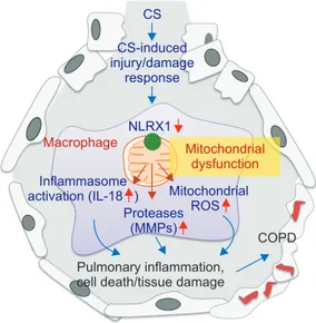
44. Overall, these studies highlight NLRX1 as a critical inhibitor in the pathogenesis of COPD (Figure 1) and other studies suggest additional ways that it is involved in mitochondrial regulation, including inter- actions with mitochondrial Tu translation elongation factor to promote autophagy
51, with UQCRC2, a subunit of ETC com- plex III to regulates ROS generation
47and tumor suppressor activity
52that could be relevant to the disease.
2. MAVS protein
As noted above, MAVS was the first mitochondria-localized protein causally linked to antiviral innate immune responses
42. It is now recognized that in addition to induction of antiviral and inflammatory responses, MAVS interacts with many mol- ecules that have roles in antiviral responses, inflammation, apoptosis, mitochondrial dynamics, autophagy and protea- some degradation. In this regard, it is interesting that MAVS appeared to have a critical role in the exaggerated production of IL-18 production as well as pulmonary inflammatory and remodeling responses that are observed after CS and respi-
ratory virus or viral PAMP co-exposures in a murine COPD model
43. Furthermore, the exaggerated CS-induced COPD- like phenotype observed in NLRX1 null mutant (
–/–) mice was significantly ameliorated in NLRX1 and MAVS double- mutant (NLRX1
–/–/MAVS
–/–) mice, suggesting that MAVS is functioning as a critical downstream molecule during NLRX1 signaling in a murine COPD model
44. Taken together, these observations allow us to speculate that NLRX1 is a critical in- hibitor of CS-induced pulmonary inflammation and remodel- ing responses via its regulation of MAVS.
3. Is NLRX1 a key inhibitor to keep macrophage in a quiescent homeostatic state?
In the lung, pulmonary macrophages are major cell type that expresses NLRX1 and this is suppressed after CS expo- sure
44. In addition, the known markers of CS-induced acti- vation of macrophages are markedly enhanced in alveolar macrophages from CS-exposed NLRX1
–/–lungs compared to those from WT controls (M.J.K, unpublished data). These re- sults raise an exciting hypothesis that NLRX1 is an important inhibitor of CS-induced activation of macrophages. Macro- phages are essential for pulmonary host defense through their capacity to survey the exposed airways and regulate innate and adaptive immunity
53-55. It is generally believed that alveo- lar macrophages are kept in a relatively quiescent state with active suppression of inflammation in response to harmless antigens to prevent collateral damage to lung tissue
56. Thus, NLRX1 might keep macrophages in a quiescent homeostatic state that is disrupted by CS, which suppresses NLRX1 leading to macrophage activation (Figure 1).
A Mitochondrial Perspective of COPD Pathogenesis
We have endeavored here to illuminate connections be- tween mitochondria and COPD that we argue could unify the- ories on COPD pathogenesis. Mitochondria are critical play- ers in inflammasomes activation, ROS imbalance, apoptosis and regulation of immune responses, all of which have been implicated in COPD. The mitochondrial NRLX1 pathways we have delineated (Figure 1) might represent one type of unified hypothesis that can begin to explain the complex processes involved in COPD from a mitochondrial perspective. As such we hope it will spur further research in this rich vein to better understand and treat this currently undermet health problem.
Conflicts of Interest
No potential conflict of interest relevant to this article was reported.
Figure 1. Mitochondrial perspective of chronic obstructive pulmo- nary disease (COPD) pathogenesis. Nucleotide binding domain and leucine-rich-repeat-containing protein X1 (NLRX1), which might have a crucial inhibitory role to keep alveolar macrophages (AMs) in a quiescent homeostatic status, is suppressed in patients with COPD. The suppression of NLRX1 in AMs is associated with mitochondrial dysfunction and leads to the increase of inflamma- some activation, protease burden as well as production of mito- chondrial reactive oxygen species (ROS), which culminate in the development of COPD. CS: cigarette smoke; IL-18: interleukin 18;
MMP: matrix metalloproteinase. Please see the main text for the ex- planation in detail. Modified from Yoon CM, et al. J Innate Immun 2016;8:121-8
35, with permission of S. Karger AG, Basel.
CS CS-induced injury/damage
response
Macrophage Mitochondrial dysfunction
COPD Pulmonary inflammation,
cell death/tissue damage NLRX1
Mitochondrial ROS Inflammasome
activation (IL-18 ) Proteases
(MMPs)