ORIGINAL ARTICLE
Histone deacetylase inhibitor CG200745 ameliorates high-fat
diet-induced hypertension via inhibition of angiotensin II production
Ga-Eun Yoon 1 & Jin Ki Jung 1 & Yun-Han Lee 2 & Byeong-Churl Jang 2 & Jee In Kim 1
Received: 25 June 2019 / Accepted: 3 October 2019 / Published online: 26 October 2019
#
Abstract
Obesity is growing rapidly worldwide due to consumption of westernized diet and lack of exercise. Obesity is one of the major risk factors of hypertension. The novel histone deacetylase (HDAC) inhibitor CG200745 was originally developed to treat various cancers. Previous studies showed that CG200745 attenuated hypertension through inhibition of cardiac hypertrophy and fibrosis in deoxycorticosterone acetate-induced hypertensive rat. The purpose of this study is to investigate the role and underlying mechanism of CG200745 in high-fat diet (HFD)-induced hypertension. Nine-week old C57BL/6 mice were fed a normal diet (ND) or HFD for 17 weeks. Each group of mice was treated with vehicle or CG200745 by intraperitoneal injection for 9 days. HFD group showed higher body weight, blood pressure (BP), HDAC activities, angiotensinogen and renin expres- sions in kidney, angiotensin-converting enzyme (ACE) expression in the lung, serum angiotensin II (Ang II) concentration, and myosin light chain 20 (MLC 20 ) phosphorylation in mesenteric artery compared with ND group. CG200745 lowered BP, HDAC activity, renin and angiotensinogen in the kidney, ACE in the lung, serum Ang II level, and phosphorylation of MLC 20 in HFD group. In conclusion, CG200745 ameliorated HFD-induced hypertension through inhibition of HDAC/Ang II/vascular contrac- tion axis. Our results offer CG200745 as a novel therapeutic option for HFD-induced hypertension.
Keywords HDAC inhibitor . Obesity . Hypertension . Ang II . HDAC activity
Introduction
The obese and overweight population is increasing rapidly worldwide due to excessive energy intake from a western diet and lack of physical activity (Popkin and Gordon-Larsen 2004). In 2016, 39% adults of 18 years and older were over- weight, and 13% were obese (World Health Organization 2018). If current trends continue, by 2030, up to 57.8% of the adult population could be either of overweight or obese (Kelly et al. 2005). Obesity/overweight often accompanies disorders including diabetes, liver and kidney disease, and cardiovascular disease (Pi-Sunyer 2009), and is one of the major risk factors for hypertension (Mark et al. 1999).
Obesity is known to induce hypertension through activat- ing renin-angiotensin-aldosterone system in the kidney (Thethi et al. 2012). Angiotensin II (Ang II) induces vasocon- striction and consequent blood pressure elevation (Satou et al.
2018). Our previous study showed that increased basal myo- sin light chain 20 (MLC 20 ) phosphorylation and consequent vascular smooth muscle hyper-contractility by enhanced Ang II in high-fat diets (HFD) induced hypertension in rat (Kim 2017). Vascular contraction/relaxation is controlled by various pathways that are calcium-dependent and -independent (Touyz et al. 2018). In aorta smooth muscle cells, Ang II activated RhoA, a member of the Rho family of small GTPase-binding proteins (Seko et al. 2003). Ca 2+ -indepen- dent chronic activation of RhoA-activated coiled coil kinase (ROCK) phosphorylates myosin phosphatase target subunit1 (MYPT1) of myosin light chain phosphatase (MLCP) resulting in reduced activity of MLCP and consequent MLC 20 phosphorylation and vascular contraction (Wirth 2010).
Even though inhibitors for the production of Ang II such as angiotensin-converting enzyme (ACE) inhibitors and Ang II receptor blockers (ARBs) are currently used to treat
* Jee In Kim
[email protected]
1 Department of Molecular Medicine and Medical Research Center, Keimyung University School of Medicine, 1095 Dalgubeol-daero, Dalseo-gu, Daegu 42601, Republic of Korea
2 Department of Molecular Medicine, Keimyung University School of Medicine, Daegu, Republic of Korea
https://doi.org/10.1007/s00210-019-01749-5
The Author(s) 2019
hypertension (Food and Drug Administration 2011), a variety of side effects limit the usage particularly in obesity-induced hypertension, which demonstrates higher drug resistance (Setaro and Black 1992). Thus, more specific and effective inhibitor is needed to treat obesity-mediated hypertension (Delaney 2009).
Epigenetics suggests new approaches for several diseases including obesity and related disorders (Arguelles et al. 2016).
A number of studies showed that histone deacetylase (HDAC) inhibitors are effective to treat cancer, inflammation, fibrosis, and cardiovascular diseases (Tang et al. 2013; Wang et al.
2014). Our previous study reported that pan-HDAC inhibitor valproic acid (VPA) has a protective effect in the HFD- induced hypertension through inhibition of angiotensinogen expression in the kidney (Choi et al. 2017). CG200745 is a recently developed HDAC inhibitor that inhibits class I and class II HDACs (Hyun et al. 2009) and is being tested in phase II clinical trials for its anti-cancer effects (CrystalGenomics 2019). Previous studies showed that CG200745 attenuated hypertension through inhibition of cardiac hypertrophy and fibrosis in deoxycorticosterone acetate (DOCA)-induced hy- pertensive rat (Lee et al. 2016, 2018) suggesting that CG200745 may also be effective in other hypertension models. Therefore, we investigated the effect and underlying mechanism of CG200745 in HFD-induced hypertension to test the possibility of usage of CG200745 in obesity- mediated hypertension.
Feeding mice HFD recapitulates metabolic, neurohumoral, renal, and cardiovascular alterations observed in obese person (Hall 2003). Thus, the effect and underlying mechanism of CG200745 in high-fat diet-induced hypertension will provide the novel and potent therapeutic option for obese hypertension.
Materials and methods Animal preparation
All animal experiments were conducted in accordance with the guidelines of the National Institutes of Health for the care and use of laboratory animals. The experimental protocol (KM-2017-34R1) was approved by the Institutional Animal Care and Use Committee at Keimyung University. All the ethical regulations were complied. Nine-week-old C57BL/6 male mice (Koatech, Inc., Gyounggido, Korea) were used in this study. Mice were randomly assigned to receive either an HFD containing 60% Kcal from fat (TD.06414, Harlan Laboratories, Inc., Madison, WI, USA) or normal diet (ND) containing 10% Kcal from fat (TD.94048, Harlan Laboratories, Inc., Madison, WI, USA). When the HFD group reached a hypertensive phase, which is over 140 mm Hg sys- tolic blood pressure, mice were administered with CG200745
(0.2 mg kg −1 body weight per day by intraperitoneal injection (i.p.); CrystalGenomics, Inc., Gyeonggido, Korea) or vehicle for 9 days.
Measurement of body weight and consumption of food and water
Body weights were measured using a scale (AND KOREA, Inc., Seoul, Korea) once per week while the mice were fed an ND or HFD. During administration of vehicle or CG200745, body weights were measured daily and consumption of food and water were measured every other day.
Measurement of blood pressure
Blood pressure was measured using a noninvasive tail-cuff system according to the manufacturer’s instruction. Briefly, mice were preheated on a hot plate at 35 °C for 10 min and then placed in a restrainer. A cuff with a pneumatic pulse sensor was attached to the tail. Blood pressure values were recorded on a CODA High Throughput Noninvasive Blood Pressure system (Kent Scientific, Torrington, CT, USA) on a 35 °C heating pad and were averaged from 10 consecutive readings obtained from each mouse.
HDAC activity assay
Kidney HDAC activity was determined using HDAC ac- tivity fluorometric assay kit (no. K330, BioVision, Inc., Milpitas, CA, USA) according to the manufacturer’s in- struction. Briefly, mouse kidney lysate was placed in each black plate well. Ten microliters of 10X HDAC assay buff- er and 5 μL of HDAC fluorometric substrate were added to each well, and then the mixture was incubated at room temperature (RT) for 30 min. Ten microliters of Lysine Developer was added, and the excitation/emission at 380/
440 nm was detected using a microplate reader (Tecan, Seestrasse, Männedorf, SUI) detecting deacetylated lysin.
The HDAC activity was presented as deacetylated lysine (μM/kidney 50 μg) and analyzed using the SigmaPlot (Systat Software, Inc., San Jose, CA, USA).
Quantitative real-time PCR analysis
RNA was extracted using PureHelix RNA extraction solution
(Nanohelix, inc., Daejeon, Korea) from kidney and lung ly-
sate. One microgram of RNA was used for cDNA synthesis
using the DiaStar RT Kit (DR23-R10k, SolGent, Inc.,
Daejeon, Korea). Quantitative real-time PCR (qRT–PCR)
was performed using LightCycler 480 SYBR Green I
Master mix and the LightCycler machine (Roche Applied
Sciences, Basel, Switzerland). Murine qRT–PCR primer se-
quences were 5 ′-ACAAACGCATTGCCTGTGAGG AAG-3′
and 5′-TTTGGCTTCTGGCTTCTCCTCCTT-3′ for HDAC1;
5′-TAGGCCTCATAAAGCCACTGCTGA-3′ and 5′-ACCG GACAATCTTCTCCGACGTTA-3 ′ for HDAC2; 5′-TTCG A G T T C T G C T C C C G T TA C A C A - 3′ and 5′-TAGC AGAAGCCAGAGGCCTCAAAT-3′ for HDAC3; 5′-AACC C T G A G A C A A G A G T G C C A G T T- 3 ′ and 5′-TCAG TTGCTCTCTGATGGCATGGA-3′ for HDAC6; 5′-CTCG A A C T C A A A G C A G G A G A G - 3 ′ a n d 5 ′ - G TA G ATGGCGAACAGG-AAGG-3′ for angiotensinogen; 5′- GAACGAATCCCGCTC-AAGA A-3′ and 5′-AGGA AGGCCTCTTTGTGAATAC-3′ for renin; 5′-GACAGGTT- CGTGGAAGAGTATG-3′ and 5′-TTGCTGCCCTCTAT GGTAATG-3′ for ACE; 5′-GTAACCCGTTGAAC CCCATT-3′ and 5′-CCATCCAATCGGTAGTA-GCG-3′ for 18S rRNA, sense and antisense, respectively.
Ang II enzyme immunoassay
Serum Ang II concentration was measured using Ang II en- zyme immunoassay kit (EK-002-12, Phoenix Pharmaceutical, Inc., Burlingame, CA, USA) following the manufacturer’s instruction. Briefly, 50 μL of mouse serum was mixed with 25 μL of primary antibody, 25 μL of biotinylated peptide in the immunoplate precoated with secondary antibody. The mixture was incubated at RT for 2 h. After incubating, the plate was washed, and then 100 μL of streptavidin- horseradish peroxidase solution was added and the mixture was incubated at RT for 1 h. After washing the plate, 100 μL of the TMB (3,3′,5,5′-tetramethylbenzidine) substrate was added and incubated at RT for 1 h. To terminate reaction, 100 μL of 2 N HCl was added, and the optical density at 450 nm was detected using a microplate reader (Bio-Rad, Hercules, CA, USA). The Serum Ang II concentration was analyzed using the SigmaPlot (Systat Software, Inc., San Jose, CA, USA).
Western blot analysis
Protein samples were prepared by lysis of the kidney and mesenteric artery with lysis buffer containing 5 mM HEPES (pH 7.4), 5 mM EGTA, 1 mM Na 3 VO 4 , 1%
Triton X-100, 10% Glycerol, 1 mM DTT, 5 mM NaF, 1 mM PMSF, 5 μg/mL Leupeptin, 2 μg/mL Aprotinin, and 1% sodium deoxycholate. Lysate was centrifuged at 13,000 rpm at 4 °C. Supernatant was taken and the con- centration of proteins was measured using BCA kit (23225, Thermo Fisher Scientific, San Diego, CA, USA). Lysates mixed with 5X SDS-PAGE Loading Buffer containing 60 mM tris-HCl (pH 6.8), 50% glycerol, 2% SDS, 0.1%
BPB, and 5% 2-mercaptoetanol, and heated at 98 °C for 5 min. SDS samples were stored at − 20 °C until use. Protein samples were electrophoresed on 8~15% polyacrylamide gel with 0.1% SDS and transferred to PVDF or NC
membranes, and then subjected to an immunoblotting with antibodies against angiotensinogen (orb10088, Biorbyt, Cambridge, UK), p-MYPT1 at Thr853 (sc-17432 Santa Cruz Biotechnology, Inc., Dallas, TX, USA), p-MLC at Thr18 and Ser19 (PA5-17727, Thermo Fisher Scientific, San Diego, CA, USA), and beta-actin (sc-47778, Santa C r u z B i o t e c h n o l o g y, I n c . , D a l l a s , Te x a s , U S A ) . Horseradish peroxidase-conjugated secondary antibodies (anti-rabbit: A120-101P, bethyl laboratories, Inc., mont- gomery, TX, USA; anti-goat: A50-101P, bethyl laborato- ries, Inc., montgomery, TX, USA; anti-mouse: A90-116P, bethyl laboratories, Inc., montgomery, TX, USA) were ap- plied, and immunoblots were visualized using chemilumi- nescence reagent (NEL104001EA, PerkinElmer Life Sciences, Inc., Waltham, MA, USA). Densities of immu- noblots were quantified using image analysis software ImageJ (National Institutes of Health, Inc., Bethesda, ML, USA).
Statistical analysis
The results are expressed as the mean ± SE. Statistical signif- icance were evaluated with an analysis of variance using Student ’s t test. Differences between groups were considered statistically significant with a p value of < 0.05.
Results
HFD increased body weight and blood pressure
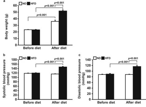
To induce obesity-mediated hypertension, mice were random- ly divided into two groups and fed a ND or HFD for 17 weeks.
Before feeding different diets, there was no difference be- tween the body weights of the two groups. After 17 weeks of consuming different diets, both groups showed increased body weights, while HFD significantly accelerated the body weight increase (from 22.8 ± 0.2 to 34.7 ± 0.9 g in the ND group, from 23.0 ± 0.3 to 48.1 ± 1.4 g in the HFD group) (p <
0.001 ND vs. HFD) (Fig. 1a). Before feeding different diets, there was no difference between the systolic blood pressures of the two groups. The ND did not affect systolic blood pres- sures (from 118.4 ± 1.4 to 115.4 ± 1.2 mm Hg), but the HFD significantly increased the systolic blood pressures (from 119.8 ± 1.3 to 147.3 ± 2.2 mm Hg) ( p < 0.001 before HFD vs. after HFD) (Fig. 1b). The diastolic blood pressures were also not different between groups before feeding different di- ets. The ND did not affect diastolic blood pressure (from 88.0
± 1.3 to 88.2 ± 1.0 mm Hg), but the HFD increased diastolic blood pressure (from 89.6 ± 2.3 to 116.3 ± 3.2 mm Hg) (p <
0.001 before HFD vs. after HFD) (Fig. 1c).
Treatment of CG200745 ameliorated HFD-induced hypertension
To investigate the effect of CG200745 on HFD-induced hy- pertension, each diet-fed group was administered with vehicle or CG200745 (0.2 mg kg −1 day −1 , i.p.). The blood pressure of ND-fed mice did not change in response to either vehicle or CG200745 (Fig. 2a, b). While the vehicle-administered HFD group maintained high systolic and diastolic blood pressure, the CG200745 treatment lowered the blood pressure to the normal state (from 149.1 ± 2.5 to 121.0 ± 1.2 mm Hg of systolic blood pressure, from 119.2 ± 3.5 to 89.3 ± 1.2 mm Hg of diastolic blood pressure) (p < 0.001 vehicle-HFD vs.
CG200745-HFD for both systolic and diastolic blood pres- sures) (Fig. 2a, b). The body weights and consumption of food and water of ND- and HFD-fed mice did not change with the CG200745 treatment (Fig. 2c–e).
CG200745 reversed HFD-induced increase in HDAC activity and expression in mouse kidney
HDAC activity in mouse kidney was higher in HFD group (22.6 ± 0.9 μM/kidney 50 μg) than in ND group (19.0 ± 0.9 μM/kidney 50 μg) (p = 0.011 vehicle-ND vs. vehicle-HFD).
The CG200745 treatment to HFD group reversed the HDAC activity (19.1 ± 0.6 μM/kidney 50 μg) to the level in the ND group (p = 0.005 vehicle-HFD vs. CG200745-HFD).
CG200745 did not affect HDAC activity in the ND group (Fig. 3a). mRNA expressions of HDAC1, 2, 3, and 6 in the kidney were higher in HFD group than in ND group.
However, CG200745 administration did not significantly downregulated the expressions of HDACs in the HFD group
(Fig. 3b–e) indicating that 200 μg/kg of CG200745 could reduce HDACs activities but not expressions.
CG200745 downregulated the expressions
of renin-angiotensin system components and serum Ang II in HFD-fed group
mRNA expression of angiotensinogen in the kidney was higher in HFD group (1.5 ± 0.0-fold) than in ND group (1.0
± 0.1-fold) (p < 0.001 vehicle-ND vs. vehicle-HFD).
CG200745 administration downregulated the expression of angiotensinogen in the HFD group to the ND group level (0.8 ± 0.1-fold) (p < 0.001 vehicle-HFD vs. CG200745- HFD) (Fig. 4a). Protein angiotensinogen level was also upreg- ulated in HFD group in the kidney (1.6 ± 0.2-fold) than in ND group (1.0 ± 0.1-fold) (p = 0.028 vehicle-ND vs. vehicle- HFD). CG200745 administration downregulated the protein angiotensinogen level in the HFD group (0.8 ± 0.1-fold) (p = 0.008 vehicle-HFD vs. CG200745-HFD) (Fig. 4b). Serum Ang II concentration was higher in the HFD group (15.2 ± 2.6 ng/μL) than in the ND group (1.7 ± 0.2 ng/μL) (p < 0.001 vehicle-ND vs. vehicle-HFD). CG200745 administration de- creased serum Ang II (6.5 ± 2.1 ng/μL) in the HFD-fed mice (p = 0.030 vehicle-HFD vs. CG200745-HFD) (Fig. 4c).
mRNA expression of renin in the kidney was higher in the HFD group (1.4 ± 0.0-fold) than in ND group (1.0 ± 0.1-fold) ( p < 0.001 vehicle-ND vs. vehicle-HFD). CG200745 admin- istration downregulated the expression of renin in the HFD group to the ND group level (1.1 ± 0.0-fold) (p = 0.001 vehicle-HFD vs. CG200745-HFD) (Fig. 4d). mRNA expres- sion of ACE in the lung was higher in the HFD group (1.6 ± 0.1-fold) than in ND group (1.0 ± 0.1-fold) (p = 0.006 vehicle- N D v s . v e h i c l e - H F D ) . C G 2 0 0 7 4 5 a d m i n i s t r a t i o n
0 10 20 30 40 50 60
Body w e ight (g)
p<0.001
0 20 40 60 80 100 120 140 160 180
S y stolic blood pr essur e (mmHg)
p<0.001 p<0.001
After diet Before diet
b c
0 20 40 60 80 100 120 140
Diastolic blood pr essur e (mmHg)
p<0.001
After diet Before diet
a
p<0.001
After diet Before diet
p<0.001
ND HFD ND HFD p<0.001
ND HFD
Fig. 1 Increased body weight and
blood pressure by HFD. Mice
were fed either ND or HFD for 17
weeks. Blood pressure was
measured using the tail-cuff
method. Graphs summarize body
weight (a), systolic blood pres-
sure (b), and diastolic blood
pressure (c). HFD accelerated in-
crease in body weight and blood
pressure. Results are expressed as
the mean ± SE ( n = 5–8 mice per
group). ND, normal diet; HFD,
high-fat diet
downregulated the expression of ACE in the HFD group to the ND group level (1.0 ± 0.1-fold) (p = 0.006 vehicle-HFD vs.
CG200745-HFD) (Fig. 4e).
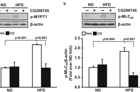
CG200745 decreased the phosphorylation of MLC 20 via phosphorylation of MYPT1 in mouse mesenteric artery
To investigate the mechanism by which HFD-induced hyper- tension was ameliorated by CG200745, we measured phos- phorylation of MYPT1, which increases phosphorylation of MLC 20 resulting in vascular contraction. MYPT1 phosphory- lation was higher in the HFD group (2.2 ± 0.0-fold) than in ND group (1.0 ± 0.0-fold) (p < 0.001 vehicle-ND vs. vehicle- HFD). MLC 20 phosphorylation was higher in the HFD group (1.8 ± 0.2-fold) than in ND group (1.0 ± 0.1-fold) (p = 0.004 vehicle-ND vs. vehicle-HFD). CG200745 administration downregulated the phosphorylation of MYPT1 in the HFD group to the ND group level (1.0 ± 0.2-fold) (p = 0.003 vehicle-HFD vs. CG200745-HFD). CG200745 administra- tion downregulated the phosphorylation of MLC 20 in the
HFD group (0.6 ± 0.1-fold) (p = 0.001 vehicle-HFD vs.
CG200745-HFD) (Fig. 5a, b).
Discussion
The most important finding of the current study is that novel HDAC inhibitor CG200745, which was developed for cancer treatment, is useful for ameliorating HFD-induced hyperten- sion and the major target is Ang II. These data offer CG200745 as a novel therapeutic option for the obese hypertension.
HDAC inhibitors have been studied primarily in the field of cancer and recently studied in cardiovascular diseases (Yoon and Eom 2016; Eckschlager et al. 2017). Various HDAC inhibitors have shown anti-hypertensive effects through various mechanisms. Although it has been generally accepted that acetylation of histones loosens the chromosomes and recruits transcription factors to the promoter region resulting in activation of gene expression, recent studies showed that this is not always the case. For example, VPA-
0.0 0.5 1.0 1.5 2.0
0 2 4 6 8
ND-Veh ND-CG
HFD-Veh HFD-CG
0.0 0.5 1.0 1.5 2.0
0 2 4 6 8
ND-Veh ND-CG
HFD-Veh HFD-CG
40 60 80 100 120 140
0 1 2 3 4 5 6 7 8 9
ND-Veh ND-CG
HFD-Veh HFD-CG
*** ***
*** *** *** *** *** *** *** **
### ###
###
###
###
###
##
##
&& &
+
&
&&
&&
&&&
&&& &&&
0 20 40 60
0 1 2 3 4 5 6 7 8 9
ND-Veh ND-CG
HFD-Veh HFD-CG
*** *** *** *** *** *** *** *** *** ***
### ### ### ### ### ### ### ### ### ###
*** *** *** *** *** *** *** *** *** **
### ### ### ###
### ### ###
###
&
&&
##
&&
&&
&&&
&&&
&&& &&&