Ⅰ. 서 론
발치는 개개의 치아에 있어서 비가역적인 최후의 처치가 되므로 발치가 필요하다는 판정을 내리기까지는 신중을 기하지 않으면 안된다. 일반적인 원칙은 치아가 기능을 제 대로 못하는 경우 발치의 적응증이 되는데, 종종 부적당한
악궁 길이나 맹출 공간의 부족에 의해 매복되는 경우가 있 다. 매복 하악 제3대구치처럼 맹출이상 등으로 치아의 존 재 자체가 장애의 원인이 되고 있는 경우 발치의 적응증이 된다. 매복치아를 남겨둔다면 주위 치아나 골의 손실등 건 전한 조직들이 손상받을 가능성이 높아진다.
한편, 매복 제3대구치 발치는 인접 제2대구치에 여러 가 지 치주적 문제를 일으킬 수 있다1-3)
. 발치 후 일차치유가 일
어나는 동안 제2대구치 주위로 dehiscence가 발생할 수 있 으며 이것은 이차적으로 치유될 수 있다. 이차 치유는 부착 소실을 야기할 수 있고 제2대구치 원심부에 치주 결손을 일으킬 수 있다4). 또한 발치 도중 골 삭제나 발치와로 인한
골결손을 보이기도 한다. 이러한 문제들은 환자의 나이가 김 경 욱330-716, 충남 천안시 신부동 산 7-1 단국대학교 치과대학 부속병원 구강외과
Kyung-Wook KimDept. of OMFS, College of Dentistry, Dankook University, San 7-1 Sinbudong, Choenan, Chungnam, 330-716, Korea Tel: 82-41-550-1991 Fax: 82-41-551-8988
E-mail: [email protected]
매복 하악 제3대구치 발치와에 Atelo-collagen Sponge 삽입이 제2대구치 예후에 미치는 영향
남진우∙김경욱
단국대학교 치과대학 구강악안면외과학교실
THE EFFECTS OF ATELO-COLLAGEN SPONGE INSERTION ON THE PERIODONTAL HEALING OF SECOND MOLARS AFTER IMPACTED MANDIBULAR THIRD MOLAR EXTRACTION
Jin-Woo Nam, Kyung-Wook Kim
Department of Oral and Maxillofacial surgery, College of Dentistry, Dankook University
Extracellular matrix (ECM) is known to function as a reservoir of endogenous growth factors, can be an effective delivery system of growth factor that easily lost bioactivity in solution.
Fibrillar collagens like type I collagen, are the major constituent of the ECM and structural protein of bone. Also, it can be a scaffold for osteoblast migration.
The purpose of this study was to compare the effects of absorbable Atelo-collagen Sponge(Teruplug
�) insertion in tooth extraction sites on peri- odontal healing of the mandibular second molar after the extraction of the impacted third molar.
The study population comprised 31 cases who had been scheduled for surgical removal of impacted mandibular third molars. All patients were in good general health and were not using any medication that would influence wound healing after surgery. In 15 cases control group, none was inserted into the tooth extraction site. In 16 cases experimental groups, Teruplug
�was inserted into the tooth extraction site.
We evaluated tooth mobility, pocket depth, gingival margin level preoperatively and 1 week, 2 weeks, 4 weeks, and 3 months postoperatively. The change was compared with two groups using Mann-Whitney test.
The results were as follows.
1. There was no significant change of tooth mobility on both groups.
2. There was tendency of decreasing of previous pocket depth causing tooth extraction on both groups.
3. On gingival margin level, there was various change according to initial swelling and loss of attachment on both groups.
4. There was tendency of decreasing of gingival margin level on both groups because of removal of inflammation and decreasing of previous pock- et depth.
5. There was large change of pocket depth on buccal middle, distal, lingual distal area because of tooth extraction and bone reduction. Compared with the control group and experimental group, we observed significant difference during some periods.
The results of this study suggest that absorbable atelo-collagen sponge (Teruplug
�) is relatively favorable bone void filler with prevention of tissue collapse, food packing and enhance periodontal healing.
Key words: Extracellular matrix, Collagen, Teruplug
�Abstract
(J. Kor. Oral Maxillofac. Surg. 2009;35:112-119)많을수록, 발치 전 치주적 문제가 있었을 경우 더 빈번히 나타날 수 있다5,6)
.
치아를 발치한 후 생기는 발치창은 치은에서 치조심부에 달하는 실질 결손창으로서 골의 개방창이다. 그러나 치조 벽은 혈병으로 보호되고 치조벽에는 치근막의 일부가 잔존 하고 있어서 여기에서 증식되는 섬유아세포가 발치와 내의 혈병을 신속하게 기질화시킨다. 술 후 7일경까지 혈병은 육 아조직에 의해 치환된다. 좀 더 성숙된 결합조직에 의한 육 아조직의 치환은 술 후 3~4일에 시작되어 약 20일 후에 완 성된다. 발치와의 상피화는 치은연에서 술 후 4일경부터 시 작되지만 약 24~35일 경과 후에도 종결되지는 않는다. 술 후 38일경까지 발치와의 ⅔가 거친 원섬유성 골로 채워지 지만 이 과정은 술 후 6주~8주까지도 종결되지는 않는다.
여러 연구에 의하면 골 형성을 위해서는 성장 인자나 분 화 인자 같은 분자 형성 신호, 신호에 반응하여 조골세포가 될 수 있는 숙주 세포, 분자 형성 신호를 특정 위치로 전달 하고 숙주세포가 골 세포로 성장할 수 있도록 골격 역할을 하는 생체신호 전달 물질 및 살아있는 잘 혈관화된 숙주체 등이 필수 조건이라고 하였다7-9)
.
가장 좋은 방법으로 여겨지는 것이 자가골 이식인데 사 용 가능한 이식재 중 유일하게 골형성능이 있다. 하지만 공 여부에 또 다른 수술이 필요하고, 합병증이 생길 수 있으 며, 어떤 경우에는 충분한 양의 골을 얻지 못할 수 있다는 단점이 있다. 이런 면에서 골대체물질은 많은 장점을 가지 고 있다고 할 수 있다.
세포외 기질은 내인성 성장 인자의 저장고 역할을 한다10,11)
.
성장인자 단백질은 수용액 상태에서 쉽게 불활성화되기 때 문에 치료목적으로 사용될 때는 효과적인 전달 시스템이 필 요한데 세포외 기질이 이상적인 모델이 될 수 있다12). 단백 분
해로부터 성장 인자를 보호하고, 불활성 상태에서 격리시키 고 환경적 자극에 대해 방출하는 역할을 한다10-13). Type I col- lagen 같은 fibrillar collagen은 세포외 기질의 주요 성분이다
14).
또한 골의 주요 구성 단백질로서, 조골세포의 이주를 위한 자연적인 기질이다15). collagen은 연조직 치유를 위해 사용되
어 왔으며16), 골조직 재건에도 사용되고 있다
17,18).
Collagen 사용의 장점으로 서로 다른 종간에도 적은 면역
반응을 일으키고19,20), 유용하며, 자연에 풍부하며, 지혈 촉진
작용을 하며, 다른 형태로 만들기 쉽다는 점 등이 있다21,22).
Teruplug
�(Termo Co. Tokyo, Japan)는 흡수성 아테로-콜라
겐 스폰지로써, 송아지의 진피에서 추출한 85~95%의 type Icollagen과 5~15%의 type II collagen으로 구성되어 있다. 생
체적합성을 위해 열처리 방법으로 Cross-link된 아테로-콜 라겐으로 만들어져 항원성을 최소화하였다. 발치와에 삽 입하여 사용할 수 있는데 상처를 보호해 주고, 육아조직형 성을 촉진시켜준다. 지혈작용을 촉진하고, 주변 조직 세포 를 흡수하여 그와 동일한 조직을 생성함으로써 치유를 촉 진시킨다23).
이에 이번 연구를 통해 매복 하악 제3대구치 발치후
Atelo-collagen Sponge(Teruplug
�) 사용이 제2대구치 예후에
미치는 영향에 대해 조사해 보고자 하였다.Ⅱ. 연구 재료 및 방법 1. 연구 대상
매복 하악 제3대구치 발치를 주소로 단국대학교 치과병 원 구강악안면외과에 내원한 환자 22명(남자 13명, 여자 9 명)을 대상으로 하였다.
환자의 연령대는 10대 1명, 20대 19명, 30대 1명, 40대 1명 으로 대부분 20대였다.
1) 방사선 사진상 매복치의 깊이를 Pell & Gregory 분류에
따라 Level A, B, C로 분류하였다(Table 1).Level A : 매복지치 치관이 인접된 제2대구치 치경부 상
방 존재Level B : 매복지치 교합면이 제2대구치 치경부에 근접 Level C : 매복치 전체 치관이 제2대구치 치근첨 근처에
위치
2) 제2대구치에 대한 매복 제3대구치의 방사선학적인 공
간 관계는 다음과 같았다(Table 2).2. 연구 방법
술전 모든 환자에게 재료의 사용과 술후 치주조직 평가 에 대해 설명하고 동의를 얻었다.
임의로 대조군(Control, Teruplug�를 사용하지 않은 그룹)
15 cases와 실험군(Experimental, Teruplug
�를 사용한 그룹)16 cases로 나누었다.
발치 전, 발치 후 1주, 2주, 4주, 3개월로 나누어 치주상태 를 평가하였다.
모든 술식 및 평가는 오차를 줄이기 위해 동일한 술자에 의해 시행되었다.
치주 상태 평가시 William's periodontal probe를 사용하였다.
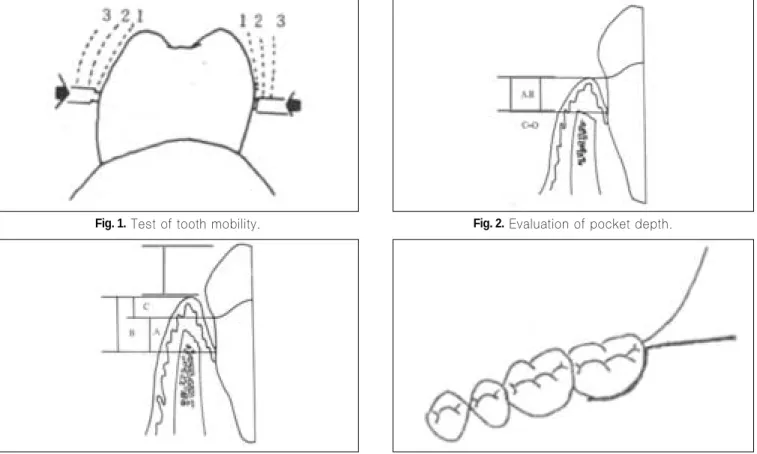
1) 치아동요도는 다음과 같이 4가지로 분류하였다(Fig. 1.).
∙ 생리적 동요도
∙ 1도 : 생리적 동요도보다 근원심 및 협설측으로 약간 증가된 동요도 (1mm이내)
Table 2.
Angulation of each tooth.mesioangulation horizontal vertical distoangulation Total 7 cases 19 cases 4 cases 1 case 31 cases Table 1.
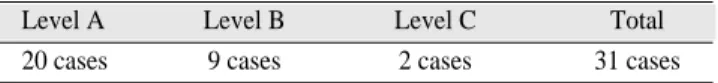
Level of each tooth.Level A Level B Level C Total
20 cases 9 cases 2 cases 31 cases
∙ 2도 : 중등도의 근원심 및 협설측으로의 치아 동요도
(1mm 이상)
∙ 3도 : 심한 치아 동요도를 보이고 수직으로도 동요도 를 보이는 경우
2) Pocket depth는 Free gingival margin에서 Pocket bottom
까지 거리를 측정하였다(Fig. 2.).제2대구치의 협측 근심, 중간, 원심, 설측 근심, 중간, 원 심 6부분으로 나누어 Pocket depth를 측정하였다.
3) Gingival margin level은 각 Cusp tip에서 Free gingival margin까지 거리를 측정하였다.
근심 협설측, 원심 협설측 교두부위에서 Gingival margin
level을 측정하여 치은 퇴축여부를 평가하였다(Fig. 3.).
4) 발치 술식 (Fig. 4.)
(1) 점막골막 피판의 설계와 형성 (2) 치조골의 삭제
(3) 치아의 분할 및 발치 (4) 창상의 봉합
1:100,000 epinephrine을 함유한 2% Lidocaine 국소마취하
에 full-thickness muco-periosteal flap을 형성하여 매복 하악 제3대구치를 발치하였다. flap 절개는 retromolar trigone부 위에서 시작해 하악 제2대구치 원심부까지 연장한 후 제2대구치 협측 열구 절개를 시행하였다. 고속(high speed)
round bur로 치관절제술(odontectomy)를 시행하고, 하악 매
복 제3대구치 주위로 골삭제술을 시행하였으며, 필요한 경 우 주수하에 low Lindemann bur를 사용해 치관절제술(odontectomy)를 시행하여 발치를 하고 대조군의 경우 아
무것도 넣지 않았으며, 실험군의 경우 발치와의 크기에 따 라 medium, small size의 Teruplug를 삽입 후 black silk로 봉 합하였다.5일 간의 항생제(amoxicillin and clavulanic acid) 및 진통
제를 처방하였다.Ⅲ. 연구 결과 1. 치아 동요도
모든 환자에서 술 전, 술 후 생리적 동요도를 보였다.
2. Pocket Depth
Window용 SPSS 14.0을 이용
95% 유의 수준에서 비모수통계 표본검정 중 Mann-Whitney Test를 시행하였다.
초기 치주상태 평가 후 각 시기에 따른 변화량을 측정하 였다 (Fig. 5,6,7,8,9 & 10, Table 3,4,5,6,7 & 8).
Fig. 3.
Evaluation of gingival margin level.Fig. 4.
Flap design.Fig. 1.
Test of tooth mobility.Fig. 2.
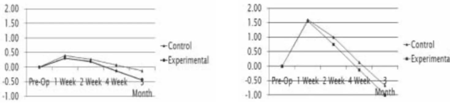
Evaluation of pocket depth.Fig. 5.
Change of pocket depth (Mesio-buccal).Fig. 6.
Change of pocket depth (Mid-buccal).Fig. 7.
Change of pocket depth (Disto-buccal).Fig. 8.
Change of pocket depth (Mesio-lingual).Table 6.
Change of pocket depth (Mesio-lingual).Mesio-lingual Pre-OP Post 1 Week Post 2 Weeks Post 4 Weeks Post 3 Months
Control 2.73±0.59 -0.07±0.46 -0.07±0.59 -0.47±0.64 -0.47±0.74
Experimental 2.94±0.25 0.00±0.00 -0.19±0.40 -0.31±0.48 -0.38±0.50
P value 0.538 0.194 0.085 0.590
Degree : mm, Mean±SD(range) *Significant difference at P<.05 Table 5.
Change of pocket depth (Disto-buccal).Disto-buccal Pre-OP Post 1 Week Post 2 Weeks Post 4 Weeks Post 3 Months
Control 4.87±2.03 4.67±2.23 3.80±2.21 2.00±1.96 -0.40±2.47
Experimental 5.94±3.02 4.56±2.78 3.38±3.30 2.25±3.44 -1.38±3.61
P value 0.858 0.307 0.187 0.031*
Degree : mm, Mean±SD(range) *Significant difference at P<.05 Table 4.
Change of pocket depth (Mid-buccal).Mid-buccal Pre-OP Post 1 Week Post 2 Weeks Post 4 Weeks Post 3 Months
Control 3.20±1.32 1.60±1.92 1.00±2.00 0.13±0.99 -0.67±1.18
Experimental 4.00±2.19 1.56±1.71 0.75±1.81 -0.13±2.06 -1.00±1.75
P value 0.918 0.048* 0.763 0.746
Degree : mm, Mean±SD(range) *Significant difference at P<.05 Table 3.
Change of pocket depth (Mesio-buccal).Mesio-buccal Pre-OP Post 1 Week Post 2 Weeks Post 4 Weeks Post 3 Months
Control 2.60±0.51 0.40±0.74 0.27±0.59 0.07±0.46 -0.13±0.52
Experimental 3.06±0.57 0.31±0.70 0.19±0.75 -0.13±0.62 -0.44±0.63
P value 0.687 0.644 0.482 0.467
Degree : mm, Mean±SD(range) *Significant difference at P<.05
Fig. 9.
Change of pocket depth (Mid-lingual).Fig. 10.
Change of pocket depth (Disto-lingual).Table 8.
Change of pocket depth (Disto-lingual).Disto-lingual Pre-OP Post 1 Week Post 2 Weeks Post 4 Weeks Post 3 Months
Control 5.27±1.87 0.93±1.94 0.27±1.91 -0.60±1.96 -1.60±1.96
Experimental 5.31±2.18 1.88±2.36 1.25±2.72 -0.19±2.76 -1.75±2.08
P value 0.206 0.117 0.399 0.645
Degree : mm, Mean±SD(range) *Significant difference at P<.05 Table 7.
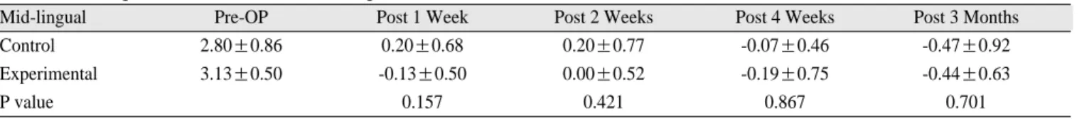
Change of pocket depth (Mid-lingual).Mid-lingual Pre-OP Post 1 Week Post 2 Weeks Post 4 Weeks Post 3 Months
Control 2.80±0.86 0.20±0.68 0.20±0.77 -0.07±0.46 -0.47±0.92
Experimental 3.13±0.50 -0.13±0.50 0.00±0.52 -0.19±0.75 -0.44±0.63
P value 0.157 0.421 0.867 0.701
Degree : mm, Mean
±SD(range) *Significant difference at P<.05Fig. 11.
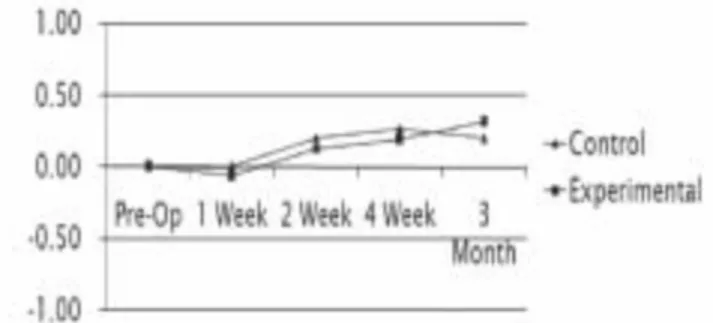
Change of gingival margin level (Mesio-buccal).Fig. 12.
Change of gingival margin level (Disto-buccal).Table 10.
Change of gingival margin level (Disto-buccal).Disto-buccal Pre-OP Post 1 Week Post 2 Weeks Post 4 Weeks Post 3 Months
Control 5.47±1.06 0.13±1.06 0.33±0.98 0.53±0.99 0.73±0.88
Experimental 5.44±1.21 -0.69±1.20 -0.13±1.09 0.31±1.40 0.44±1.03
P value 0.057 0.123 0.347 0.268
Degree : mm, Mean±SD(range) *Significant difference at P<.05 Table 9.
Change of gingival margin level (Mesio-buccal).Mesio-buccal Pre-OP Post 1 Week Post 2 Weeks Post 4 Weeks Post 3 Months
Control 6.87±0.64 -0.07±0.80 0.13±0.83 0.20±0.77 0.33±0.72
Experimental 7.19±1.17 -0.38±0.89 -0.25±0.77 -0.13±0.81 0.13±0.50
P value 0.539 0.684 0.725 0.614
Degree : mm, Mean±SD(range) *Significant difference at P<.05
3. Gingival Margin Level
Window용 SPSS 14.0을 이용
95%
유의 수준에서 비모수통계 표본검정 중 Mann-Whitney Test를 시행하였다.
초기 치주상태 평가 후 각 시기에 따른 변화량을 측정하 였다 (Fig. 11,12,13 & 14, Table 9,10,11 & 12).
Ⅳ. 총괄 및 고찰
매복치아와 인접한 치아는 치주질환에 걸리기 쉬워 매복 된 제3대구치가 존재함으로써 인접 제2대구치의 원심측 골의 양이 감소될 수 있다.
발치창의 치유과정을 살펴보면 보통 술후 40일경 발치와 가 원섬유골로 채워진다고 한다. 이때 신생골 지주의 숫자 와 배열은 치조골에 가해지는 기능적 응력에 의존한다.
매복 제3대구치 발치가 제2대구치에 미치는 영향이나 치 주 치유를 촉진시키고자 하는 여러 방법들에 대한 상반된 결과를 나타내는 여러 연구들이 있어왔다4,24-26)
.
Cunqueiro 등
24)은 paramarginal flap이 marginal flap에 비해 발치 후 5일에서 10일 사이에 probing depth를 줄여준다고 하였으나 3달 후 두 flap간 probing depth의 차이는 없다고 하였다. Tugrul Kirtiloglu 등27)은 Szmyd flap이 3-corneredflap에 비해 초기 치유에는 좋은 영향을 준다고 하였으나
12달 후 치주상태는 큰 차이가 없다고 하며, 초기 적은 외
상을 가하는 주의 깊은 발치가 초기 치유에 중요하다고 하 였다. Jakse 등4)은 flap 디자인이 일차 치유에 영향을 준다 고 하였다. Quee 등26)은 제2대구치 치은을 보존하는 flap 디 자인을 하더라도 부착소실을 예방하지는 못한다고 하였 다. Stephens 등28)은 발치 12주 후의 치주상태가 발치 전보 다 좋아진다고 하였으나 flap 디자인간 차이는 없다고 하였 다.반면 Rosa, Quee 등26,29)은 발치 6개월 후의 치주상태가 발 치 전보다 나빠진다고 하였다.
Platelet-rich plasma(PRP)는 platelet-derived growth fac- tors(PDGF)나 transforming growth factor-β같은 성장인자를
함유해 제3대구치 발치 후 제2대구치 발치창의 치유와 치 주적 합병증 예방에 사용될 수 있었다30-32).
생체내 삽입 물질은 염증반응을 일으킬 수 있으나 colla-
gen은 친수성, 지혈작용, 상처 보호 작용 등
34-36)과 생체친화 성, 생체내 분해능력 및 다른 거대분자와의 상호작용 등33) 이 탁월하여 염증반응을 최소화하고 골전도성이 좋아 치 유기간을 단축시킬 수 있다15).
Collagen은 골전도 능력이 있어 숙주 세포가 재구성하는
데 필요한 구조적 골격을 제공하는 역할을 하는데, 중요한 구조적 성질은 다공성(porocity), 소공크기(pore size), 소공 연결성(pore connectivity), 표면거칠기(surface roughness) 등 이다. 그러나 골전도성은 결손부의 크기, 수혜부의 세포성,Fig. 13.
Change of gingial margin level (Mesio-lingual).Fig. 14.
Change of gingival margin level (Disto-lingual).Table 12.
Change of gingival margin level (Disto-lingual).Disto-lingual Pre-OP Post 1 Week Post 2 Weeks Post 4 Weeks Post 3 Months
Control 4.80±1.57 0.00±0.93 0.20±1.01 0.27±0.96 0.20±1.08
Experimental 4.81±1.42 -0.06±0.68 0.13±0.89 0.19±0.83 0.31±0.89
P value 0.685 0.85 0.981 0.279
Degree : mm, Mean±SD(range) *Significant difference at P<.05 Table 11.
Change of gingival margin level (Mesio-lingual).Mesio-lingual Pre-OP Post 1 Week Post 2 Weeks Post 4 Weeks Post 3 Months
Control 5.67±1.40 0.00±0.85 0.20±0.56 0.07±0.59 0.27±0.80
Experimental 5.38±1.09 0.13±0.62 0.19±0.75 0.25±0.86 0.13±0.89
P value 0.477 0.423 0.364 0.062
Degree : mm, Mean±SD(range) *Significant difference at P<.05
공여조직과의 접촉, 수혜부로의 수동적 신생골 성장, 흡수 와 재성형에 대한 숙주의 조절성 등의 요소들에 의해 좌우 될 수 있다.
최근 유전공학, 생화학 및 생물공학의 발전과 더불어
rhBMP
또는 성장인자 등 인공적으로 합성된 폴리펩티드들이 주목받고 있다. Collagen 기질에 성장인자나 proteo-
glycan
같은 세포외 구성요소를 첨가함으로써, 또는 화학적 변형을 통해서 생체물질로서의 적용 가능성을 증가시 키고 치유를 촉진시킬 수 있다40-45)
. Basic fibroblast growth factor(bFGF)는 angiogenesis와 미분화간엽세포의 증식을
촉진시키는 성장인자이다. 세포외 기질의 heparan sulfateproteoglycans와 결합함으로써 기능하게 된다
10-13,44-46). Collagen은 상처의 neovascularization에 의한 remodeling이
일어날 때까지 bFGF의 일시적 저장소 역할을 할 수 있다14).
또한 collagen의 전기적 전하 분포가 세포외 기질의 분포에 영향을 줄 수 있다. 조골세포는 integrin-RGD(-arg-gly-asp-)site의 상호작용에 의해 직접 collagen에 부착한다고 알려져
있다43). 또한 세포 부착에 음전하를 띤 site가 밀접하게 관련
되어 있다고 알려져 있다. 따라서 collagen의 전기적 전하 를 변화시킴으로써 성장인자의 부착에 영향을 줄 수 있고 세포의 이주와 분화에 영향을 미쳐 결과적으로 새로운 골 형성에 영향을 줄 수 있다15).
현재 각 성장요소들의 작용은 아직 확실히 규명되지는 않았고, 상호 반응이 복잡하다. 많은 학자들은 각 요소들이 창상 치유에 미치는 역할이나 상호 반응이 규명되어, 향후 인공 합성된 각 성장인자들이 임상에 이용될 수 있을 것으 로 생각되고 있다.
Ⅴ. 결 론
1. 두 군 모두 치아 동요도에는 큰 변화를 보이지 않았다.
2. 두 군 모두 발치로 인해 기존에 존재하던 Pocket depth
가 감소하는 양상을 보였다.3. Gingival margin level의 경우 두 군 모두 초기에 swelling
으로 증가하거나 부착 상실로 감소하는 경우 등 다양 한 변화를 보였다.4. 조사기간 동안 염증 소멸, pocket depth 감소 등의 치유
과정을 거쳐 두 군 모두 전체적 gingival margin level은 감소하는 양상을 보였다.5. 제2대구치 협측 중간, 원심, 설측 원심 pocket depth의
경우 발치창, bone 삭제 등으로 큰 변화를 보였으며, 대 조군과 실험군을 비교한 결과 치유 속도에 있어 일부 기간에서 유의할 만한 차이를 보였다.이상의 결과를 종합하여 볼 때, 임상적으로 Teruplug�
(Termo Co. Tokyo, Japan) 사용시 발치와의 packing으로 함
몰 및 food packing 방지 효과가 있었으며, 3개월간의 조사 기간동안 특히 제2대구치 원심부에서 치주조직 치유 촉진 효과가 있는 것으로 생각되었다.이번 조사 기간은 3개월로 향후 bone healing에 따른 장기 간의 예후 관찰 및 추가적인 평가가 필요할 것으로 사료되 었다.
참고문헌