집중치료실에서 치료한 중첩성 경련 환자의 신경생리학적 결과 분석
동남보건대학 임상병리과1, 서울아산병원 신경과2
김대식1․김천식2
The Analysis of Neuro-Physiological Outcome of Patients with Status Epilepticus in an Intensive Care Unit
Dae-Sik Kim1, and Cheon-Sik Kim2
Department of Clinical Laboratory Science, Dongnam Health College, Suwon 440-714, Korea1 Department of Clinical Neurosciences, Asan Medical Center, Seoul 138-736, Korea2
Status epilepticus is a medical emergency, so that rapid and vigorous treatment is required to prevent neuronal damage and systemic complication. Status epilepticus is generally defined as a continuous or intermittent seizure or an unconscious condition after the onset of seizure, lasting for 30 minutes or more. We report here the outcome of status epilepticus. We retrospectively reviewed medical record of 15 patients who were diagnosed with status epilepticus at the Asan Medical Center from January 2003 to February 2004. This outcome was evaluated considering various factors such as age of patients, history of seizures, neurologic impairment, etiology, mortality, return to baseline and initial electroencephalogram (EEG) findings. The range of age was between 1 to 79 years old and the longest duration of treatment was 118 days. Most patients were treated by using pentobarbital, midazolam, phenobarbital and other antiepileptic drugs. The overall mortality was 5 (33%) out of 15 patients. The mortality was related to etiology, underlying other medical conditions and initial EEG findings. 5 (55%) out of the 9 patients with acute etiology, 5 (71%) out of the 7 patients with a multifocal or burst-suppression EEG activity, and 3 (60%) out of the 5 patients with other medical disease were related to mortality. This data demonstrate high mortality due to status epilepticus. Mortality is related to etiology, other medical conditions and abnormalities on the initial EEG.
Key Words : Status epilepticus, Electroencephalogram, Etiology 대한임상검사학회지 : 37권 제2호, 96-101, 2005
1)
I. 서 론
중첩성 경련(status epilepticus)은 신경학적 응급질환으 로 빠른 시간에 적절한 조치를 취하지 않으면 높은 사망
교신저자 : 김천식, (우)138-736 서울특별시 송파구 풍납동 388-2, 서울아산병원 뇌신경센터
Tel : 02-3010-4831, 010-2282-5492 E-mail : [email protected]
본 연구는 2005년도 동남보건대학 연구비 지원에 의해서 시행되 었음.
률과 신경학적 장애가 발생하는 질환이다. 중첩성 경련은 30분 또는 그 이상동안 계속해서 경련발작을 하거나, 의 식회복 없이 반복되는 경련발작 상태를 말한다(Working Group, 1993). 영국에서는 매 년 대략 14,000명 정도의 중 첩성 경련 환자가 발생하고(Walker 등, 1995), 미국에서 는 약 150,000명 정도의 환자가 발생하였다(DeLorenzo 등, 1995).
동물실험 결과에 의하면 경련이 오래 지속되는 경우 첫 30분내에는 고혈압, 뇌혈류 증가 및 뇌 대사량 증가,
산혈증, 고혈당 소견을 보이나(Benowitz 등, 1986;
Lothman, 1990), 그 이후로는 저혈당, 저칼륨혈증, 저체 온, 저혈압 및 저산소증 소견을 보인다. 이러한 변화는 결 과적으로 뇌부종을 초래하고 뇌의 자율조절(autore- gulation) 능력한계를 지나칠 때 비가역적 신경 손상을 유 발하기 때문에 가능한 빨리 경련을 치료해야 되는 이유 가 된다(Terrence 등, 1981; Simon, 1985; Treiman 등, 1990). 중첩성 경련은 나이가 어릴수록 흔하고 경련이 오 래 지속될수록 후유증 빈도가 높다는 보고가 있다(오와 고, 1993; 차와 고, 1996). 특히, 1시간 이상 지속되거나 노인인 경우 예후가 나쁜 것으로 알려져 있다(Towne, 1994).
중첩성 경련 환자의 치료목표는 경련발작을 최대한 빨 리 종료시키는 것이며, 이에 중첩성 경련 환자에게 24시 간 집중감시 뇌파와 약물 농도에 의한 뇌파검사에서 주 기적 간질파(burst-suppression)를 확인하여 임상적인 완전 한 경련 조절을 유도하였다.
본 연구는 환자의 나이, 치료기간, 중첩성 경련원인, 다 른 질병을 가지고 있으면서 중첩성 경련이 함께 동반된 경우, 처음 실시한 뇌파검사 소견 등이 중첩성 경련을 치 료하는 데 있어 중요성과 임상적 유용성을 알아보고자 하였다.
Ⅱ. 재료 및 방법
2003년 1월부터 2004년 4월까지 서울아산병원 집중치 료실에서 치료한 15명의 중첩성 경련 환자를 대상으로 하 였고, 모든 환자는 뇌파 집중감시(EEG monitoring)를 시 행했다. 24 시간 뇌파 집중감시는 21채널 뇌파기기 (EEG-4421A, Nihon Koden, Tokyo, Japan)를 사용하였다.
환자의 나이, 성별, 내과적․신경과적 질병 유무, 특히 경련의 유무를 확인했고, 경련을 일으킨 원인(etiology), 중첩성 경련 치료기간, 첫번째 시행한 뇌파검사 소견, 뇌 영상 이미지 등을 차트를 통하여 정보를 수집했다. 뇌파 와 뇌영상 이미지는 신경과․소아과․방사선과 전문의가 판독하였다.
고용량(high-dose)의 phenobarbital, pentobarbital, midazolam 및 그 외의 항 경련제를 지속적으로 사용하여 혼수요법(coma therapy) 후 회복상태를 뇌파를 통하여 확 인하였다(Rashkin, 1987; Lowenstein, 1988; Krishnamurt 와 Drislane, 1999). 중첩성 경련의 치료 순서는 lorazepam
(0.1 mg/kg IV at 2mg/min)을 사용하여 뇌파 검사상 경련 이 멈추지 않으면 phenytoin(20 mg/kg IV at 50 mg/min) 또는 fosphenytoin(20 mg/kg PE IV at 150 mg/min)을 사 용하고, 이후 경련이 멈추지 않으면 추가로 phenytoin (5~10 mg/kg IV at 50 mg/min) 또는 fosphenytoin(5~10 mg/kg PE IV at 150 mg/min)을 사용하였다. Phenytoin과 fosphenytoin을 사용하여 경련이 멈추지 않을 경우 phenobarbital(20 mg/kg IV at 50~75 mg/min)을 사용하고, 이후 경련이 멈추지 않으면 추가로 phenobarbital(5~10 mg/kg IV at 50~75 mg/min)을 사용하였다. 이후에도 경 련이 멈추지 않는다면 midazolam, propofol 또는 pentobarbital 등을 사용하여 혼수요법을 하였다.
Midazolam, propofol 또는 pentobarbital은 첫 투여 후 약 12-24시간 유지 후 서서히 약물 투여량을 줄이면서 임상 적 경련발작이나 뇌파의 간질파를 관찰하였다.
원인에 따른 중첩성 경련의 분류는 Maytal 등 (1989)에 의해 분류되어진 5가지 형태로 분류하였다. (a) 급성 증상 성(acute symptomatic, AS) : 급격한 신경과적 손상이나 시스템 방해가 있으면서 동시에 경련이 발생한 경우, (b) 원발성 증상성(remote symptomatic, RS) : 경련을 일으킬 수 있는 신경과적 손상은 가지고 있으나 급격한 유발원 인(provocation) 없이 경련이 발생한 경우, (c) 급성원발성 증상성(remote symptomatic with acute precipitant, RSAP) : 경련을 일으킬 수 있는 신경과적 손상을 가지고 있으면 서 급격한 유발원인(provocation)과 함께 경련이 동반된 경우, (d) 진행성 뇌병증(progressive encephalopathy, PS) : 진행성 신경질환을 가지고 있으면서 경련을 일으킨 경우, (e) 열성경련(febrile) : 열과 힘께 동반되는 경련으로 분류 하였다.
Ⅲ. 결 과
고용량의 혼수요법를 통하여 15명의 환자를 치료하였 고, 각 환자의 임상적 특징은 Table 1과 2에 자세하게 설 명하였다. 중첩성 경련 환자는 남자 7명, 여자 8명으로 남 여 비율은 비슷하였고, 연령은 1세부터 79세까지 광범위 하였다. 사망률은 나이가 많을수록, 중첩성 경련이 1시간 이상 지속될수록 높았으며, 남여 비율은 남자 2명, 여자 3 명으로 여자가 높았다.
중첩성 경련의 치료기간은 10일 이내 8명, 10-30일 4 명, 30일 이상이 3명 이었다. 10일 이내 치료 환자 8명 중
Table 1. Etiology, duration and outcome of SE in 15 patients
Pat. Age(yrs) / Sex
H/O medical illness
H/O neuro illness
H/O
epilepsy Treatment
Treatment duration
(days)
Etiology Outcome Etiology group
1 1/F None Mental
retardation No PHB, DPH,
PTB 7
Previous traumatic
SDH
RTB RS
2 6/M None None No PHB, DPH,
PTB, MDL 75 Seizure Seizure AS
3 8/M None Hypoxic brain No PHB, DPH,
PTB, MDL 118 Viral
encephalitis Seizure RSAP
4 10/M None Hypoxic brain No DPH, PHB,
VPA, MDL 43 Seizure RTB RS
5 17/F None None No PHB, DPH,
PTB, MDL 11 Viral
encephalitis Died AS
6 23/F None None No DPH 23 MCAD
deficiency Seizure PE
7 24/F None Encephalitis,
febrail seizure Yes DPH, PHB,
ORFIL, TOPA 21 Viral
encephalitis Died RSAP
8 26/F HBV None No DPH,
ATIVAN 5 Viral
encephalitis RTB AS
9 27/M None None Yes DPH 7 Encephalitis RTB AS
10 28/M None None No DPH, VAL,
PTB 5 HSV
encephalitis RTB AS
11 41/M PCKD, CVA Mutiple brain
infartion No MDL 1
PCKD, CVD, Viral encephalitis
Died RSAP
12 42/F Liver failure None No Phenytoin 5 Seizure Died AS
13 48/M HBV LC
(Child C) None No DPH,
ATIVAN 6 Seizure Died AS
14 67/F Hepatitis None No NO MEDI 1 Hepatic
encephalitis RTB AS
15 79/F None None No Ativan 11 Encephalitis RTB AS
Pat., patient; DPH, dephenylhydration; PTB, pentobarbital; PHB, phenobarbital; MDL, midazolam; AVL, valprate; TOPA, topamax;
H/O, history of; HSV, herpes simplex virus; AS, acute symptomatic; RS, remote symptomatic; RSAP, remote symptomatic with acute precipitant; RTB, return to baseline; POKD, polycystic kidney disease; MCAD, medium-chain acyl-CoA dehydrogenase
3 명, 10-30일 이내의 4명 중 2명이 사망하였고, 30일 이 상 치료한 환자에서 사망자는 없었다.
중첩성 경련을 일으킨 환자의 원인은 급성 증상성이 9 명으로 가장 많았고, 원발성 증상성 2명, 급성원발성 증상 성 3명, 진행성 증상성 1명으로 나타났다. 중첩성 경련을 일으킨 원인에 따른 결과는 Table 3에 기재하였다. 15명 의 환자 중 5명이 사망하여 33%의 사망률을 보였으며, 이 는 중첩성 경련이 매우 심각한 질환임을 의미하였다. 원 인 분류에 의한 사망률은 다음과 같다. 급성 증상성 9명 중 3명, 원발성 증상성 3명 중 2명이 사망하여 각각 33%,
66%의 사망률을 나타내었다.
처음 실시한 뇌파 검사의 소견이 주기적 간질파 (burst-suppression)나 다초점(multifocal) 형태를 나타낸 환 자는 15명 중 7명, 이들 중 5명이 사망하여 처음 실시한 뇌파 검사의 소견이 사망률과 연관성이 깊음을 나타내었 다(Table 4).
다른 질병과 중첩성 경련이 동시에 동반된 경우 5명 중 3명, 단지 중첩성 경련만 나타낸 경우10명 중 2명이 사망 하였다(Table 5).
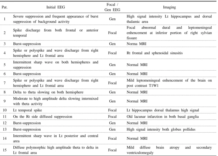
Table 2. Electroencephalogram and imaging of patients with status epilepticus
Pat. Initial EEG Focal /
Gen EEG Imaging
1 Severe suppression and frequent appearance of burst
suppression of background activity Gen High signal intensity Lt hippocampus and dorsal thalamic area
2 Spike discharge from both frontal or anterior
temporal Focal
Focal abnormal dural and leptomeningeal enhencement at inferior portion of right sylvian fissure
3 Burst-suppression Gen Norma MRI
4 Spike or polyspike and wave discharge from right
hemisphere and Lt frontal area Focal Rt frontal and sphenoidal sinusitis 5 Intermittent sharp wave on both hemispheres and
suppression Gen Normal MRI
6 Burst-suppression Gen Normal MRI
7 Spike or polyspike and wave discharge from right
hemisphere and Lt frontal area Focal Mild leptomeningeal enhencement of the brain on post contrast T1W1
8 Delta to theta slowing on both hemisphere Gen Normal MRI 9 Moderate to high amplitude delta slowing intermixed
with theta activity Gen Normal MRI
10 Lt temporal spike Focal Lt hippocampus dorsal thalamus high signal
11 On the Rt side diffused suppression Focal Old lacunar infarction in both basal ganglia
12 Burst-suppression Gen Normal MRI
13 Burst-suppression Gen High signal intensity both globus pollidus
14 Intermittent sharp wave in Lt posterior and central
area Focal Normal MRI
15 Diffuse polymorphic high amplitude theta to delta in
Lt frontal area Focal Mild diffuse brain atropy and secondary
ventriculomegaly
Table 3. Etiology versus outcome Etiology AS
(n=9) RS (n=2)
RSAP (n=3)
PE (n=1)
Total (n=15)
Died 3 - 2 - 5
New deficit 2 - 1 1 4
RTB 4 2 - - 6
AS, Acute sysmptomatic; RA, Remote symptomatic; RSAP, Remote symptomatic with acute precipitant; PE, Progressive encephalopathy
Table 4. Initial EEG versus outcome EEG Burst suppression
(n=7)
Non-burst- suppression
(n=8)
Total (n=15)
Died 5 - 5
New deficit 1 2 3
RTB 1 6 7
Table 5. Underlying desease versus outcome Outcome Underlying
desease (n=5)
Only status epilepticus (n=10)
Total (n=15)
Died 3 2 5
New deficit - 3 3
RTB 2 5 7
Ⅳ. 고 찰
중첩성 경련은 오랫동안 지속되는 경련발작 또는 의식 회복 없이 반복되는 경련을 말한다. 중첩성 경련이 30분 이상 지속 시 신경계 손상을 유발하기 때문에 가능한 빨 리 치료를 하는 것이 중요하다. 또한 중첩성 경련 기간이 길면 길수록 약물치료에 반응을 하지 않는 난치성으로 변하기 때문에 신경계 손상도 더욱 심해진다.
집중치료실에서 치료한 중첩성 경련 환자에 대한 결과 는 15명 환자 중 5명이 사망하여 사망률은 33%였고, 이 는 중첩성 경련을 보인 환자들의 예후가 매우 나쁘다는 것을 증명해 주었다. 중첩성 경련의 원인과 처음 실시한 뇌파 검사에서 주기적 간질파(burst-suppression)나 다초점 (multifocal) 모양을 나타낸 뇌파, 다른 질병과 중첩성 경 련이 동반된 경우 예후를 결정하는 중요한 인자로 작용하 였다. 중추신경성 감염, 뇌졸중, 저산소증, 다른 질병과 중 첩성 경련이 동반된 경우, 약물중독 및 뇌파검사에서 주 기적 간질파를 보이는 경우 예후가 나쁜 것으로 알려져 있고(Cross-Tsur와 Shinnar, 1993), 알코올과 관련된 중첩 성 경련은 예후가 좋은 것으로 알려져 있다.
중첩성 경련의 평균 치료 기간은 성인이 38-62시간, 소 아가 31.4일로 소아가 훨씬 긴 것으로 보고되어 있다 (Osorio 등, 1989; Krishnamurthy와 Orislane, 1996). 대상 환자들의 평균치료기간은 22.6일이고, 성인 8.5일, 소아 50.8일로 소아가 훨씬 길게 나타났다. 이러한 원인은 소아 의 경우 성인에 비해 약물에 대한 부작용이 높고, 발작을 일으키는 병인론적 차이점이 있을 것으로 생각되어 진다.
Mustafa 등(2001)의 보고에 의하면 치료 기간이 길수록 사망률이 높았으나 본 연구의 결과는 10일 이내 단기간 치료한 환자의 사망률이 더 높았다. 전체 15명 환자 중 5 명이 사망했고, 그 중 10일 이내 기간에 사망한 환자는 3 명, 10-30일 이내 2명, 30일 이상 치료한 환자의 사망자는 없었다. 10일 이내 사망자 3명은 다른 질병이 있으면서 동시에 중첩성 경련이 동반된 경우로, 이 경우 환자의 대 사성 이상과 동시에 신경계 손상이 초래되었기 때문에 환 자의 상태가 급속하게 나빠졌으리라 생각되어 진다.
원인별 사망률은 15명 환자 중 급성 증상성은 9명, 이 중 3명이 사망하고 2명은 새로운 신경학적 질환을 나타냈 고, 4명은 주기적 간질파는 없어졌으나 정상적인 생활을 할 수 없는 정신적 장애를 가지게 되었다. 급성 원인은 바 이러스성 뇌염(viral encephalitis) 진단이 6명, 원인모를 경 련이 3명이었다. 원발성 증상성은 2명으로 모두 정신적 장애를 나타내었고, 급성 원발성 증상성은 3명 중 2명이 사망하고 1명은 새로운 신경학적 질환을 가지게 되었다.
진행성 증상성은 1명으로 medium-chain acyl-CoA dehy- drogenase의 결핍을 나타냈고 지속적인 경련증상을 보였다.
뇌파검사에서 전반적 또는 다초점 이상파를 나타낸 환 자는 8명, 부분적 이상 뇌파를 보인 환자는 7명이었다. 이 들 중 주기적 간질파를 보인 7명 중 5명이 사망했고,
Cross-Tsur와 Shinnar 등(1993)이 발표한 결과와 동일하 게 주기적 간질파를 보인 경우 예후가 나쁜 것으로 나타 났다.
내과적 질환이 동반된 환자에서 경련의 발생은 요독증 뇌증(uremic encephalopathy), 간장 질환, 심장 질환 및 결 체 조직성 질환에서 경련이 발생하는 것을 볼 수 있었다.
급만성 신부전 환자의 35%가 발작을 유발하는 것으로 알 려져 있다(Rodriguez 등, 2004). 이런 경우 발작의 원인이 되는 대사성 이상을 교정하여야 하나 특별한 이유 없이 발작 발생 시 항경련제 치료가 필요하다. 만약 대사성 조 정이나 항경련제의 치료가 실시되지 않을 경우 중첩성 경련으로 진행될 수 있다. 우리의 결과에서 다른 질병과 동시에 중첩성 경련을 일으킨 환자는 5명이고, 그 중 4명 은 간부전(hepatic failure), 1명은 다낭성 신장질환 (polycystic kidney disease)을 가지고 있었고, 5명 중 3명 이 사망하여 사망률이 매우 높았다.
Ⅴ. 결 론
2003년 1월부터 2004년 4월까지 서울아산병원 집중치 료실에서 중첩성 경련으로 입원한 환자의 신경생리학적 결과 분석은 다음과 같다.
1. 중첩성 경련은 1세부터 79세까지 아주 광범위하게 나타났고, 성별과 관련이 없었다.
2. 치료기간은 성인 8.5일, 소아 31.4일로 소아의 치료 기간이 길었고, 이는 소아의 경우 약물에 대한 부작 용이 높고, 발작을 일으키는 병인론적 차이점 때문 으로 생각되어진다.
3. 중첩성 경련의 원인이 급성으로 나타난 환자 9명 중 5명이 사망하여 사망률은 55%였다.
4. 처음 실시한 뇌파 검사 소견이 주기적 간질파를 나 타낸 환자 7명 중 5명이 사망하여 사망률은 71%였 다.
5. 다른 질병과 동시에 중첩성 경련이 동반된 경우 5명 중 3명이 사망하여 사망률은 60%였다.
이상의 결과로서 중첩성 경련은 사망률과 연관성이 깊 고, 향후 이들과 관련된 연구들이 더 진행되어야 할 것이 다.
참 고 문 헌
1. Benowitz NL, Simon RP, Copeland JR. Status epilepticus: divergence of sympathetic activity and cardiovascular response. Annals of Neurology 19:197- 199, 1986.
2. Cross-Tsur V, Shinnar S. Convulsive status epilepti- cus in children. Epilepsia 34(Suppl. 1):S12-20, 1993.
3. DeLorenco RJ, Pellock JM, Towne AR, Boggs JG.
Epidemiology of status epilepticus. J Clinical Neurophysiology 12:316-325, 1995.
4. Krishnamurthy KB, Drislane FW. Depth of EEG suppression and outcome in barbiturate anesthetic treatment for refractory status epilepticus. Epilepsia 40:795-762, 1999.
5. Krishnamurthy KB, Drislane FW. Relapse and survival after barbiturate anesthetic treatment of refractory status epilepticus. Epilepsia 37:863-867, 1996.
6. Lothman E. The biochemical basis and pathophysio- logy of status epilepticus. Neurology 40:13-23, 1990.
7. Lowenstein DH, Aminoff MJ, Simon RP. Barbiturate anesthesia in the treatment of status epilepticus:
clinical experience with 14 patients. Neurology 38:395-400, 1988.
8. Maytal J, Shinnar S, Moshe SL, et al. Low morbidity and mortality of status epilepticus in children.
Pediatrics 13:23-26, 1989.
9. Mustafa S, Caroline C, Menache, Gregory L, Holmes, James J, Riviello Jr. Outcome of severe refractory status epilepticus in children. Epilepsia 42:1461-1467, 2001.
10. Osorio I, Reed RC. Treatment of refractory generalized tonic-clonic status epilepticus with pento-
barbital anesthesia after high-dose phenytoin.
Epilepsia 30:464-471, 1989.
11. Rashkin MC, Youngs C, Penovich P. Pentobarbital treatment of refractory status epilepticus. Neurology 37:500-503, 1987.
12. Rodriguez UJ, Franco ME, Delgado LF, Villalobos CF. Epilepticus status and valaciclovir in chronic renal failure. Med Clin(Brac) 25:123(10):399, 2004.
13. Simon RP. Physiologic consequences of status epilepticus. Epilepsia 26(Suppl. 1):S58-66. 1985.
14. Terrence CF, Rao GR, Pepper JA. Neurogenic pulmonary edema in unexpected death of epileptic patients. Annals of Neurology 9:458-464, 1981.
15. Towne AR, Pellock JM, Ko D, et al. Determinants of mortality in status epilepticus. Epilepsia 35:27-34, 1994.
16. Treiman DM, Walton NY, Kendrick C. A progressive sequence of electroencephalographic changes during generalized convulsive status epilepticus. Epilepsy Research 5:49-60, 1990.
17. Walker MC, Smith SJ, Shorvon SD. The intensive care treatment of convulsive status epilepticus in the UK. Results of a national survey and recom- mendations. Anaesthesia 50:130-135, 1995.
18. Working Group on Status Epilepticus. Treatment of convulsive status epolepticus. Recommendations of the epilepsy foundation of Americas workings group on status epilepticus. J American Medical Association 270:854-859, 1993.
19. 오세욱, 고창준. 소아 간질지속상태의 원인과 예후에 관한 분석. 대한소아신경학회 4:122-130, 1993.
20. 차병호, 고창준. 소아 간질중첩증 환아의 임상적 고 찰. 대한소아신경학회지 4:132-142, 1996.