297 책임저자:안웅식, 137-040, 서울시 서초구 반포동 505번지
가톨릭대학교 의과대학 산부인과학교실 Tel: 02-590-2405, Fax: 02-599-4120 E-mail: [email protected]
접수일:2008년 10월 21일, 게재승인일:2008년 11월 3일
Correspondence to:Woong Shick Ahn
Department of Obstetrics & Gynecology, College of Medicine, The Catholic University of Korea, 505 Banpo-dong, Seocho-gu, Seoul 137-040, Korea
Tel: +82-2-590-2405, Fax: +82-2-599-4120 E-mail: [email protected]
Serum Selenium Level in Healthy Koreans by GF-AAS and ICP-MS
Young-Jae Kim1, Oyunbileg Galindev2, Han Sei Jun3, Su-Mi Bae2, Ho-Sub Lim2, Wen Lanying2, Eun Jeong Choi2, Young Rok Seo4 and Woong Shick Ahn2
Department of Obstetrics and Gynecology, 1College of Medicine, Hanyang University, Seoul 133-791,
2College of Medicine, The Catholic University of Korea, Seoul 137-040, 3College of Medicine, Chosun University, Gwangju 501-759, 4Department of Pharmacology, Institute of Basic Medical Science,
College of Medicine, Kyung Hee University, Seoul 130-701, Korea
In the present study, the serum selenium level was determined in 100 healthy Koreans of both genders using Graphite Furnace Atomic Absorption Spectrometry (GF-AAS) and Inductively Coupled Plasma Mass Spectrometry (ICP-MS). The experimental results showed that the mean serum selenium level in healthy Koreans was 105.58±24.06μg/l, and it was 102.82±22.21μg/l among males and 108.34±25.71μg/l (p=0.25) among women by the GF-AAS (Limit of detection (LOD) was 1.12μg/l) method, while the mean was 112.05±30.42μg/l for the whole studied subjects, and 103.29±31.05μg/l for men and 120.81±27.37μg/l (p=0.0035) for women in the case of ICP-MS (LOD=0.01μg/l), respectively. To sum up, the data from the study suggested that the healthy Koreans have a quite moderate-selenium level in their blood serum when compared with the values reported in other countries. However, Se level in serum should be improved with the help of Se-rich food products for the population to prevent from the serious chronic diseases, particularly cancer. (Cancer Prev Res 13, 297-301, 2008)
Key Words: Selenium, Serum, Graphite furnace atomic absorption and inductively coupled plasma mass spectrometry
INTRODUCTION
Selenium (Se) is an essential trace element in the body and its main function is to be an antioxidant as selenoproteins protecting the body against oxidative damage caused by hydrogen peroxide, other lipid hydroperoxides and derivatives.1) Low Se status is associated with several chronic diseases such as cancer, hypertension, coronary heart disease, asthma, diabetes, and many other pathological symptoms.2) However, it was reported that Se protects the organism against the action of certain carcinogens in various animal models, at relatively high levels.3)
The major Se source is food. For instance, garlic (Allium sativum), Indian mustard (Brassica juncea), canola (Brassica napus) and some mushrooms have been recognized as Se accumulators. They have the ability to take up large amounts of Se (>1,000 mg Se/kg) without exhibiting any negative effects.4) In addition, meat, chicken, fish and eggs are protein- rich foods containing high levels of Se.5) Se bioavailability varies according to the Se source and nutritional status of the subject and organic Se compounds are more bioavailable than inorganic forms.6)
Se levels in soil generally reflect the Se levels in human populations and its presence in food. In some regions of the world such as Finland, New Zealand, the East coast of the
Table 1. GF-AAS operating parameters
Step Temp (oC) Ramp time Hold time Gas flow Drying
Drying Ashing Atomize Cleaning
110 130 1,300 1,900 2,450
1 15 10 0 1
25 25 15 5 4
250 250 250 0 250
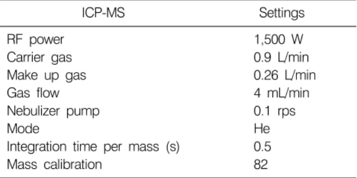
Table 2. ICP-MS operating parameters
ICP-MS Settings
RF power Carrier gas Make up gas Gas flow Nebulizer pump Mode
Integration time per mass (s) Mass calibration
1,500 W 0.9 L/min 0.26 L/min 4 mL/min 0.1 rps He 0.5 82 United States of America and China, the content of Se in soil
is remarkably low. Recently, the results obtained from the populations of the several nationalities were reported in detail by Navaro-Alarcon et al7) (2008).
So far, a few studies on the serum Se level in healthy Korean population were reported. To illustrate, an investigation reported by Kim et al8) (1998) exhibited the mean serum level of Se to be 26.4±0.41μg/dl for healthy Korean men and 24.5±0.32μg/dl for women by the ICP-MS quantification. Se level was higher in men than in women. Besides, Song et al9) (2006) reported that the mean serum Se level was 116.8 ng/ml for Korean men by ICP-MS method. In other study, Kyung-Hee et al10) (2006) conducted the study by which the dietary intakes, serum concentrations and urinary excretions of the trace elements levels including Se were determined by ICP-MS method for Korean young adult women. The study revealed that the intake of the Se was 41.93±9.28μg/dl which was almost similar to the estimated average requirement and the serum Se concentration, 3.73±0.60μg/dl, was also below its reference median and normal range.
Besides, a large number of studies have demonstrated the strong evidence that the Se deficiency can generate certain malignant tumors. Therefore, we aimed to evaluate serum levels of Se in healthy Korean population in order to prevent the population against cancer that might be caused by Se deficiency. Ideally, analytical methods should be able to accurately determine Se in a variety of sample matrices from low micrograms per kilogram concentrations to several hundred micrograms per gram. Of a variety of methods used to quantify Se, we selected the Graphite Furnace Atomic Absorption Spectrometry (GF-AAS) and Inductively Coupled Plasma Mass Spectrometry (ICP-MS) methods due to their sensitive detection of trace elements in biological samples.
MATERIALS AND METHODS
1. Materials
All chemicals used were analytical grade and all reagents were evaluated for their contribution to Se contamination in the blanks. Nitric acid (70% HNO3) was purchased from Dongwoo (Korea), 1-butanol, Triton X-100, ammonium solution, ethylenediaminetetraacetic acid (EDTA), nickelnitrate hexahydrate from Sigma Aldrich (IN, USA). Water was deionized before use. A working standard solution (10μg/ml)
for calibration was prepared from a 1,000 mg/l selenium stock solution (SPEX CertiPrepⓇ, Inc). Seronorm (Fumouze, Levallois- Perret, France), and 39th German External Quality Assessment Scheme 11A and B were used as reference materials.
2. Instrumentation
1) GF-AAS conditions: An A Analyst 800 (PerkinElmer, USA) spectrometer equipped with a transversal heated furnace atomizer (THGA), Zeeman effect background correction and pyrolitic graphite-coated tubes with L`vov platforms was used.
An Electrodeless Discharge Lamp (EDL) set at 196 nm. An AS-800 autosampler was used for the measurement of the sample. Operating parameters of the GF-AAS was shown in Table 1.
2) ICP-MS conditions: An Agilent 7500ce (Agilent Technologies, Japan) ICP-MS system was used to conduct the experiments. Operating conditions of the ICP-MS are given in Table 2. All ICP-MS operating parameters were adjusted to give a maximum signal in the selenium concentration studied.
Each day before starting analysis and after all the injections, tubing used was conditioned by rinsing with HNO3 (1%) to avoid contamination.
3. Samples and sample preparation
Serum samples were collected from 100 healthy Koreans making up of 50 males and 50 females. The age range of study
Table 4. Total and gender groups of the studied individuals with corresponding Se levels (μg/l)
Methods Mean age±S.D. n Sex Min Max Mean ± S.D. 95% CI
GF-AAS
ICP-MS
45.38±14.24 45.92±14.74 44.83±13.86
100 50 50
Both M F Both M F
47.17 47.17 58.53 32.57 32.57 57.83
170.81 170.81 169.97 192.38 190.40 192.38
105.58±24.06 102.82±22.21 108.34±25.71 112.05±30.42 103.29±31.05 120.81±27.37
4.78 6.32 7.31 6.04 8.83 7.79 M: male, F: female, SD: standard deviation, CI: confidence interval.
Table 3. Accuracy and precision of the GF-AAS and ICP-MS methods
Methods Materials Concentration of Se (μg/l) Accuracy
(%)
Precision R.S.D. (%) Certified Measured
GF-AAS
ICP-MS
Seronorm (Lot.JL4409) 39th G-EQAS 11A 39th G-EQAS 11B Seronorm (Lot.JL4409) 39th G-EQAS 11A 39th G-EQAS 11B
72.8±12.2 75.3±17.6 133.5±29
72.8±12.2 75.3±17.6 133.5±29
62.9±2.2 61.3±1.8 122.0±0.9 61.70±0.56 76.07±2.59 131.31±3.06
86.43 80.87 89.69 84.75 101.02 98.36
3.54 2.92 0.74 0.91 3.41 2.33 R.S.D: relative standard deviation.
subjects was between 21 to 69 years.
For GF-AAS, all samples were digested by introducing exactly same measured samples into a beaker containing 5 ml acid mixture. This mixed solution was heated at 200oC by hot plate and cooled to room temperature until the solution becomes colorless then turns light yellow again.
For ICP-MS, blood samples were collected in dry tubes and serum was tested after centrifugation. All serum samples and working solutions were prepared by dilution (1:19, v/v) in an acidic solution consisting of 0.7 mM ammonia, 0.01 mM EDTA, 0.07% (v/v) Triton X-100 and 1.5% 1-butanol.
RESULTS
The main purpose of this study was to determine the Se concentration in blood serum of 100 healthy Koreans. Se was detected at one atomic mass unit (m/z) 82: 82Se. The determination of serum Se levels was carried out by using the GF-AAS and ICP-MS. The mean serum Se level in the studied individuals was found to be 105.58±24.06μg/l, ranging from 47.17μg/l to 170.81μg/l when the concentration was determined by the GF-AAS. ICP-MS results however indicated that the mean level was 112.05±30.42μg/l ranging from
32.75μg/l to 192.38μg/l. Furthermore, the average serum Se level 102.82±22.21μg/l in men and 108.34±25.71μg/l in women when Se was quantified by the GF-AAS, whereas ICP-MS measurements indicated that the mean was 103.29±
31.05μg/l for men and 120.81±27.37μg/l for women, respectively. The other related data are showed in Table 4.
In addition, the standard error evaluated for each method was 2.41 for the GF-AAS and 3.04 for ICP-MS. As for detection limit from each method for the Se, it was 1.12μg/l in the case of GF-AAS and 0.01μg/l for the ICP-MS. The accuracy and the precision of the used methods were tested with the use of three serum samples with standardized con- centrations of selenium, internationally recognized: Seronorm (Fumouze, Levallois-Perret, France), and 39th German External Quality Assessment Scheme 11A and B (39th G-EQAS 11A and 11B). The results are presented in Table 3.
DISCUSSION
For the mean serum Se level, the GF-AAS method resulted in a lower value (105.58±24.06μg/l) than the ICP-MS method (112.05±30.42μg/ml) (p=0.097). Both analytical methods indicated that the mean level of Se was higher among women
Table 5. Mean serum, plasma and whole blood Se levels measured in healthy individuals from different countries
Area (country) Se (μg/l) Area (country) Se (μg/l)
Granada (South-eastern Spain) Singapore
Canary Islands (Spain) Mumbai (India)
Lower Silesian region (Poland) Bydgoszc (Poland)
Rio de Janeiro (Brazil) The United States of America Antwerp region (Belgium)
74.9±27.3 126.0 74.7±25.2
100.0 67.4±38.6 89.5±15.6 73.2±9.9
124.5±0.2 (M), 122.0 (F) 84.3
Taiwan (China) Tehran (Iran) Czech Republic Hannover (Germany) Vienna (Austria)
Asturias (Northern Spain) Taiwan (China)
Helsingborgh (Southern Sweden)
216.2±7.4 100.6±13
84.2±20.2 92.4±17.4 85.9±24.0 86.2±17.0 129.0±21.5
105.0
(108.34±25.71μg/l) than the level among men (102.82±
22.21μg/l) when the measurement was performed by the GF-AAS (p=0.25). The ICP-MS method also showed a higher value for women 120.81±27.37μg/l than for men (103.29±
31.05μg/l) (p=0.0035). There are several differences between the current study and the above-mentioned studies conducted by other Korean researchers. In comparison, the present study result showed two times lower value for the mean level of Se than that reported by Kim et al (26.4±0.41μg/dl in men and 24.5±0.32μg/dl in women by ICP-MS) study. In contrary to their result, our study also revealed that the mean Se level was higher in women than men. Additionally, Song et al (116.8 μg/dl among women) showed similar result to our study while the study of Kyung-Hee et al revealed a slightly lower value compared to the present results. As a result, our study showed somewhat different values from the studies reported by the other Korean researchers.
As mentioned earlier, Se levels in soil generally reflect the Se level in human population and food. Se level in the serum of populations throughout the world varies from 41.7μg/l in Finland to 158.2μg/l in Canada.11) A very recent survey reported by Navarro-Alarcon et al (2008), revealed the mean Se levels of the serum, plasma and whole blood in healthy adults from different countries as biomarkers of selenium nutritional status of defined population groups (Table 5). When our results were compared with those results, the level was comparable to those of India, Iran and Southern Sweden, however, it is lower than the countries such as Singapore, USA and Northern Spain. The study reported by Kostakopoulos et al12) (1990) showed that the mean serum Se level in healthy Greeks was 55±7.2μg/ml and there is no significant difference between the male and female population. Vogt et al13) (2007)
Explored Black (118.76 ng/ml or∼6% lower) and White (126.35 ng/ml) differences in serum selenium concentration using data from the Third National Health and Nutrition Examination Survey (NHANES III) for the United States population. The study conducted by Clark et al14) (2008) revealed that serum Se level of non-smoking adults living on the west coast of Canada was influenced by gender with men having a slightly higher level (1.73μmol l−1) than women (1.60μmol l−1).
Besides, the serum selenium concentrations in some European countries were reported to be low. To illustrate, serum selenium concentrations of the population living in central Bohemia were 72 ng/ml and 76 ng/ml in men and women, respectively.15) In West Germany, the serum selenium concentrations were 66 ng/ml and 67 ng/ml for men and for women, respectively.16) In connection with these countries with low serum Se levels, the data from this study for Korean populations gave substantially higher values.
CONCLUSION
In the current study, the mean serum Se level revealed a moderate level when compared with the studies carried out by other Korean researchers. In contrast, the present study showed a higher value for female than male group. For the two methods used, ICP-MS method showed high values than GF-AAS for the mean level of Se. In comparison with the results obtained from the other nationalities, Korean population has quite moderate level of Se in their blood serum. However, it is necessary to strengthen the supply of selenium-rich products for the population who still have a low-selenium level for the cancer prevention.
REFERENCES
1) Jozanov-Stankov O, Demajo M, Djujic I, Mandic M. Selenium intake as a modulator of responsiveness to oxidative stress. J Environ Pathol Toxicol Oncol 17, 251-257, 1998.
2) Kohrle J, Brigelius-Flohe R, Bock A, Gartner R, Meyer O, Flohe L. Selenium in biology: facts and medical perspectives.
Biol Chem 381, 849-864, 2000.
3) Harr JR. Effect of dietary selenium on N-2-fluorenylacetamide (FAA): Induced cancer in vitamin E-supplemented selenium depleted rat. Clin Toxicol 5, 187-194, 1972.
4) Dumont E, Vanhaecke F, Cornelis R. Selenium speciation from food source to metabolites: a critical review. Anal Bioanal Chem 385, 1304-1323, 2006.
5) Klapec T, Mandic ML, Grgic J, Primorac LG, Perl A, Krstanovic V. Selenium in selected foods grown or purchased in eastern Croatia. Food Chem 85, 445-452, 2004.
6) Thomson CD. Assessment of requirements for selenium and adequacy of selenium status: a review. Eur J Clin Nutr 58, 391-402, 2004.
7) Navarro-Alarcon M, Cabrera-Vique C. Selenium in food and the human body: A review. Sci Total Environ (2008), doi:10.1016/j.scitotenv.2008.06.024
8) Kim L, Chung YCh, Hwang EJ, Kim J, Lee MK, Park JH, Kim T, Park ST, Soo K. A study on serum concentrations of antioxidant minerals in normal Korean adults. Korean J Nutr 31, 324-332, 1998.
9) Song SH, Song K, Lee SB, Kim ChS. The serum selenium level in Korean men and its association with age and prostate cancer. Korean J Urol 47, 150-153, 2006.
10) Kyung-Hee K, Hyeon-Sook K. Dietary intakes, serum concentrations, and urinary excretions of Fe, Zn, Cu, Mn, Se, Mo, and Cr of Korean Young Adult Women. Korean J Nutr 39, 762-772, 2006.
11) Lockitch G. Selenium: clinical significance and analytical concepts. Crit Rev Clin Lab Sci 27, 483-541, 1989.
12) Kostakopoulos A, Kotsalos A, Alexopoulos J, Sofras F, Deliveliotis Ch, KAllistratos G. Serum selenium levels in healthy adults and its changes in chronic renal failure.
International Urology and Nephrology 22, 397-401, 1990.
13) Vogt TM, Ziegler RG, Patterson BH, Graubard BI. Racial differences in serum selenium concentration: Analysis of US population data from the Third National Health and Nutrition Examination Survey. Am J Epidemiol 166, 280-288, 2007.
14) Clark NA, Teschke K, Rideout K, Copes R. Trace element levels in adults from the west coast of Canada and associations with age, gender, diet, activities, and levels of other trace elements. Chemosphere 70, 155-164, 2007.
15) Korunova V, Skodova Z, Dedina J, Valenta Z, Parizek J, Pisa Z, Styblo M. Serum selenium in adult Czechoslovak population. Biol Trace Elem Res 37, 91-99, 1993.
16) Oster O, Schmiedel G, Prellwitz W. Correlations of blood selenium which hematological parameters in West Germany adults. Biol Trace Elem Res 15, 47-81, 1998.