췌장 동정맥기형은 소화기 장기에서의 매우 드문 질환이다 (1). 이 질환은 서서히 진행하며 위장관 출혈이나 문맥압 항진 증과 관련된 질환으로 나타난다. 완전한 치료를 위해서는 침범 된 장기를 모두 제거해야 하며, 문맥압 항진증이 발생하면 수 술적인 췌장 동정맥기형의 제거는 어렵다. 만약 수술이 불가능 하다면 위장관 출혈의 지혈은 경도관동맥색전술이 대체 방법 이다(2, 3). 최근 경경정맥간내문맥정맥단락술을 이용한 성공 적인 치료가 보고 되었으나 이 증례들은 경도관동맥색전술과 경경정맥간내문맥정맥단락술을 결합하여 시도되었거나 경도관 동맥색전술의 실패 후 경경정맥간내문맥정맥단락술을 시행한 경우이다(4, 5). 이에 저자들은 문맥압 항진증을 동반한 췌장 동정맥기형을 합병증없이 경경정맥간내문맥정맥단락술만으로 성공적으로 치료한 1예를 보고하고자 한다.
증례 보고
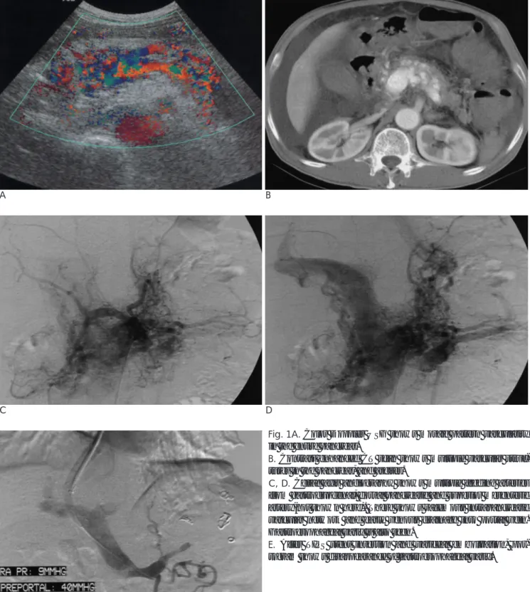
62세 남자가 복부 팽창에 대한 검사를 위해 내원하였다. 내 원 당시 간염이나, 외상의 과거력은 없었다. 이학적 검사에서 이동성 탁음계가 있었다. 회색조 초음파 검사에서 다량의 복수 가 관찰되었으며 췌장 전체에 다발성 결절과 관상의 저에코의 병변들이 보였다. 색도플러 검사에서 다발성 결절과 관상의 저 에코 병변들은 모자이크 양상의 혈류를 보이며 낮은 저항계수 를 띠는 박동성 파형을 보였다(Fig. 1A). 조영증강 전산화 단 층촬영에서 췌장에 다발성의 혈관 구조물과 복수, 식도정맥류 가 관찰되었다(Fig. 1B). 혈관 조영술에서 위십이지장 동맥, 배 측췌장 동맥, 위장간막 동맥 등으로부터 영양동맥을 받는 만상 의 혈관상(racemous vascular network)이 췌장내에 관찰되었 으며 문맥으로 조기 정맥 배출과 위식도 정맥류가 관찰되었다
(Fig. 1C, D). 입원 10일 후 토혈증이 있어 시행한 내시경에서 식도 정맥류 출혈이 있었다. 내시경적 식도 정맥류 결찰을 시 도하였으나 지혈에 실패하여 응급 경경정맥간내문맥정맥 단락 술을 시행하였다. 시술 방법은 초음파 유도하에 우측 내목 정 맥을 천자하여 Ring transjugular intrahepatic access set(Cook, Bloomington, IN, U.S.A.)내에 있는 9-F 혈관초를 삽입한 후 5-F 카테타로 우측 간정맥을 선택하였으며, 전산화 단층촬영을 기준으로 우측 간문맥이 있는 위치를 추정한 후 16-G Colapinto 침으로 우측 간문맥을 천자하였다. 0.035- inch 유도 철사(Terumo, Tokyo, Japan)를 이용하여 카테타 를 주 문맥으로 통과시킨 후 시행한 문맥 조영술에서 이간성 (hepatofugal) 혈류에 의해 위식도 정맥류가 관찰되었고 시술 전 주 문맥압은 40 mmHg이었다. 9-F 혈관초를 주문맥에 삽 입한 후 서서히 혈관초를 당기면서 지속적으로 조영제를 주입 하여 복강으로의 누출이나 담관과의 연결성이 없는 것을 확인 하였다. 직경 10 mm 풍선 카테타 (Boston Scientific, Watertown, MA, U.S.A.)로 단락 경로를 확장 시킨 후 직경 10 mm 길이 8 cm의 Niti-S 스텐트(Taewoong, Seoul, Korea)를 삽입하였다. 스텐트 삽입 후의 문맥 조영술에서 위식도 정맥류 는 감소하였으나 완전히 소실되지 않아 5-F 카테타를 이용하 여 위식도정맥류를 선택하여 5 mm 코일 5개로 색전술을 시 행하였다. 시술 후 위식도 정맥류는 더 이상 관찰되지 않았으 며 문맥과 우심방과의 압력차가 31 mmHg에서 10 mmHg로 감소되었다(Fig. 1E). 경경정맥간내문맥정맥단락술 후 정맥류 출혈은 지혈되었고 우심부전 증상이나 간성 뇌병증 소견은 관 찰되지 않았다. 4주 후 시행한 컬러 도플러 검사에서 경경정맥 간내문맥의 혈류는 300 cm/sec로 원할한 혈류를 보였으며 스 텐트의 협착소견은 보이지 않았다. 6주 후 시행한 추적 내시경 에서 위식도 정맥류는 사라졌다. 8개월 동안 복수는 잘 조절 되었고 정맥류 출혈은 없었다. 시술 8개월 후 위식도 정맥류 대한방사선의학회지 2004;50:175-178
─ 175 ─
문맥압 항진증을 동반한 췌장 동정맥기형의 경경정맥간내문맥정맥단락 치료: 1예 보고1
김 성 훈・김 영 환・김 용 주
췌장 동정맥기형은 매우 드문 질환으로서 위장관의 출혈이나 문맥압 항진증 등의 임상증상 으로 나타난다. 질환의 초기에 수술로서 췌장 동정맥기형을 완전 적출하는 것이 가장 좋은 치 료법이며 문맥압 항진증으로 수술이 불가능한 경우 위장관 출혈시 지혈을 위해 경도관동맥색 전술이 이용 될 수 있다. 저자들은 췌장 동정맥기형으로 인한 문맥압 항진증으로 복수와 식도 정맥류를 주소로 내원한 62세 남자 환자에서 경경정맥간내문맥정맥단락술을 이용해 성공적인 치료를 경험하였기에 이를 보고하고자 한다.
1경북대학교 의과대학 진단방사선과학교실
이 논문은 2003년 8월 20일 접수하여 2004년 1월 26일에 채택되었음.
출혈이 재발하여 문맥 조영술을 시행하였으며 스텐트 내부에 협착이 보여 풍선 확장술로 경경정맥간내문맥정맥단락술 교정 술을 시행하였다. 시술 후 출혈은 조절되었으나 내원 당시 심 한 출혈과 복수로 인한 전신장 질소 과잉혈증 (prerenal
azotemia)과 조영제의 신독성으로 인하여 급성 신부전을 보였 으며 응급으로 혈액투석을 권하였으나 환자와 보호자가 거부 하여 3일 후 사망하였다.
김성훈 외: 문맥압 항진증을 동반한 췌장 동정맥기형의 경경정맥간내문맥정맥단락 치료
─ 176 ─
A B
C D
E
Fig. 1A. Color Doppler USG shows mosaic pattern vasculatiry in the entire pancreas.
B. Contrast enhanced CT scan shows multiple vascular struc- tures in the pancreas, and ascites.
C, D. Celiac axis angiography shows multiple feeding arteries from gastroduodenal, dorsal pancreatic and superior mesenteric artery(not shown here). There shows racemous intrapancreatic vascular network and early venous drainage into portal vein.
Gastroesophageal varix is also seen.
E. After TIPS stent insertion and variceal embolization, por- togram shows disappearance of gastroesophageal varix.
고 찰
췌장 동정맥기형은 진단 당시의 평균나이가 48.8세(7개월 에서 67세)로 성별 차이는 없는 것으로 되어있으나 남자에게 서 약간 많다(6). 이 질환은 천천히 진행하며 때때로 심각한 위장관 출혈을 일으키는데 문맥압 항진증으로 인한 위식도 정 맥류나 동정맥 기형의 파열로 췌장관이나 담도로의 출혈(7, 8), 주위 십이지장벽으로의 파급(9), 췌장의 동정맥 기형으로 인 한 십이지장 궤양 등으로 나타난다(10, 11). 췌장 동정맥기형 은 선천성과 후천성 동정맥기형 두 가지 형태로 나누어지며 전 자의 경우 흔적 원발성 혈관망의 비정상적인 분화에 의한 것 이며 Rendu-Osler-Weber 증후군과 연관성을 가진다. 후자 는 염증이나 종양, 외상 후에 발생한다. 췌장 동정맥기형은 회 색조 초음파에서 췌장내에 다발성 결절과 관상의 저에코 병변 으로 보이며 색도플러 초음파에서 모자이크 패턴의 혈류를 보 이는데 이는 여러 방향의 혈류와 비교적 빠른 이완기 혈류로 인한 낮은 박동성에 의한 것이다(12). 역동적 전산화 단층촬 영 동맥강조기 영상에서 췌장 내에 조영 증강되는 작은 혈관 과다의 점들과 문맥이 조기에 조영 증강된다. 혈관 조영술은 췌장 동정맥기형의 진단에 가장 정확한 방법인데 확장되고 꼬 인 영양동맥이나, 만상의 췌장 내 혈관 망, 문맥으로의 빠른 배 출이 특징적이다(13). 췌장 동정맥기형의 치료로는 파급된 장 기를 수술적 방법으로 적출하는 것이 유일한 선택이며 초기의 수술은 문맥압 항진증을 막아준다. 만약 문맥압 항진증이 발생 하면 문맥압 항진증을 근치적으로 줄이는 방법은 없으며 수술 적 방법으로도 불가능하다. 또한 과다 출혈로 환자의 상태가 좋지 않거나, 췌장 동정맥기형의 범위가 광범위할 때 수술적 제거는 위험 부담이 많다. 만약 수술적 제거가 불가능 하다면 경도관동맥색전술이 이용되는데 췌장의 동정맥기형이 너무 광 범위 하면 색전술이 어려울 뿐만 아니라 장 괴사의 가능성도 많아진다. 또한 색전술 후 측부 혈관의 발달로 인한 재출혈과 문맥압 항진증의 진행이 문제가 된다(5). 본 증례의 경우 광 범위한 췌장 동정맥기형의 분포와 문맥압 항진증으로 인하여 수술적 치료는 불가능 하였으며 많은 영양공급 동맥으로 인하 여 경도관동맥색전술도 불가능하여 경경정맥간내문맥정맥단락 술을 시행하여 정맥류 출혈과 복수를 성공적으로 치료하였다.
결론적으로, 우심부전의 위험성과 간내문맥정맥단락술 경로 의 협착으로 인한 재출혈의 위험성은 있으나, 경경정맥간내문
맥정맥단락술은 수술이 불가능한 문맥압 항진증을 동반한 췌 장 동정맥기형의 환자에서 효과적인 고식적인 치료법이라 할 수 있다.
참 고 문 헌
1. Meyer C, Troncale FJ, Galloway S, Sheahan DG. Arteriovenous malformations of the bowel: an analysis of 22 cases and a review of the literature. Medicine(Baltimore) 1981;60:36-48
2. Nishiyama R, Kawanishi Y, Mitsuhashi H, et al. Management of pancreatic arteriovenous malformation. J Hepatobiliary Pancreat Surg 2000;7:438-442
3. Iwashita Y, Kawano T, Maeda T, Nagasaki S, Kitano S. Pancreatic arteriovenous malformation treated by transcatheter embolization.
Hepatogastroenterology 2002;49(48):1722-1723
4. Kodama Y, Saito H, Hiramatsu K, Takeuchi S, Takamura A. A case of pancreatic arteriovenous malformation treated by transcatheter arterial embolization and transjugular intrahepatic portosystemic shunt. Nippon Shokakibyo Gakkai zasshi 2001;98(3):320-324 5. Hayashi N, Sakai T, Kitagawa M, et al. Intractable gastrointestinal
bleeding caused by pancreatic arteriovenous malformation: suc- cessful treatment with transjugular intrahepatic portosystemic shunt. Eur J Radiol 1998;28:164-166
6. Katoh H, Kojima T, Okushiba S, Shimozawa E, Tanabe T. Bleeding esophageal varices associated with pancreatic arteriovenous mal- formation. World J Surg 1991;15:57-61
7. Mizutani N, Masuda Y, Naito N, Toda T, Yao E-I, Fukamoto M.
Pancreatic arteriovenous malformation in a patient with gastroin- testinal hemorrhage. Am J Gastroenterol 1981;76(2):141-144 8. Grannis FW, Foulk WT, Miller WE, Payne WS. Diagnosis and
management of an arteriovenous fistula of pancreas and duode- num. Mayo Clin Proc 1973;48:780-782
9. Takiguchi N, Ichiki N, Ishige H, et al. Pancreatic arteriovenous malformation involving adjacent duodenum in a patient with gas- trointestinal bleeding. Am J Gastroenterol 1995;90(7):1151-1154 10. Aida K, Nakamura H, Kihara Y, Abe S, Okamoto K, Otsuki M.
Duodenal ulcer and pancreatitis associated with pancreatic arteri- ovenous malformation. Eur J Gastroenterol Hepatol 2002;14:551- 554
11. Kato H, Tanabe T. Congenital arteriovenous malformation of the pancreas with jaundice and a duodenal ulcer: report of a case. Surg Today 1993;23:1108-1112
12. Koito K, Namieno T, Nagakawa T, Morita K. Diagnosis of arteri- ovenous malformation of the pancreas by color doppler ultra- sonography. Abdom Imaging 1998;23:84-86
13. Chuang VP, Pulmano CM, Walter JF, Cho KJ. Angiography of pan- creatic arteriovenous malformation. AJR Am J Roentgenol 1977;
129(6):1015-1018 대한방사선의학회지 2004;50:175-178
─ 177 ─
김성훈 외: 문맥압 항진증을 동반한 췌장 동정맥기형의 경경정맥간내문맥정맥단락 치료
─ 178 ─
J Korean Radiol Soc 2004;50:175-178
Address reprint requests to : Young Hwan Kim, M.D., Department of Radiology, Kyungpook National University Hospital, 50, Samduk-dong 2 Ga, Chung-gu, Daegu 700-721, Korea.
Tel. 82-53-420-5390 Fax. 82-53-422-2677 E-mail: [email protected]
A Case of Pancreatic Arteriovenous Malformation with Portal Hypertension: Treatment with Transjugular
Intrahepatic Portosystemic Shunt
1Seong Hoon Kim, M.D., Young Whan Kim, M.D., Yong Joo Kim, M.D.
1Department of Radiology, School of Medicine, Kyungpook National University
Arteriovenous malformation of the pancreas is a rare disease, and it is manifested by gastrointestinal bleed- ing and/or portal hypertension. Surgery is definitely the treatment of choice at the early stage of the disease, and a transcatheter embolization is an alternative treatment for the control of bleeding and if the lesion is surgi- cally inaccessible. We describe a 62-year-old man who had refractory ascites and esophageal variceal bleeding caused by a pancreatic arteriovenous malformation associated with portal hypertension; this was successfully treated by a transjugular intrahepatic portosytemic shunt.
Index words :Pancreas, arteriovenous malformation Portal hypertension, portosystemic shunt