Journal of Korean Arthroscopy Soc.
Volume 14, Number 2, June, 2010
서 론
전방십자인대 재건술에서 이상적인 이식건은 본래의 전방 십자인대와 유사한 생물 역학적(biomechanical) 특성을 지 니고 강한 고정이 가능하며 빠른 생물학적 결합(biologic incorporation) 및 낮은 공여부 이환률(donor site mor- bidity)이 있어야 하지만 이러한 이상적인 이식건은 아직 존 재하지 않는다. 전방십자인대 재건술에 사용되는 이식건으로 는 자가 이식건과 동종 이식건으로 크게 나누어 지는데, 자가 이식건에는 슬개건, 슬건, 대퇴 사두건 등이 있으며 동종 이식 건에는 슬개건, 아킬레스건, 슬건, 전방 및 후방 경골건 등으 로 다양하다.
현재 가장 널리 사용되는 대표적인 생물학적 이식건으로 자가 슬개건(bone patellar bone tendon autograft)과 자 가 슬건(hamstring tendon autograft)이 있다. 그 중 자가 슬개건이 가장 널리 사용되고 있으나16,22,41), 최근 자가 슬건의 사용이 증가하고 있다17). 자가 슬건의 사용방법도 한 가닥에 서 여러 가닥으로 바뀌고 있으며, 여러 가닥의 자가 슬건을 이 용한 재건술은 보다 해부학적으로 유사한 재건을 할 수 있고 보다 강한 최대 인장강도(ultimate tensile load)를 가지는 장점이 있다. 이처럼 자가 슬건의 사용이 증가하는 이유는 자 가 슬건의 높은 최대 인장 강도(ultimate tensile load)71), 낮은 공여부 이환률, 고정 방법의 발전에 있을 것으로 보인다55). 이와 더불어 자가 슬건은 채취 후 다시 재생(regeneration) 이 되며, 이는 자가 슬건 사용의 큰 장점으로 여겨지고 있다.
자가 슬건의 재생은 1992년도에 Cross 등14)에 의해 처음 보 고된 이후로 재생된 자가 슬건에 대한 재생의 기전(mecha- nism), 생물 역학적 특성(Biomechanical properties), 재 생된 슬건의 기능회복 등에 대한 다양한 연구가 있어 왔으나, 아직 논란이 되거나 밝혀지지 않은 사실이 많으며 추가적인
자
자가 가 슬 슬건 건을 을 이 이용 용한 한 재 재건 건술 술:: 장 장점 점,, 단 단점 점,, 슬 슬건 건의 의 재 재생 생
인제대학교 의과대학 서울백병원 정형외과 김진구∙최정윤
Anterior Cruciate Ligament Reconstruction Using Hamstring Tendon Autograft : Advantage, Disadvantage, Hamstring Regeneration
Jin Goo Kim, M.D, Jeong Yoon Choi, M.D.
Department of Orthopedic Surgery, Seoul Paik Hospital, College of Medicine, Inje University, Seoul, Korea
The Hamstring autograft and the bone patellar bone tendon autograft have been widely used for anterior cruciate ligament recon- struction. In recent years, use of hamstring autograft for ACL reconstrution has been increased. The reason seems to be the advan- tages of the hamstring tendon such as high ultimate tensile load, low donor site morbidity and development of graft fixation method.
These theoretical advantages have been increased as studies have shown that hamstring tendons actually regenerate after harvesting for ACL reconstruction. However, the concerns have arisen regarding the disadvantages of hamstring harvest, which were weakness of tibial internal rotation, the loss of flexion strength. The flexion strength loss has been controversial, therefore it needs to study whether restoration of flexion strength after hamstring regeneration is or not. In this study, we reviewed the current research of con- cerns on the advantage and disadvantage of hamstring tendon autograft and the hamstring regeneration. Furthermore, we compared the earlier studies and experiences regarding Hamstring regeneration with our research.
KEY WORDS: Hamstring regeneration, Donor site morbidity, Knee flexor strength, Anterior cruciate ligament
�Address reprint request to Jin Goo Kim, M.D.
Department of Orthopedic Surgery, Sports Medical Center, Seoul Paik Hospital, Inje University
85, 2Ka Jur-dong Chung-gu, Seoul, Korea Tel: 82-2-2270-0025, Fax: 82-2-2270-0048 E-mail: [email protected]
연구가 필요하다. 또한 최근 사용이 증가하고 있는 자가 슬건 의 장점 및 단점에 대한 이해는 환자의 특성에 따라 적절한 이 식물을 선택하고 성공적인 재건술을 시행하기 위해 필요할 것으로 생각된다.
본 종설에서는 전방십자인대 재건술에서 최근 많이 사용되 는 자가 슬건에 대한 여러 장점과 단점 및 자가 슬건의 큰 장 점 중 하나인 슬건의 재생에 대해 문헌을 고찰하였고, 본 저자 들의 연구와 비교해 보았다.
본 론 1. 자가 슬건 이식
1) 자가 슬건의 장점 및 단점
자가 슬건의 일반적인 장점으로 대퇴 슬개 관절면의 동통, 무릎 전방 통증, 무릎 꿇기 통증, 신전력의 감소, 방사선학적 관절염, 공여부 이환률 등과 같은 합병증이 적으며, 이식물의 생물역학 및 고정 방법이 우수하다54,71). 단점으로 술 후 안정 성의 감소, 경골 내회전력의 약화, 굴곡력의 감소 등이 알려져 있다. 이러한 장점과 단점은 자가 슬건과 자가 슬개건의 비교 연구에서 오래 전부터 다뤄지고 있으나7,8,24,56)아직도 어떤 이 식물이 더 우수한가에 대한 논란이 있다.
최근 연구에서 Dequin 등17)은 미국 정형외과 스포츠 의학 회(AOOSM, American Orthopaedic Society for Sports Medicine) 회원들을 대상으로 이식건의 사용빈도를 조사한 결과, 최근 5년간 자가 슬건의 사용이 25%에서 32%
로 증가하였다고 한다. 그리고 전방십자인대 재건술에서 이 식물을 선택할 때 운동 선수, 남자, 노동자(Heavy laborer) 들은 자가 슬개건을 많이 사용하고, 성장판이 열려 있는 청소 년기 환자와 대퇴 슬개 관절 증상이 있는 환자들은 자가 슬건 을 많이 사용하고 있었다17). 이식물을 선택할 때 환자의 활동 도, 직업, 나이, 증상, 성별 등 환자의 여러 상황을 고려하여 선택하는 것이 보다 중요할 것으로 보인다.
2) 이식물의 생물 역학 및 고정방법
과거 이식물의 생물 역학에 대한 연구에서 자가 슬개건은 인장강도가 전방십자인대의 159~168%로 더 강하고, 단일 반건양건은 전방십자인대의 70%, 단일 박건은 49%의 낮은 인장강도를 보여 자가 슬개건이 자가 슬건 보다 더 강하다고 하였다46). 하지만 두 가닥 이나 네 가닥으로 만들면 강성이나 인장강도가 증가하게 되는데, 이에 대한 최근 연구에서 최대 인장 강도(ultimate tensile load)와 강성(stiffness)이 전 방십자인대에서 각각 2160 N, 242 N/m , 자가 슬개건에서 1784 N, 210 N/m으로 서로 비슷하며, 네 가닥 자가 슬건은 4090 N, 776 N/m로 전방십자인대에 비해 높은 인장강도와 강성을 지닌다고 보고하였다71). 이와 유사한 연구에서도 네 가닥 자가 슬건은 4140N, 807N/m로 자가 슬개건에 비해 뛰
어난 최대 인장 강도와 강성을 보인다고 하였다11). 또한 자가 슬건은 원통형 모양으로 단면적(cross-sectional area)이 좀더 넓어 9 mm의 자가 슬건은 10 mm의 자가 슬개건보다 1.6배 더 강하다는 장점이 있다. 이처럼 자가 슬개건은 전방 십자인대와 비슷한 생물역학적 특성을 가지고 있으며, 네 가 닥 자가슬건이 자가 슬개건에 비해 좀더 뛰어난 생물 역학적 특성을 보인다.
3) 이식물의 고정
최근 전방 십자 인대 재건술 후 조기 체중부하와 적극적인 재활을 시행하고 있으며, 술 후 초기에 이식건의 고정력이 약 해 지기 때문에 이식물을 강하게 고정하는 것이 중요하다11). 과거 전방십자인대 재건술에서 자가 슬개건의 골편(bone block)에 대한 고정으로 간섭나사(interference screw)를 사용하여 성공적인 결과를 보였다고 한다32,33). 일상 활동을 위 한 이식건 고정력은 450N으로46)자가 슬개건에서 경골에 사 용되는 간섭나사의 고정력은 718N이고30)대퇴골 부위를 고정 하는 간섭나사의 고정력은 416~640N으로3,30,73)비교적 강한 고정을 할 수 있다. 이에 비해 자가 슬건은 과거에 강한 골 고 정을 할 수 없었으며 건과 골의 결합 또한 천천히 이루어져 조 기에 체중부하가 어려웠다59). 이러한 문제를 해결을 위해 강 한 고정력을 지닌 기구들이 개발되었고, 최근 들어 개발된 Femur Endobutton CL, Mulch screw, Tibia tandem washer, WasherLoc Device 등은 고정력이 매우 강하여 자가 슬건의 고정력 문제를 해결하였다.
이식건 고정을 보다 효과적으로 하기 위해 Dargel 등15)은 골 터널에 순차적으로 확장기(dilater)를 사용하여 확장하는 방법을 사용하였다. 이는 이식건의 직경보다 1 mm 작은 확 공 드릴(reamer)로 터널을 만든 후에 순차적으로 확장하는 방법으로 골터널과 건 사이에 압박 밀착 (press-fit)을 시킴 으로써 술 후 초기의 고정력 (pull out strength)를 증가시킬 수 있다고 보고하였다. Cain 등12)은 생물 역학적 사체연구에 서 이식건보다 2 mm 가량 작게 경골에 터널을 만들고 확장 기로 점차적으로 확장하는 방법으로 터널 확장이 반건양건과 박건의 pull out strength에 미치는 영향을 연구하였다. 연 구결과 평균 최대 부하(mean peak load)가 드릴로 확공 (reaming)한 방법 보다 확장기(dilator)로 확장하는 방법이 보다 높다고 보고하였다. 이러한 확장을 통한 압박 밀착 방식 은 초기의 pull out strength를 증가시키는 장점 외에 활액 이 터널로 들어오는 것과 windshield-wiper effect를 막고, 터널의 벽과 강한 밀착을 가능하게 하여 이식물의 결합을 높 일 수 있을 것으로 보인다50). 본 저자들은 2 mm 적은 크기의 전동 확공기(motorized reamer)로 터널을 만든 후 1 mm 큰 확공기를 손으로 확공하고, 0.5 mm간격으로 확장기를 이 용하여 점차적으로 터널 확장하는 방법을 사용하고 있다.
4) 인대화(Ligamentization)
전방십자인대 재건술을 시행 후 이식물은 인대화 된다. 이 는 생물역학적 조직학적 재건으로 1986년 Amiel 등4)에 의해 처음으로 보고되었으며4), 생물학적 이식물의 실패(biologi- cal failure)의 한 요소로 생각되고 있다40). 최근까지 이러한 인대화의 과정과 기전 및 조직학적 특성 등에 대한 다양한 연 구가 진행되어 왔다. Scheffler 등63)에 따르면 인대화 과정은 4주까지 초기 치유기(early graft healing phase), 4~12주 까지 증식기 (proliferation phase), 이후 인대화기(liga- mentation phase)를 거치나 인대화기가 끝나는 명확한 시 점에 대해서는 알 수 없다고 하였다63). 초기 치유기에는 이식 물의 괴사가 발생하고, 이로 인해 방출되는 여러 가지 싸이토 카인(cytokine)들이 세포 이동과 증식을 유도한다28,31). 증식 기에는 세포 활동도가 높고 세포 외기질에 변화가 일어난다.
인대화기 과정에는 6~12개월에 전반적인 혈관의 분포가 나
타나고68,70), 현미경으로 관찰 시 정상 십자인대의 모양이 나타
난다고 하였다69). Maumo 등39)은 자가 이식건이 정상 전방십 자인대와 생물역학적 특성이 유사하며, 자가 슬건과 자가 슬 개건이 술 후 1년 내에 인대화된다고 보고 하였다. 하지만 인 대화기가 끝나는 시점에 대해서는 논란이 있으며, 1~3년 까 지 저자들 마다 다양한 보고들이 있다10,44,52,69)
.
전방십자인대 재건술 후 자가 슬개건과 자가 슬건 모두 인 대화 되나4,6,25,34,61)여러 문헌들을 비교해볼 때 자가 슬건이 자 가 슬개건 보다 인대화가 우수한 것으로 보인다. 이에 대한 연 구로 Hadjicosta 등26)은 자가 슬건이 자가 슬개건 보다 원섬 유/간질조직의 비율(fibril/interstitium ratio)이 반건양건 에서 20% ,박건에서 30% 더 높으며, 섬유아세포(fibrob- last)의 밀도 역시 반건양건에서 50%, 박건에서 35% 더 높 다고 보고한 바 있다. 이처럼 인대화 과정을 거친 후 재생된 건은 조직학적, 생물역학적, 형태학적으로 본래의 십자인대 와 유사하나, 자가 슬건이 자가 슬개건보다 재건(remodel- ing)과 재생에서 더 우수한 이점이 있을 것으로 보인다.
5) 공여부 이환률
자가 슬개건을 사용한 전방십자인대 재건술에서 전방 슬관 절통, 무릎 꿇을 때 통증, 대퇴사두근 약화, 슬개골 골절, 슬개 대퇴 관절 면의 탄발음, 굴곡 구축, 신전력 약화, 방사선학적 관절염 등의 합병증이 발생할 수 있다2,13,27,49,53,60,62)
. 이러한 공 여부 이환률은 자가 슬건에서 낮게 보고되고 있다9-14). 특히 대 부분의 연구에서 자가 이식건을 채취 후 초기에 자가 슬건이 자가 슬개건보다 등속성 무릎 신전력(isokinetic knee extension)이 우수하다고 하였다35). 자가 슬건을 이용한 전 방십자인대 재건술에서는 굴곡력의 약화에 대한 논란이 있어 왔다. 자가건 채취 후 근력 회복에 대한 초창기 연구는 1990 년대부터 시작되었으며 Lipscomb 등37)은 자가 슬건 채취 후 99%의 근력 회복을 이루었다고 보고하였고, 그밖의 연구에 서도 굴곡력에 대해 차이가 없다고 하였다38,47,72). 하지만 2000 년대 이후 최근 연구들에서는 슬건 채취 후 등속성 굴곡력 (isokinetic flexor torque)의 감소1,9,35), 내 회전력(internal rotator torque)의 감소 등을 많이 보고하고 있다5,64,67).
2. 자가 슬건의 재생
1) 슬건의 재생
자가 슬건의 재생에 대한 연구로 2001년에 Eriksson 등18)은 16명의 만성전방십자인대 손상 환자에서 자가 슬건을 채취 후 78%에서 슬건이 재생 되었다고 하였고, 2004년에 Tadokoro 등66)은 28명의 환자에 대해 반건양건에서 79%, 박건에서 46%
로 슬건이 재생되었다고 하였다. 2007년에 Nikolaou 등45)은 이전에 보고된 문헌에 기초하여 89%에서 슬건의 재생이 일어 나고, 동물 모델에서는 87%에서 재생이 일어 난다고 하였다.
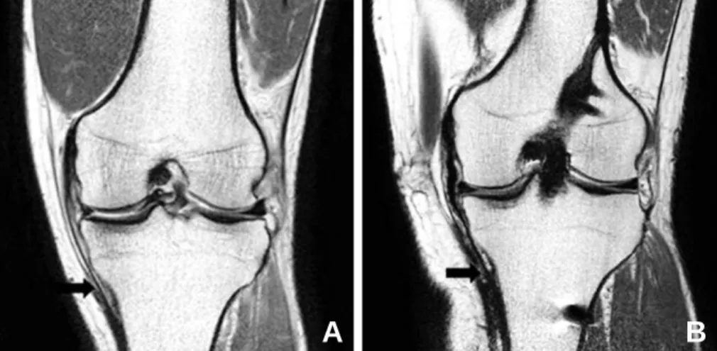
저자들은 24명의 환자에 대해 자가 슬건을 이용하여 전방십자 인대 재건술을 시행 후 1년 경과 관찰한 결과 반건양건에서 83%(20명), 박건에서 78%(17명)가 재생되었고, 박건의 재생 률은 Tadokoro 등66)이 보고한 46% 보다 높은 결과를 얻었다 (Fig. 1). 이와 같이 여러 연구에서 자가 슬건은 채취 후에 높은 비율로 재생되며 박건의 재생이 비교적 낮게 일어난다고 한다.
Fig. 1. (A) Regeneration of semitendinosus tendon and gracilis tendon on axial MRI at joint level. (B) Both tendons were regenerated . (C) The semitendinosus tendon (block arrow) was absent. The gracilis tendon (open arrow) was absent.
A B C
2) 슬건 재생의 해부학적 특징
재생된 건의 원위부 부착 부위에 대한 연구로 Simonian 등65)은 9명의 환자 중 6명의 환자에서 수술 후 좀 더 근위부로 이동하였다고 보고 하였다. 또한 Papandrea 등51)은 재생된 모든 경우에서 관절선 수준에서 medial popliteal fascia에 붙는다고 하였다. 하지만 Eriksson 등20)은 8명의 환자 중 3명 의 환자에서 거위발건(pes anserinus)과 결합 건(con- joined tendon)을 이루기 전인 관절선의 약 3 cm아래에서 닿았다고 보고하였다. 이에 반해 저자들의 연구결과는 재생 된 건의 부착부가 수술 전과 거의 유사하게 원위부로 0.43±
0.76 cm가량 이동한 결과를 얻은 바 있다(Fig. 2).
재생된 건의 부착부위가 변하기도 하지만 근건 이행부 (Musculotendinosus junction)의 위치도 변하게 되는데 근건 이행부는 근위부로 이동하게 된다. Nakamae 등42)은 3D MRI 연구에서 반건양건의 재생에 대해 근건 이행부의 근 위부로의 이동과 슬건의 굴곡력에 대한 상관관계를 보고하였 는데 술 후 6개월에는 근건 이행부의 근위부로의 이동함에 따
라 피크 토크가 낮았으나 술 후 12개월에는 피크 토크에 영향 을 미치지 않았다는 결과를 얻었다. 저자들은 MRI를 이용하 여 재생된 반건양건에서 근건이행부가 근위부로 평균 4.43±
2.68의 이동한다는 결과를 얻은 바 있다(Fig. 3).
3) 재생의 기전(mechanism of regeneration)
Eriksson 등19)은 2001년에 네가닥 반건양건을 이용한 전 방십자인대 재건술 후 MRI로 검사한 결과 6명중 5명에서 재 생된 건을 관찰할 수 있었고, 생체검사(biopsy)를 시행하여 조직학적 검사를 시행하였다. 연구 결과 이식물을 채취한 결 손부위에 혈종이 생기고, 혈종은 섬유세포 이동의 scaffold 역할을 하여 점차적으로 재생된다고 보고 하였다. Rispoli 등
57)은 슬건의 재생방향에 대한 연구를 보고하였다. 21명의 환 자를 대상으로 전방십자인대 재건술 후 2~32주간 관찰하였 으며 술 후 6주에 슬개골의 상단에서, 술 후 3~32개월에 관절 선 수준에서 건 모양의 조직이 MRI상 확인 되었고, 슬건은 근막을 따라 근위부에서 원위부로 마치 신경재생과 같이 재
Fig. 2. Coronal MRIs demonstrated distal insertion of hamstring tendons. More proximal insertion of harvest hamstring tendon (B) than on the non-operated side (A).
A B
Fig. 3. Saggital MRIs showed that the musculotendinous junction of the regenerated semitendinosus tendon (B) was more proximally shifted than those of preoperative semitendinosus tendon (A).
A B
생이 된다고 하였다. 이처럼 근막을 따라 원위부에서 근위부 로 재생되는 과정을 Leis 등36)은“도마뱀 꼬리 현상(Lizard tail phenomenon)”이라 명명하기도 하였다.
4) 초음파 연구
MRI 및 CT 이외에 초음파에서도 슬건의 재생에 대한 연 구가 있었으며, Papandarea 등51)은 40명의 환자에 대해 술 후 2주~24개월까지 초음파를 시행하여 슬건의 재생에 관해 연구 하였다. 술 후 1개월에 hypoechoic mass가 보였고 술 후 2~12개월까지 점차적으로 성숙하였으나 정상의 반건양건 에 비해 hypoechoic하고 비후되어 있었다고 하였다. 또한 술 후 18~24개월이 되어서야 정상적인 반건양건과 매우 유사 한 echogenicity를 보였고, 관절선 수준에서 medial popliteal fascia에 부착되었다고 하였다. 초음파 연구는 간 편하고 비용이 저렴하므로 동일 환자에게 연속적인 검사를 통하여 슬건의 재생 여부, 기전, 재생의 위치 및 성숙 정도 등 많은 정보를 얻을 수 있어 최근 유용한 연구로 이용되고 있다.
5) 생물역학적 특성
재생된 슬건의 생물역학적 특성에 대해 Leis 등36)은 본래
건의 강도(strength)에 비해 술 후 16주에 23%, 28주에 62%로 하였고, Gill 등23)은 토끼 모델에서 본래 건의 최대 부 하(maximal load)에 비해 25%로 감소하고 인장력은 20~75%(평균 32%)로 감소한다고 하였다. Tadokoro 등 66)은 다양한 각도에서 굴곡력 검사를 시행하였고 굴곡력과 슬건이 재생된 정도와의 상관관계를 분석하였다. 굴곡력은 앉은 상태에서 86.2%(90도), 복와위에서 54.6%(90도), 49.1%(120도)로 감소한다고 하였고 형태학적 재생과 상관없 이 굴곡력은 감소한다고 하였다.
6) 재채취 조직학(Rehavest histology)
재생된 슬건에 대한 조직학적인 연구도 이루어져 왔다.
Okahashi 등48)은 전방십자 인대 재건 후 1년 후에 11명의 환 자 중 9명에서 재생됨을 확인하였고, 재생된 슬건은 조직학적 면역학적으로 정상과 유사하다고 보고하였다. Ferretti 등21) 등은 3명의 환자에 대해 전방십자인대 재건술 후 에 재생된 조직의 조직학적인 검사를 시행하였다. 그 결과 6개월에 섬유 아세포가 증식되고 주위에 몇 개의 혈관이 둘러싸게 되며, 2 년 후에 건의 중앙부위에는 교원 섬유가 평행하게 주행하고 변연부에 섬유 아세포의 증식이 두드러짐을 관찰할 수 있다.
7) 근전도 연구
전방십자인대 재건술에서 슬건을 채취 후에 근전도 검사를 시행한 결과 1992년에 Cross 등14)은 근전도 활동도의 감소가 뚜렷하지 않다고 하였으나, Ristanis 등58)은 2009년에 슬관 절의 굴곡근에서 electromechanical delay가 증가한다는 상반된 결과을 보고하였다.
3. 재생된 슬건의 굴곡력 회복
전방십자인대 재건술 후 슬건이 재생되지만 굴곡력이 이전 에 비해 약해진다는 것에 대해서 논란이 있다. Ohkoshi 등47) 은 자가 슬건의 채취 후 수술 전후의 근력의 변화는 없다고 하 였다. 또한 많은 연구에서 슬건의 채취가 등속 최대 근력 (isokinetic peak torque)에 영향을 미치지 않는다고 하였
다37,38,72). 하지만 이러한 과거의 연구들은 굴곡력을 측정할 때
앉은 상태, 즉 작은 각도에서 등속 최대 근력을 측정하였다.
Fig. 4. Isokinetic hamstring strength measurement in prone position at 120 degree of knee flexion with maximum plantar flexion of ankle. To evaluate the hamstring mus- cle function, the measurement of deep flexor strength would be required.
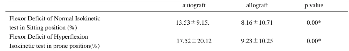
Table 1. Comparisons of flexor deficit between in sitting position and in prone position
autograft allograft p value
Flexor Deficit of Normal Isokinetic
test in Sitting position (%) 13.53±9.15.0 8.16±10.71 0.00*
Flexor Deficit of Hyperflexion
Isokinetic test in prone position(%) 17.52±20.12 9.23±10.25 0.00*
Values are mean±SD
* p<0.05
복와위(prone position) 자세로 슬관절을 과 굴곡 상태에서 등속 최대 근력을 측정하면 굴곡력의 감소을 볼 수 있는데 (Fig. 4), Nakamura 등43)은 90도에서 등속 최대 근력과 굴 곡력(flexion torque)을 측정하여 반대편에 비해 각각 91.3%, 78.8%로 감 소 한 다 는 결 과 를 얻 었 다 . 또 한 Tadokoro 등66)도 과 굴곡 상태에서 슬건의 굴곡력이 반대쪽 에 비해 49%로 감소한다는 결과를 보고하였다. 하지만 이러 한 굴곡력의 감소와 형태학적인 재생과 상관관계는 발견할 수 없었다. 본 저자들의 전방십자인대 재건술에서 자가 슬건 을 채취 후 굴곡력의 변화에 대해 연구하였으며, 앉은 자세에 서 등속 최대 근력은 93.8%, 과굴곡 상태에서 등속 최대 근력
은 89.8%로 감소한다는 결과를 얻었다. 이러한 결과는 Nakamura 등43)의 연구와 유사한 결과였다.
이처럼 등속 최대 근력의 측정 결과가 측정 각도에 따라 차 이가 나는 이유는 슬건이 높은 굴곡각도에서 최대 근력을 발 휘한다는데 있다. 15도에서 30도 사이의 낮은 굴곡 각도에서 최대 근력(peak torque)은 대퇴 이두근(biceps femoris)에 의해 발휘되고, 슬건은 75도 이상의 높은 굴곡 각도에서 굴곡 및 내회전 기능을 발휘한다. 결국 낮은 굴곡 각도에서는 등속 최대 근력을 측정 하면 대퇴 이두근과 같은 여러 근육의 보상 작용이 있어 수술 전후 근력의 변화가 없게 측정되게 된다. 따 라서 굴곡력을 측정할 때 복와위에서 높은 굴곡 각도의 등속
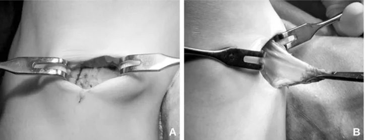
Fig. 5. Our modified technique for harvest hamstring. Reversed L-shaped incision to the sartorius fascia was made (A). The flap was reflected (B).
A B
Table 3. The correlations between flexor deficit between flexor deficit and proximal shift of musculotendinosus junctions
PSMJ PSMJ
semitendinosus grasilis
Flexor Deficit of Normal Isokinetic test in Sitting position. -.104 -.043 Flexor Deficit of Hyperflexion Isokinetic test in prone position 0-.365* 0-.332*
Values are correlation coefficient.
PSMTJ: Proximal shift of musculotendinous junction.
* p<0.05
Table 2. The correlations between flexor deficit and functional performance test in hamstring autograft group
Co-contraction Carioca Shuttle run One leg hop Flexor Deficit of Normal Isokinetic
test in Sitting position 0.364* .442* .336*
0-.431*
Flexor Deficit of Hyperflexion
Isokinetic test in prone position .209 -.026 .228
0 -.213
Values are correlation coefficient.
* p<0.05
최대 근력을 측정하는 것이 바람직할 것으로 보이며, 재생된 슬건의 굴곡력은 높은 굴곡 각도에서 감소할 것으로 보인다.
4. 슬건의 재생, 굴곡력, 기능 수행검사의 상관관계
본 저자들은 자가 슬건을 이용한 전방십자인대 재건술 후 낮은 각도뿐만 아니라 높은 각도의 굴곡에서 등속 최대 근력 을 평가였다. 또한 MRI상의 슬건의 재생, 기능적 결과와 굴 곡력 사이의 상관 관계에 대해 연구하였다. 자가 슬건을 사용 한 39례 와 대조군으로 동종 이식물(allograft)를 사용한 34 례를 대상으로 하였고, 근력검사는 앉은 자세(0~90도)와 복 와위(60~120도)에서 각각의 등속 최대 근력을 측정하였다.
그 결과 자가 슬건을 사용한 군에서 낮은 각도와 높은 각도 모 두에서 굴곡력의 감소가 보였다(Table 1). 또한 자가 슬건을 사용한 군에서 기능적 검사 결과 와 굴곡력 감소는 낮은 각도 에서만 유의한 상관관계를 보였다(Table 2). 근건 이행부의 근위부 이동과 굴곡력의 상관관계는 높은 각도에서만 유의한 상관관계를 보였다(Table 3).
결국 자가 슬건을 이용한 전방십자인대 재건술에서 낮은 굴 곡 각도와 높은 굴곡 각도 모두에서 굴곡력의 감소는 발생하 며, 과굴곡 상태에서의 뚜렷한 굴곡력의 감소가 발생하나 기능 적 결과와 상관관계가 없으며, 근건 이행부가 근위부로 이동 할수록 높은 각도에서 굴곡력 감소가 발생함을 알 수 있었다.
5. 굴곡력 감소에 대한 해결방안
전방십자 인대 재건술에 자가 슬건을 사용 후 발생하는 굴 곡력의 감소를 극복할 수 있는 방법에 대한 연구가 향후에 필 요할 것으로 보인다. 이에 대해 제시할 수 있는 몇 가지 방법 으로 먼저, 원위부로의 재생을 돕고 재생을 안내하는 통로을 만들기 위해 봉공근 근막(satorius fascia)를 봉합하는 것이 필요할 것으로 보인다. 저자들은 특징적으로 봉공근 막의 근 위부에서부터 reversed L-shape 절개를 가한 후 거위발 건 전체를 골막하로부터 박리하여 뒤집고 심부에서부터 박건과 반건양건을 확인하는 변형된 자가 슬건 채취하는 방법을 사 용하고 있으며, 건의 확인과 분리가 용이하고 봉공근의 보존 이 가능하며 신경 손상을 방지할 수 있는 장점이 있다29). 이러 한 방법으로 재생된 슬건이 이전의 연구보다 원위부에 부착 하는 결과를 얻을 수 있었다(Fig. 5). 또한 자가 슬건 채취 시 반건양건 만을 채취하여 굴곡력의 감소를 줄일 수 있을 것으 로 보이며, 슬건에 특성화된 운동(hamstring specific exercise)를 시행하여 자가슬건의 근력을 강화 시키는 방법 도 유용할 것으로 보인다.
결 론
자가 슬건은 최근 사용이 증가하는 추세이고 이에 대한 장
점, 단점에 대해 정확한 이해가 필요하며 여러 요인을 고려하 여 적절한 이식건을 선택해야 한다. 과거의 문헌을 고찰하고 저자들의 연구와 비교 분석한 결과 슬건은 채취 후에 재생이 잘 일어나지만 굴곡력 감소가 발생할 것으로 보인다. 또한 과 굴곡에서의 굴곡력과 기능적 수행능력과 상관관계는 없었지 만 굴곡력의 감소는 기능수행능력을 감소시킬 것으로 보인 다. 이러한 굴곡력의 감소를 낮추기 위해 꾸준한 재활이 하나 의 방법이 될 수 있을 것으로 생각된다.
REFERENCES
01) Adachi N, Ochi M, Uchio Y, Sakai Y, Kuriwaka M, Fujihara A: Harvesting hamstring tendons for ACL recon- struction influences postoperative hamstring muscle perfor- mance. Arch Orthop Trauma Surg, 123: 460-465, 2003.
02) Aglietti P, Buzzi R, Zaccherotti G, De Biase P: Patellar tendon versus doubled semitendinosus and gracilis ten- dons for anterior cruciate ligament reconstruction. Am J Sports Med, 22: 211-217; discussion 217-218, 1994.
03) Ahmad CS, Gardner TR, Groh M, Arnouk J, Levine WN: Mechanical properties of soft tissue femoral fixation devices for anterior cruciate ligament reconstruction. Am J Sports Med, 32: 635-640, 2004.
04) Amiel D, Kleiner JB, Roux RD Harwood FL, Akeson WH: The phenomenon of “ligamentization”: anterior cru- ciate ligament reconstruction with autogenous patellar ten- don. J Orthop Res, 4: 162-172, 1986.
05) Armour T, Forwell L, Litchfield R, Kirkley A, Amendola N, Fowler PJ: Isokinetic evaluation of inter- nal/external tibial rotation strength after the use of ham- string tendons for anterior cruciate ligament reconstruc- tion. Am J Sports Med, 32: 1639-1643, 2004.
06) Arnoczky SP, Tarvin GB, Marshall JL: Anterior cruci- ate ligament replacement using patellar tendon. An evalua- tion of graft revascularization in the dog. J Bone Joint Surg Am, 64: 217-224, 1982.
07) Biau DJ, Katsahian S, Kartus J, et al: Patellar tendon versus hamstring tendon autografts for reconstructing the anterior cruciate ligament: a meta-analysis based on indi- vidual patient data. Am J Sports Med, 37: 2470-2478, 2009.
08) Biau DJ, Katsahian S, Nizard R: Hamstring tendon auto- graft better than bone-patellar tendon-bone autograft in ACL reconstruction - a cumulative meta-analysis and clin- ically relevant sensitivity analysis applied to a previously published analysis. Acta Orthop, 78: 705-707; author reply, 707-708, 2007.
09) Bizzini M, Gorelick M, Munzinger U, Drobny T: Joint laxity and isokinetic thigh muscle strength characteristics after anterior cruciate ligament reconstruction: bone patel-
lar tendon bone versus quadrupled hamstring autografts.
Clin J Sport Med, 16: 4-9, 2006.
10) Bosch U, Kasperczyk WJ: Healing of the patellar tendon autograft after posterior cruciate ligament reconstruction-- a process of ligamentization? An experimental study in a sheep model. Am J Sports Med, 20: 558-566, 1992.
11) Brand J, Jr., Weiler A, Caborn DN, Brown CH, Jr., Johnson DL: Graft fixation in cruciate ligament recon- struction. Am J Sports Med, 28: 761-774, 2000.
12) Cain EL, Phillips BB, Charlebois SJ, Azar FM: Effect of tibial tunnel dilation on pullout strength of semitendi- nosus-gracilis graft in anterior cruciate ligament recon- struction. Orthopedics, 28: 779-783, 2005.
13) Corry IS, Webb JM, Clingeleffer AJ, Pinczewski LA:
Arthroscopic reconstruction of the anterior cruciate liga- ment. A comparison of patellar tendon autograft and four- strand hamstring tendon autograft. Am J Sports Med, 27:
444-454, 1999.
14) Cross MJ, Roger G, Kujawa P, Anderson IF : Regeneration of the semitendinosus and gracilis tendons following their transection for repair of the anterior cruci- ate ligament. Am J Sports Med, 20: 221-223, 1992.
15) Dargel J, Schmidt-Wiethoff R, Bruggemann GP, Koebke J: The effect of bone tunnel dilation versus extraction drilling on the initial fixation strength of press- fit anterior cruciate ligament reconstruction. Arch Orthop Trauma Surg, 127: 801-807, 2007.
16. Delay BS, Smolinski RJ, Wind WM, Bowman DS:
Current practices and opinions in ACL reconstruction and rehabilitation: results of a survey of the American Orthopaedic Society for Sports Medicine. Am J Knee Surg, 14: 85-91, 2001.
17) Duquin TR, Wind WM, Fineberg MS, Smolinski RJ, Buyea CM: Current trends in anterior cruciate ligament reconstruction. J Knee Surg, 22: 7-12, 2009.
18) Eriksson K, Hamberg P, Jansson E, Larsson H, Shalabi A, Wredmark T: Semitendinosus muscle in ante- rior cruciate ligament surgery: Morphology and function.
Arthroscopy, 17: 808-817, 2001.
19) Eriksson K, Kindblom LG, Hamberg P, Larsson H, Wredmark T: The semitendinosus tendon regenerates after resection: a morphologic and MRI analysis in 6 patients after resection for anterior cruciate ligament reconstruction. Acta Orthop Scand, 72: 379-384, 2001.
20) Eriksson K, Larsson H, Wredmark T, Hamberg P.
Semitendinosus tendon regeneration after harvesting for ACL reconstruction. A prospective MRI study. Knee Surg Sports Traumatol Arthrosc, 7: 220-225, 1999.
21) Ferretti A, Conteduca F, Morelli F, Masi V : Regeneration of the semitendinosus tendon after its use in anterior cruciate ligament reconstruction: a histologic
study of three cases. Am J Sports Med, 30: 204-207, 2002.
22) Francis A, Thomas RD, McGregor A: Anterior cruciate ligament rupture: reconstruction surgery and rehabilita- tion. A nation-wide survey of current practice. Knee, 8:
13-18, 2001.
23) Gill SS, Turner MA, Battaglia TC, Leis HT, Balian G, Miller MD: Semitendinosus regrowth: biochemical, ultra- structural, and physiological characterization of the regen- erate tendon. Am J Sports Med, 32: 1173-1181, 2004.
24) Goldblatt JP, Fitzsimmons SE, Balk E, Richmond JC:
Reconstruction of the anterior cruciate ligament: meta- analysis of patellar tendon versus hamstring tendon auto- graft. Arthroscopy, 21: 791-803, 2005.
25) Goradia VK, Rochat MC, Kida M, Grana WA: Natural history of a hamstring tendon autograft used for anterior cruciate ligament reconstruction in a sheep model. Am J Sports Med, 28: 40-46, 2000.
26) Hadjicostas PT, Soucacos PN, Paessler HH, Koleganova N, Berger I: Morphologic and histologic comparison between the patella and hamstring tendons grafts: a descriptive and anatomic study. Arthroscopy, 23:
751-756, 2007.
27) Kartus J, Movin T, Karlsson J: Donor-site morbidity and anterior knee problems after anterior cruciate ligament recon- struction using autografts. Arthroscopy, 17: 971-980, 2001.
28) Kawamura S, Ying L, Kim HJ, Dynybil C, Rodeo SA:
Macrophages accumulate in the early phase of tendon- bone healing. J Orthop Res, 23: 1425-1432, 2005.
29) Kim JG, Moon HT, Kim JY: Modified technique for harvesting the hamstring tendons. J of Korean Orthop Soc Sports Med, 3(1): 56-59, 2004.
30) Kousa P, Jarvinen TL, Kannus P, Jarvinen M: Initial fixation strength of bioabsorbable and titanium interfer- ence screws in anterior cruciate ligament reconstruction.
Biomechanical evaluation by single cycle and cyclic load- ing. Am J Sports Med, 29: 420-425, 2001.
31) Kuroda R, Kurosaka M, Yoshiya S, Mizuno K : Localization of growth factors in the reconstructed anteri- or cruciate ligament: immunohistological study in dogs.
Knee Surg Sports Traumatol Arthrosc, 8: 120-126, 2000.
32) Kurosaka M, Yoshiya S, Andrish JT: A biomechanical comparison of different surgical techniques of graft fixa- tion in anterior cruciate ligament reconstruction. Am J Sports Med, 15: 225-229, 1987.
33) Lambert KL. Vascularized patellar tendon graft with rigid internal fixation for anterior cruciate ligament insuf- ficiency. Clin Orthop Relat Res, 85-89, 1983.
34) Lane JG, McFadden P, Bowden K, Amiel D: The liga- mentization process: a 4 year case study following ACL reconstruction with a semitendinosis graft. Arthroscopy, 9:
149-153, 1993.
35) Lautamies R, Harilainen A, Kettunen J, Sandelin J, Kujala UM: Isokinetic quadriceps and hamstring muscle strength and knee function 5 years after anterior cruciate ligament reconstruction: comparison between bone-patel- lar tendon-bone and hamstring tendon autografts. Knee Surg Sports Traumatol Arthrosc, 16: 1009-1016, 2008.
36) Leis HT, Sanders TG, Larsen KM, Lancaster-Weiss KJ, Miller MD: Hamstring regrowth following harvesting for ACL reconstruction: The lizard tail phenomenon. J Knee Surg, 16: 159-164, 2003.
37) Lipscomb AB, Johnston RK, Snyder RB, Warburton MJ, Gilbert PP: Evaluation of hamstring strength following use of semitendinosus and gracilis tendons to reconstruct the anterior cruciate ligament. Am J Sports Med, 10: 340-342, 1982.
38) Maeda A, Shino K, Horibe S, Nakata K, Buccafusca G:
Anterior cruciate ligament reconstruction with multi- stranded autogenous semitendinosus tendon. Am J Sports Med, 24: 504-509, 1996.
39) Marumo K, Saito M, Yamagishi T, Fujii K: The “liga- mentization”process in human anterior cruciate ligament reconstruction with autogenous patellar and hamstring ten- dons: a biochemical study. Am J Sports Med, 33: 1166- 1173, 2005.
40) Menetrey J, Duthon VB, Laumonier T, Fritschy D:
“Biological failure”of the anterior cruciate ligament graft.
Knee Surg Sports Traumatol Arthrosc, 16: 224-231, 2008.
41) Mirza F, Mai DD, Kirkley A, Fowler PJ, Amendola A:
Management of injuries to the anterior cruciate ligament:
results of a survey of orthopaedic surgeons in Canada.
Clin J Sport Med, 10: 85-88, 2000.
42) Nakamae A, Deie M, Yasumoto M, et al: Three-dimension- al computed tomography imaging evidence of regeneration of the semitendinosus tendon harvested for anterior cruciate lig- ament reconstruction: a comparison with hamstring muscle strength. J Comput Assist Tomogr, 29: 241-245, 2005.
43) Nakamura N, Horibe S, Sasaki S, et al: Evaluation of active knee flexion and hamstring strength after anterior cruciate ligament reconstruction using hamstring tendons.
Arthroscopy, 18: 598-602, 2002.
44) Ng GY, Oakes BW, Deacon OW, McLean ID, Eyre DR: Long-term study of the biochemistry and biomechan- ics of anterior cruciate ligament-patellar tendon autografts in goats. J Orthop Res, 14: 851-856, 1996.
45) Nikolaou VS, Efstathopoulos N, Wredmark T : Hamstring tendons regeneration after ACL reconstruction:
an overview. Knee Surg Sports Traumatol Arthrosc, 15:
153-160, 2007.
46) Noyes FR, Butler DL, Grood ES, Zernicke RF, Hefzy MS: Biomechanical analysis of human ligament grafts used in knee-ligament repairs and reconstructions. J Bone Joint Surg Am, 66: 344-352, 1984.
47) Ohkoshi Y, Inoue C, Yamane S, Hashimoto T, Ishida R: Changes in muscle strength properties caused by har- vesting of autogenous semitendinosus tendon for recon- struction of contralateral anterior cruciate ligament.
Arthroscopy, 14: 580-584, 1998.
48) Okahashi K, Sugimoto K, Iwai M, et al: Regeneration of the hamstring tendons after harvesting for arthroscopic anterior cruciate ligament reconstruction: a histological study in 11 patients. Knee Surg Sports Traumatol Arthrosc, 14: 542-545, 2006.
49) Otero AL, Hutcheson L: A comparison of the doubled semitendinosus/gracilis and central third of the patellar tendon autografts in arthroscopic anterior cruciate liga- ment reconstruction. Arthroscopy, 9: 143-148, 1993.
50) Paessler HH, Mastrokalos DS: Anterior cruciate liga- ment reconstruction using semitendinosus and gracilis ten- dons, bone patellar tendon, or quadriceps tendon-graft with press-fit fixation without hardware. A new and inno- vative procedure. Orthop Clin North Am, 34: 49-64, 2003.
51) Papandrea P, Vulpiani MC, Ferretti A, Conteduca F:
Regeneration of the semitendinosus tendon harvested for anterior cruciate ligament reconstruction. Evaluation using ultrasonography. Am J Sports Med, 28: 556-561, 2000.
52) Petersen W, Laprell H: Insertion of autologous tendon grafts to the bone: a histological and immunohistochemi- cal study of hamstring and patellar tendon grafts. Knee Surg Sports Traumatol Arthrosc, 8: 26-31, 2000.
53) Pinczewski LA, Lyman J, Salmon LJ, Russell VJ, Roe J, Linklater J: A 10-year comparison of anterior cruciate ligament reconstructions with hamstring tendon and patel- lar tendon autograft: a controlled, prospective trial. Am J Sports Med, 35: 564-574, 2007.
54) Prodromos C, Joyce B, Shi K: A meta-analysis of stabili- ty of autografts compared to allografts after anterior cruci- ate ligament reconstruction. Knee Surg Sports Traumatol Arthrosc, 15: 851-856, 2007.
55) Prodromos CC, Han YS, Keller BL, Bolyard RJ:
Stability results of hamstring anterior cruciate ligament reconstruction at 2- to 8-year follow-up. Arthroscopy, 21:
138-146, 2005.
56) Prodromos CC, Joyce BT, Shi K, Keller BL: A meta- analysis of stability after anterior cruciate ligament recon- struction as a function of hamstring versus patellar tendon graft and fixation type. Arthroscopy, 21: 1202, 2005.
57) Rispoli DM, Sanders TG, Miller MD, Morrison WB:
Magnetic resonance imaging at different time periods fol- lowing hamstring harvest for anterior cruciate ligament reconstruction. Arthroscopy, 17: 2-8, 2001.
58) Ristanis S, Tsepis E, Giotis D, Stergiou N, Cerulli G, Georgoulis AD: Electromechanical delay of the knee flex- or muscles is impaired after harvesting hamstring tendons
for anterior cruciate ligament reconstruction. Am J Sports Med, 37: 2179-2186, 2009.
59) Rodeo SA, Arnoczky SP, Torzilli PA, Hidaka C, Warren RF: Tendon-healing in a bone tunnel. A biome- chanical and histological study in the dog. J Bone Joint Surg Am, 75: 1795-1803, 1993.
60) Roe J, Pinczewski LA, Russell VJ, Salmon LJ, Kawamata T, Chew M: A 7-year follow-up of patellar tendon and hamstring tendon grafts for arthroscopic ante- rior cruciate ligament reconstruction: differences and simi- larities. Am J Sports Med, 33: 1337-1345, 2005.
61) Rougraff B, Shelbourne KD, Gerth PK, Warner J:
Arthroscopic and histologic analysis of human patellar tendon autografts used for anterior cruciate ligament reconstruction. Am J Sports Med, 21: 277-284, 1993.
62) Sajovic M, Vengust V, Komadina R, Tavcar R, Skaza K: A prospective, randomized comparison of semitendi- nosus and gracilis tendon versus patellar tendon autografts for anterior cruciate ligament reconstruction: five-year fol- low-up. Am J Sports Med, 34: 1933-1940, 2006.
63) Scheffler SU, Unterhauser FN, Weiler A: Graft remod- eling and ligamentization after cruciate ligament recon- struction. Knee Surg Sports Traumatol Arthrosc, 16: 834- 842, 2008.
64) Segawa H, Omori G, Koga Y, Kameo T, Iida S, Tanaka M: Rotational muscle strength of the limb after anterior cruciate ligament reconstruction using semitendinosus and gracilis tendon. Arthroscopy, 18: 177-182, 2002.
65) Simonian PT, Harrison SD, Cooley VJ, Escabedo EM, Deneka DA, Larson RV: Assessment of morbidity of semitendinosus and gracilis tendon harvest for ACL reconstruction. Am J Knee Surg, 10: 54-59, 1997.
66) Tadokoro K, Matsui N, Yagi M, Kuroda R, Kurosaka M, Yoshiya S: Evaluation of hamstring strength and ten- don regrowth after harvesting for anterior cruciate ligament reconstruction. Am J Sports Med, 32: 1644-1650, 2004.
67) Torry MR, Decker MJ, Jockel JR, Viola R, Sterett WI, Steadman JR: Comparison of tibial rotation strength in patients’status after anterior cruciate ligament reconstruc- tion with hamstring versus patellar tendon autografts. Clin J Sport Med, 14: 325-331, 2004.
68) Unterhauser FN, Bail HJ, Hoher J, Haas NP, Weiler A:
Endoligamentous revascularization of an anterior cruciate ligament graft. Clin Orthop Relat Res, 276-288, 2003.
69) Weiler A, Forster C, Hunt P, et al: The influence of locally applied platelet-derived growth factor-BB on free tendon graft remodeling after anterior cruciate ligament reconstruction. Am J Sports Med, 32: 881-891, 2004.
70) Weiler A, Peters G, Maurer J, Unterhauser FN, Sudkamp NP: Biomechanical properties and vascularity of an anterior cruciate ligament graft can be predicted by contrast-enhanced magnetic resonance imaging. A two- year study in sheep. Am J Sports Med, 29: 751-761, 2001.
71) Woo SL, Wu C, Dede O, Vercillo F, Noorani S : Biomechanics and anterior cruciate ligament reconstruc- tion. J Orthop Surg Res, 1: 2, 2006.
72) Yasuda K, Tsujino J, Ohkoshi Y, Tanabe Y, Kaneda K: Graft site morbidity with autogenous semitendinosus and gracilis tendons. Am J Sports Med, 23: 706-714, 1995.
73) Zantop T, Weimann A, Rummler M, Hassenpflug J, Petersen W: Initial fixation strength of two bioabsorbable pins for the fixation of hamstring grafts compared to inter- ference screw fixation: single cycle and cyclic loading. Am J Sports Med, 32: 641-649, 2004.
전방십자인대 재건술에 자가 슬건과 자가 슬개건이 널리 사용되고 있으나 최근 자가 슬건의 사용이 증가하고 있다. 그 이유는 높은 최대 인장 강도, 낮은 공여부 이환률, 이식건의 고정 방법의 발전과 같은 장점 때문으로 보인다. 전방십자인 대 재건술을 위해 슬건을 채취 후 슬건이 실제로 재생된다는 연구는 이러한 이론적 장점을 크게 부각시켰다. 그러나 내 회 전력의 약화와 굴곡력의 감소와 같은 단점에 대한 우려가 있다. 이러한 굴곡력의 감소에 대해서 논쟁이 있으며, 과연 슬건 의 재생 후에 굴곡력을 회복할 수 있는지에 대해 연구가 필요하다. 본 종설에서는 자가슬건의 장점과 단점, 슬건의 재생에 관한 최근의 연구를 고찰하고 저자들의 연구와 경험을 정리하여 기존의 연구와 비교하였다.
색인 단어: 슬건 재생, 공여부 이환, 슬관절 굴곡력, 전방십자인대 초 록