Posterior Spinal Artery Aneurysm Presenting with Leukocytoclastic Vasculitis
Travis C. Hill1, Omar Tanweer2, Cheddhi Thomas3, John Engler2,5, Maksim Shapiro4, Tibor Becske4, Paul P. Huang2
1New York University School of Medicine, New York, NY, USA
2Department of Neurosurgery, New York University School of Medicine, New York, NY, USA
3Department of Pathology, New York University School of Medicine, New York, NY, USA
4Department of Radiology, New York University School of Medicine, New York, NY, USA
5Department of Neurosurgery, Naval Medical Center Portsmouth, Portsmouth, VA, USA
Rupture of isolated posterior spinal artery (PSA) aneurysms is a rare cause of subarachnoid hemorrhage (SAH) that presents unique diagnostic chal- lenges owing to a nuanced clinical presentation. Here, we report on the diagnosis and management of the first known case of an isolated PSA aneurysm in the context of leukocytoclastic vasculitis. A 53-year-old male presented to an outside institution with acute bilateral lower extremity paralysis 9 days after admission for recurrent cellulitis. Early magnetic res- onance imaging was read as negative and repeat imaging 15 days after presentation revealed SAH and a compressive spinal subdural hematoma.
Angiography identified a PSA aneurysm at T9, as well as other areas sus- picious for inflammatory or post-hemorrhagic reactive changes. The pa- tient underwent a multilevel laminectomy for clot evacuation and aneur- ysm resection to prevent future hemorrhage and to establish a diagnosis.
The postoperative course was complicated by medical issues and led to the diagnosis of leukocytoclastic vasculitis that may have predisposed the patient to aneurysm development. Literature review reveals greater mortal- ity for cervical lesions than thoracolumbar lesions and that the presence of meningitic symptoms portents better functional outcome than symp- toms of cord compression. The outcome obtained in this case is con- sistent with outcomes reported in the literature.
J Cerebrovasc Endovasc Neurosurg.
2016 March;18(1):42-47 Received : 16 December 2014 Revised : 27 June 2015 Accepted : 5 July 2015
Correspondence to Paul P. Huang
Bellevue Hospital Center 462 First Ave, Room 7S4 New York, NY 10016, USA
Tel : 1-212-263-6414 Fax : 1-212-263-8225
E-mail : [email protected]
ORCID : http://orcid.org/0000-0002-7995-9315
This is an Open Access article distributed under the terms of the Creative Commons Attribution Non- Commercial License (http://creativecommons.org/li- censes/by-nc/3.0) which permits unrestricted non- commercial use, distribution, and reproduction in any medium, provided the original work is properly cited.
Keywords Aneurysm, Subarachnoid hemorrhage, Leukocytoclastic vasculitis, Hypersensitivity vasculitis, Posterior spinal artery syndrome, Spinal cord vascular diseases
INTRODUCTION
Spinal artery aneurysms are most commonly asso- ciated with high-flow states secondary to either arte- riovenous shunting or occlusive disease of larger ar- teries causing increased flow through spinal arteries recruited as collateral circulation.1)2)4) An aneurysm is considered "isolated" when it occurs in the absence of
such a high flow state. Aneurysms of the spinal vas- culature are more common along the anterior axis than the posterior axis, and isolated aneurysms ac- count for only a small fraction of these. Histologically, isolated spinal artery aneurysms have features shared with cerebral artery aneurysms,7) suggesting that at least some may be the product of a congenital predis- position to aneurysm formation. However, isolated
A B C
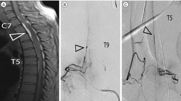
Fig. 1. Diagnostic imaging, 15 days (MR) and 16 days (Angiography) post-presentation. (A) Sagittal T1 MR with compressive hyper-in- tense ventral lesion at T2-level (open arrowhead) and axial extension from C7-T8. (B) Masked angiogram of the T9 radiculopial artery with 1.5mm ectasia (open arrowhead). (C) Masked angiogram of the T5 radiculopial artery with lesion suspicious for vascular pathol- ogy (open arrowhead).
anterior spinal artery and cerebral artery aneurysms have been associated with both central nervous sys- tem-specific and systemic vasculitis.5)13)17)20) Here, we present the first case to our knowledge documenting a posterior spinal artery (PSA) aneurysm in the con- text of a hypersensitivity vasculitis (leukocytoclastic vasculitis).
CASE REPORT
History and presentation
A 53-year-old male with a history of hepatitis C, hepatitis B, and poly-substance abuse presented to an outside hospital for bilateral lower-extremity recurrent cellulitis, hypoalbuminemia and acute kidney injury.
On the ninth day of admission, the patient developed acute paraplegia associated with a T5/6 sensory level deficit. A thoracic spine magnetic resonance imaging (MRI) obtained on the day of presentation was read
as negative for evidence of compressive cord lesion and the patient was medically managed with broad-spectrum antibiotics and steroids.
The patient's neurological symptoms did not im- prove with medical management and 15 days later a second thoracic spine MRI was obtained. A hyper-in- tense intradural lesion was noted (Fig. 1A) with com- pressive effects at the level of C7-T1 with associated increase in T2 signal intensity in the cord extending from C7-T5, suggesting edema. The patient was sub- sequently transferred to our institution for further evaluation. Thoracic spinal angiography revealed a 1.5 mm ectasia of the right T9 segmental artery's radi- culopial branch, consistent with an aneurysm of the posterior spinal artery (Fig. 1B). In addition to the right T9 aneurysm, a segment of the left T5 radi- culopial artery revealed vascular pathology along the posterior aspect of the spinal cord suggestive of either an inflammatory process or post-hemorrhagic reactive
A B C
Fig. 2. Intraoperative images and photomicrographs stained with hematoxylin and eosin. (A) Intraoperative image of the aneurysm in situ with the T9 nerve root retracted. (B) Adjacent segments of the parent vessel wall appear normal (filled arrowhead) compared to the aneurysm wall (open arrowhead; 100 ×). (C) Myxoid degeneration (open arrowhead) and medial necrosis with fibrin deposition (filled arrowhead), both typical features of an aneurysm, were present in the aneurysm wall (400 ×).
changes (Fig. 1C). Vascular bleeding and lumen irreg- ularities consistent with an acute inflammatory proc- ess were noted throughout the cervical and thoracic spine, and the angiogram also revealed a 3 mm right middle cerebral artery (MCA) aneurysm.
Intervention
A coil was placed in the segmental artery supplying the T9 aneurysm for subsequent intra-operative local- ization during the open resection. The patient under- went a multilevel laminectomy (C7-T2 and T8-10) for hematoma evacuation and microsurgical aneurysm resection. The lesion at T9 (Fig. 2A) was resected and submitted to pathology. The histology was consistent with a dissecting aneurysm with localized regions of lymphocytic infiltration of the intima, and fibrosis and necrotic changes in regions of the tunica media (Fig.
2B, C). No histological abnormalities were noted in the adjacent, non-aneurysmal vessel wall. Samples of the hematoma from the T2-level had histological fea- tures consistent with an organizing intradural hematoma.
He was returned to the medical service on post-oper- ative day 2.
Postoperative course
Multiple medical issues complicated the postoperative course. Neurologically, the patient's pre-operative def- icits remained unchanged for the duration of his hospitalization. Medically, the patient deteriorated over the next 60 days. He developed anuric renal fail-
ure and hypercapnic respiratory distress. Rheumatologic work-up revealed low C3 and C4 levels. A skin biop- sy was performed which was positive for leukocyto- clastic vasculitis. Renal biopsy demonstrated mem- branoproliferative glomerulonephritis secondary to hepatitis C virus-related cryoglobulinemic vasculitis and he underwent treatment with rituximab, plasma- pheresis and prednisone with improvement in his re- nal function. The patient subsequently developed a urinary tract infection with multiple-drug resistant Klebsiella and was started on imipenem. Several days later the patient became hypoxic and hypotensive.
The family declined further interventions, and the pa- tient expired.
DISCUSSION
Very little is known about the pathophysiology of isolated PSA aneurysms. Here, we present the case of a ruptured PSA aneurysm in the context of a leukocy- toclastic vasculitis. Spinal angiography demonstrated evidence of diffuse inflammatory process affecting multiple levels, histology confirmed the presence of a leukocytic infiltrate at the site of the resected aneur- ysm and subsequent skin biopsy confirmed the diag- nosis of leukocytoclastic vasculitis. Leukocytoclastic vasculitis is a small-vessel hypersensitivity vasculitis resulting from immune complex deposition in the vessel wall, leading to lymphocytic infiltration. This
Reference Age, Sex Lesion site Treatment Symptoms before
intervention Outcome (concise)
Henson and Croft, 1956 51, M C1 None SAH Death
Cavusoglu et al., 2010 27, F C1 Clipping SAH Good
Goto et al., 1988 53, M C2 Resection SAH Good
Handa et al., 1992 3, F C2 Resection Quadriparesis Good
Kocak et al., 2006 54, F C2 None SAH Death
Johnson et al., 2014 19*, M C5 Resection SAH Good
Geibprasert et al., 2010 43, M T4 Resection SAH, radiculopathy Good
Berlis et al., 2005 62, F T5 Resection SAH, paraparesis Good
Kim et al., 2012 52, M T7 Embolization failure,
coiling Nuchal rigidity, myelopathy, radiculopathy
Good
Present report 53, M T9 Resection ASIA grade A Stable
Tanweer et al., 2012 67, F T11 Embolization ASIA grade A Stable
Nemecek et al., 2006 55, M T12 Resection ASIA grade A Stable
Massand et al., 2005 54, M T12 Resection LBP Good
Massand et al., 2005 69, M L1 Resection LBP Good
Shankar et al., 2012 72, F L2 Coiling LBP Good
PSA = posterior spinal artery, SAH = subarachnoid hemorrhage, LBP = lower back pain.
*age estimated from text
Table 1. Literature review of isolated PSA aneurysms
inflammatory process may have predisposed our pa- tient to aneurysm formation, or it may have compro- mised the wall integrity of a pre-existing PSA aneur- ysm, precipitating rupture. We identified 14 cases of isolated PSA aneurysms, which are summarized in Table 1. None occurred in the context of a confirmed vasculitis but it is not clear that vasculitis was ruled out. For the current case, an important question re- mains whether diagnosis and treatment of the vasculi- tis could have prevented aneurysm rupture.
The natural history of spinal aneurysms likely mir- rors that of intracranial aneurysms, although pub- lished natural history data of spinal aneurysms is sparse. Most presented after an initial rupture and re- hemorrhage occurred in 2 of 15 cases (13%). The aver- age age at presentation was 48.6 (95% confidence in- terval: 37.8-59.5) years. When cervical and thor- acolumbar lesions were considered separately, cer- vical lesions tended to present at a younger age (Fig.
3A). Three of 15 patients (20%) exhibited multiple aneurysms; 2 of the 14 reported cases harbored other spinal artery aneurysms,14) while our patient had an
additional MCA artery aneurysm. This rate is similar to the rate of multiple aneurysms observed in patients with cerebrovascular aneurysms, where 20-30% of pa- tients with at least one cerebrovascular aneurysm will have multiple aneurysms.19) Our analysis of the lim- ited data available is consistent with the assumption that PSA aneurysms and cerebral aneurysms share similar epidemiology, natural history and re-hemor- rhage risk.
The clinical presentation of ruptured PSA aneur- ysms depends on the level of the lesion. Eighty-three percent of cervical-level PSA aneurysms present with signs and symptoms of intracranial SAH, including severe headache, nuchal rigidity, and nausea and vomiting in the absence of myelopathy. For in- fra-cervical PSA aneurysms, the presentation is more suggestive of spinal SAH (myelopathy, localized back pain, and radicular pain). Cervical lesions are more likely to present suddenly than infra-cervical lesions (66% vs. 33%, respectively). In summary, ruptured cervical PSA aneurysms are more likely to present in younger patients with symptoms of intracranial SAH
A B C
Fig. 3. Literature review on posterior spinal artery (PSA) aneurysm presentation, management and outcomes. (A) Age of presentation for cervical and thoracolumbar PSA aneurysms. (B) Outcomes observed in cases of cervical and thoracolumbar PSA aneurysms.
Outcomes observed were not significantly different between cervical and thoracolumbar lesions (p > 0.5, Fisher's Exact test). (C) Cases that presented with pain or meningitic symptoms ("SAH") had significantly better outcomes after intervention than those that presented with symptoms of cord compression (p < 0.05, Fisher's Exact test). SAH = subarachnoid hemorrhage.
(including blood in the basal cisterns) without any re- cent history of progressive symptom. In contrast, rup- tured infra-cervical PSA aneurysms presented in older patients with symptoms of spinal SAH and a one-week history of lower back or abdominal pain.
Once identified, PSA aneurysms are amenable to re- section, endovascular intervention, or watchful waiting.
For ruptured PSA aneurysms, our literature analysis suggests that more aggressive management should be considered for cervical PSA aneurysms, as the only reported deaths have followed re-hemorrhage in these lesions prior to intervention. Indeed, the mortality rate of cervical lesions is higher than that of thor- acolumbar lesions (33% vs. 0% mortality; Fig. 3B).
Treatment modalities include either microsurgical re- section or endovascular embolization. While newer microcatheters have made it possible to embolize these lesions, surgeons have historically chosen re- section because the operation is curative and the su- perficial nature of the PSA aneurysm facilitates an open surgical approach. In addition, surgical decom- pression with a laminectomy can relieve compression caused by a hematoma. However, outcome is in- dependent of approach (endovascular vs. open). Of
patients who underwent intervention, those present- ing with pain or meningitic symptoms were sig- nificantly more likely to have a positive outcome than those who presented with symptoms of cord com- pression (100% vs. 40% improvement; Fig. 3C). Thus, outcome now appears more dependent on symptoms at presentation, leaving the physican to choose inter- vention based on case-specific factors and personal preference.
For the present case, endovascular intervention was not pursued because occlusion of the proximal seg- mental vessel of the T9 aneurysm would not provide definitive cure, and there was concern that direct liq- uid embolic injection could result in additional spinal cord infarction. Since the patient required an evacua- tion of his hematoma to decompress his spinal cord, we felt that the aneurysm could be resected surgically at the same time and would provide a tissue sample to help establish a diagnosis for his suspected sys- temic vasculitis. Postoperatively, the patient was neu- rologically stable with negligible evidence of recovery.
However, the patient's postoperative course was com- plicated by medical factors that resulted in the death of the patient after a protracted hospital course.
CONCLUSION
In general, isolated PSA aneurysms are difficult to diagnose but are amenable to surgical intervention with good results. Cervical lesions have a higher mor- tality than thoracolumbar lesions, providing justifica- tion for more aggressive management. Patients who present with pain or meningitic symptoms are more likely to recover than those who present with symp- toms of cord compression, a trend consistent with the results of the presented case. Once diagnosed, both open and endovascular approaches have been used successfully with high cure rates and low morbidity.
Consequently, the choice of management strategy will ultimately depend on the specific anatomic pathology, the medical status of the patient, the patient's person- al preference and the surgeon.
Disclosure
The authors report no conflict of interest concerning the materials or methods used in this study or the findings specified in this paper.
REFERENCES
1. Berlis A, Scheufler KM, Schmahl C, Rauer S, Götz F, Schumacher M. Solitary spinal artery aneurysms as a rare source of spinal subarachnoid hemorrhage: potential eti- ology and treatment strategy. AJNR Am J Neuroradiol.
2005 Feb;26(2):405-10.
2. Biondi A, Merland JJ, Hodes JE, Aymard A, Reizine D.
Aneurysms of spinal arteries associated with intramedullary arteriovenous malformations. II. Results of AVM endo- vascular treatment and hemodynamic considerations. AJNR Am J Neuroradiol. 1992 May-Jun;13(3):923-31.
3. Cavusoglu H, Ozdilmac A, Sahin Y, Aydin Y. Isolated posterior spinal artery aneurysm causing intracranial acute subarachnoidal hemorrhage. Acta Neurochir (Wien). 2010 Apr;152(4):721-4.
4. Doppman JL, Di Chiro G, Glancy DL. Collateral circu- lation through dilated spinal cord arteries in aortic coarc- tation and extraspinal arteriovenous shunts. An arterio-
graphic study. Clin Radiol. 1969 Apr;20(2):192-7.
5. Fody EP, Netsky MG, Mrak RE. Subarachnoid spinal hem- orrhage in a case of systemic lupus erythematosus. Arch Neurol. 1980 Mar;37(3):173-4.
6. Geibprasert S, Krings T, Apitzsch J, Reinges MH, Nolte KW, Hans FJ. Subarachnoid hemorrhage following poste- rior spinal artery aneurysm. A case report and review of the literature. Interv Neuroradiol. 2010 Jun;16(2):183-90.
7. Goto Y, Kamijyo Y, Yonekawa Y, Kikuchi H. Ruptured aneurysm of the posterior spinal artery of the upper cervical spinal cord: case report. Neurosurgery. 1988 Mar;22(3):558-60.
8. Handa T, Suzuki Y, Saito K, Sugita K, Patel SJ. Isolated intramedullary spinal artery aneurysm presenting with quadriplegia. Case report. J Neurosurg. 1992 Jul;77(1):148-50.
9. Henson RA, Croft PB. Spontaneous spinal subarachnoid haemorrhage. Q J Med. 1956 Jan;25(97):53-66.
10. Johnson J, Patel S, Saraf-Lavi E, Aziz-Sultan MA, Yavagal DR. Posterior spinal artery aneurysm rupture after 'Ecstasy' abuse. J Neurointerv Surg. 2015 Jul;7(7):e23.
11. Kim HJ, Choi IS. Dissecting aneurysm of the posterior spinal artery: case report and review of the literature.
Neurosurgery. 2012 Sep;71(3):E749-56; discussion E756.
12. Kocak A, Ates O, Cayli SR, Sarac K. Isolated posterior spinal artery aneurysm. Br J Neurosurg. 2006 Aug;20(4):241-4.
13. Kossorotoff M, Touze E, Godon-Hardy S, Serre I, Mateus C, Mas JL, et al. Cerebral vasculopathy with aneurysm formation in HIV-infected young adults. Neurology. 2006 Apr;66(7):1121-2.
14. Massand MG, Wallace RC, Gonzalez LF, Zabramski JM, Spetzler RF. Subarachnoid hemorrhage due to isolated spinal artery aneurysm in four patients. AJNR Am J Neuroradiol. 2005 Oct;26(9):2415-9.
15. Nemecek AN, Sviri G, Hevner R, Ghodke B, Britz GW.
Dissecting aneurysm of the thoracic posterior spinal artery.
Case illustration. J Neurosurg Spine. 2006 Dec;5(6):555.
16. Shankar JJ, terBrugge K, Krings T. Subarachnoid hemor- rhage following posterior spinal artery aneurysm rupture.
Can J Neurol Sci. 2012 Jul;39(4):531-2.
17. Takahashi JC, Sakai N, Iihara K, Sakai H, Higashi T, Kogure S, et al. Subarachnoid hemorrhage from a ruptured anterior cerebral artery aneurysm caused by polyarteritis nodosa. Case report. J Neurosurg. 2002 Jan;96(1):132-4.
18. Tanweer O, Woldenberg R, Zwany S, Setton A. Endovascular obliteration of a ruptured posterior spinal artery pseu- doaneurysm. J Neurosurg Spine. 2012 Oct;17(4):334-6.
19. Weir B. Unruptured intracranial aneurysms: a review. J Neurosurg. 2002 Jan;96(1):3-42.
20. Yoong MF, Blumbergs PC, North JB. Primary (granulomatous) angiitis of the central nervous system with multiple aneurysms of spinal arteries. Case report. J Neurosurg.
1993 Oct;79(4):603-7.