유방암환자에서
전체 글
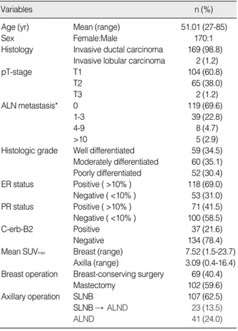
수치
관련 문서
Kameoka, “Sentinel lymph node biopsy for breast cancer patients using fluorescence navigation with indocyanine green,” World Journal of Surgical Oncology, Vol..
In the present study, as a preliminary study, a LACC-based Compton CT was developed to estimate the activity of the spot inside the radioactive waste drum. To
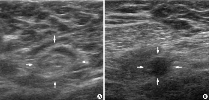
Relationship between lymph node metastasis and lymphatic invasion, diagnosed by immunohistochemical staining and H&E staining in gastric
For my study, this being diagnosed with prostate cancer receiving treatment in patients with complementary and alternative therapies for their experience
F18-FDG uptake in thyroid from PET for evaluation in cancer patients: high prevalence of malignancy in thyroid PET incidentaloma.. Thyroid incidentalomas:
Near-infrared fluorescent type II quantum dots for sentinel lymph node mapping,.. Sungjee Kim, Yong Taik
Near-infrared fluorescent type II quantum dots for sentinel lymph node mapping,.. Sungjee Kim, Yong Taik
Epilepsy surgery outcome in children with tuberous sclerosis complex evaluated with alpha-[11C]Methyl-L-Tryptophan positron emission tomography (PET). Neuronuclear
