Purpose: The axillary lymph node status is an important prognostic factor for recurrence and survival of patients who have primary breast cancer. This study determined the accu- racy of ultrasonography and 18F-FDG positron emission tomo- graphy (PET)/computed tomography (CT) in preoperative staging in axilla in patients with breast cancer. Methods: One hundred seventy-one patients with primary breast cancer were recruited from January 2007 to August 2008. All the patients underwent axillary ultrasonography and 18F-FDG PET/CT for the axillary staging before their operation. Results:
The overall sensitivity, specificity, and the positive and negative predictive values and the accuracy of axillary ultrasonography for making the diagnosis of axillary metastasis were 73.07%, 84.87%, 67.85%, 87.82%, and 81.28%, respectively. On a
visual assessment of 18F-FDG PET/CT, the diagnostic accu- racy was 85.38% with 69.23% sensitivity, 92.43% specificity, a positive predictive value of 80.00%, and a negative predic- tive value of 87.30%. By the combined use axillary ultraso- nography and 18F-FDG PET/CT to the axilla, the sensitivity, specificity, the positive and negative predictive values and the diagnostic accuracy were 82.35%, 97.91%, 93.33%, 94.00%, and 93.84%, respectively. Conclusion: The combination of
18F-FDG PET/CT and ultrasonography improves preoperative axillary staging in breast cancer that are often not found if only one imaging modalities are applied.
Key Words: Breast neoplasms, Positron-emission tomography, Ultrasonography 중심단어: 액와 림프절, 양전자방출단층촬영, 초음파검사
서 론
유방암은 최근 유방촬영술을 이용한 유방암 검진과 초음파검 사의 이용으로 조기 유방암이 증가하는 추세이며, 항암화학요법 및 표적치료의 발달로 인하여 생존율이 향상되고 있지만 아직까 지 완치할 수 없다. 유방암으로 인한 사망률을 줄이기 위해서는 조기 진단뿐 아니라 정확한 병기의 진단이 필요하다. 그 중에서도 유방암의 재발과 생존을 예측할 수 있는 가장 의미 있는 예후인자
는 액와림프절 전이 여부이다.(1-3) 과거에는 액와림프절 전이의
진단과 치료 목적으로 액와곽청술이 시행되었다. 하지만 액와곽 청술은 팔저림, 운동 제한, 림프부종, 삶의 질 감소 등 많은 합병 증을 유발한다.(4) 최근에 액와림프절 전이 여부에 대한 최소 침
습적인 방법으로 감시림프절 생검술이 소개되었다.(5) 감시림프
절 생검술은 액와곽청술의 합병증을 최소화하면서도 액와림프절 병기를 비교적 정확히 예측할 수 있어 액와림프절 전이 상태를 평 가할 수 있는 또 하나의 방법으로 자리 매김하고 있다. 그러나 감 시림프절 생검술은 액와곽청술에 비해 수술 중 동결 절편 조직검 사를 시행하므로 더 많은 노력과 시간 및 비용이 필요하다. 따라서 수술 전 임상적인 액와림프절 전이 여부를 정확히 평가할 수 있다 면 최소한의 액와 수술로 액와림프절 전이 여부를 진단할 수 있을
Preoperative Axillary Staging Using
18F-FDG PET/CT and Ultrasonography in Breast Cancer Patients
Yong Hun Kim, Jin Sun Lee, Chul Joo Lee, Je Ryong Kim, Eil Sung Chang
Department of Surgery, Research Institute for Medical Science, Chungnam National University School of Medicine, Daejeon, Korea
Breast Cancer
O R I G I N A L A R T I C L E
김용훈ㆍ이진선ㆍ이철주ㆍ김제룡ㆍ장일성
충남대학교병원 유방∙내분비외과, 충남대학교 의학전문대학원 의학연구소
유방암환자에서 18 F-FDG PET/CT와초음파검사를 이용한 수술 전 액와 림프절 전이 평가
책임저자: 장일성
301-721 대전광역시 중구 대사동 640, 충남대학교병원 외과 Tel: 042-280-7175, Fax: 042-257-8024
E-mail: [email protected]
접수일: 2009년 3월 31일 게재승인일: 2009년 6월 10일
163
것이다.
수술 전 임상적인 액와림프절 전이 여부를 평가하는데 있어 가장 손쉽게 접할 수 있는 방법은 초음파검사이다. Nori 등(6)과 Sato 등(7)은 감시림프절 생검술 여부 결정에 있어 수술 전 액와 초음 파검사가 적절하다고 보고하고 있다. 그러나 초음파검사는 시술 자에 의해 결과가 달라질 수 있다. 최근에 개발된18F-FDG를 사 용한 양전자방출단층촬영(positron emission tomography, PET)은 기존의 장비들이 물리적 성질에 따른 해부학적인 영상을 제공하는 것과 달리 주입된 물질의 생화학적 특성과 분포에 따라 영상을 제공한다. 이론적으로18F-FDG/PET는 단 한 번의 검사 로 원발종양, 전이 림프절, 원격전이 부위, 모두에서 증가된 당대 사를 검출할 수 있다. 초기에 몇몇 연구에서 수술 전 액와림프절 전이 평가에 대한18F-FDG/PET의 유용성을 보고하였다.(8-10) 그러나 연구가 진행됨에 따라 점차 낮은 민감도를 나타내고 다양 한 결과를 보인다는 보고가 나왔다.(11,12) 그리고18F-FDG/PET 는18F-FDG 흡수의 해부학적 위치 판단이 용이하지 않다. 이러 한 문제점을 극복하기 위해, 전산화단층촬영(computed tomo- graphy, CT)과 PET를 융합한18F-FDG PET/CT가 등장했다.
Tatsumi 등(13)은18F-FDG PET/CT가 기존의 PET나 CT에 비해 병변의 위치를 더 잘 평가할 수 있다고 보고하고 있다.
이에 본 저자는 수술 전 임상적인 액와림프절 전이 여부를 초음 파검사와18F-FDG PET/CT로 평가하고, 수술 후 병리학적 소 견에 따른 초음파검사 및18F-FDG PET/CT의 민감도, 특이도, 정확도를 비교하여 수술 전 액와림프절 전이 평가에 대한 유용성 을 살펴보고자 하였다.
방 법
대상
2007년 1월부터 2008년 8월까지 충남대학교병원에서 유방암 으로 진단 후 수술을 시행 받은 171명의 환자를 대상으로 하였다.
대상기간 동안 상피내암이나 수술 전 항암화학요법을 받은 환자 는 제외하였다. 모든 환자에서 액와림프절 전이에 대하여 수술 전 초음파검사 및18F-FDG PET/CT를 시행하였다. 연구 대상의 병리 진단 보고서를 검토하여 조직학적 유형과 림프절 전이 유무 를 조사하였고, 대상 환자의 의무기록과 영상기록을 통하여 임상 양상과 병리학적 병기, 임상 병기를 조사하였다. 호르몬 수용체 상태는 면역조직화학검사(immunohistochemistry, IHC)에서 10% 이상일 경우에만 양성으로 하였다. C-erb-B2의 상태는 IHC 3+와 IHC 2+일 경우 형광제자리부합법(fluorescence in situ hybridization, FISH) 양성일 경우에 양성으로 하였다. 병기 분 류는 American Joint Committee on Cancer의 분류(2002년
제6판)에 따랐다.
감시림프절생검술
수술 시 0.8% indigocarmine (Korea United Pharm Inc., Seoul, Korea) 5 mL를 유륜하 피하조직으로 주입한 뒤 3분간 부드럽게 마사지를 시행하였다. 절개는 액와부에 털이 없는 부위 를 택해 대흉근의 외연에서 활배근의 전연까지 비스듬히 3 cm 가 량 절개를 가하였다. 빗장가슴근막(clavipectoral fascia)을 향해 절개를 계속해 들어가다가 염색된 림프관을 발견한 뒤 이를 따라 추적하여 염색된 림프절을 찾아내었다. 그리고 감시림프절의 동결 절편검사를 시행하여 림프절 전이 양성인 경우에는 액와곽청술을 시행하였다. 수술 전 초음파 검사 및18F-FDG PET/CT에서 각 각 액와림프절 전이 양성으로 예측한 경우와, 초음파 검사와18F- FDG PET/CT 중 한 검사에서만 액와림프절 전이 양성을 저명하 게 예측한 경우에는 감시림프절 생검술을 시행하지 않고 액와곽 청술을 시행하였다.
초음파검사
초음파검사는 12 MHz의 고주파 선형 탐촉자를 가진 장비(Phi- lips Medical Systems, Bothell, USA)를 사용하였다. 수술 전 한 명의 영상의학과 전문의가 시행하고 판독하였다(Figure 1).
18F-FDG PET/CT
18F-FDG PET/CT를 시행하기 전 6시간 이상 금식을 하였으 며, 정맥주사를 통한 포도당 주입도 금하였다. 촬영 전 원활한 이 뇨를 통해 신장 신우와 방광의18F-FDG 배출을 용이하게 하기 위해서 약 500 mL 이상의 수분 섭취를 권장하였고18F-FDG 정 맥 주사 후 이뇨제를 정맥주사하였다. GE Discovery LS 스캐너 (GE Healthcare, Milwaukee, USA)를 이용하여 영상을 획득 하였다. 몸무게 1 kg당 8.14 MBq (0.22 mCi/kg)의18F-FDG를 정맥주사하고 침대에 누워 50분 경과 후 조영증가가 되지 않은 CT 영상을 얻었다. 이어서 1 bed당 3분씩 2D-모드로 방출영상 을 얻었다. 스캔의 범위는 두개골 기저부에서 허벅지의 중간까지 로 하였다. PET 데이터는 CT 데이터를 이용하여 감쇠보정한 후 ordered subset expectation maximization algorithm을 이 용하여 재구성하였다. PET/CT 영상의 분석은 워크 스테이션을 이용하여 재구성된 PET 영상과 감쇠보정용 CT 영상, 그리고 PET 영상과 CT 영상의 융합영상을 핵의학과 전문의가 판독하였 다. 판독 결과는 액와 림프절 부위에 따른 전이 양성 또는 음성으 로 판독하였고, 육안판독를 통하여18F-FDG 섭취가 증가된 경우 에 전이 양성으로 판독하였다. 각 병소의18F-FDG의 섭취 정도를 평가하기 위한 매개변수로 최대 표준화섭취계수(maximum stan-
dardized uptake value, SUVmax)를 이용하였다(Figure 2).
통계분석
액와 림프절에 대하여 초음파검사 및18F-FDG PET/CT 검사 에 따른 민감도(sensitivity), 특이도(specificity), 양성예측도 (positive predictive value), 음성예측도(negative predictive value) 그리고 정확도(accuracy)를 계산하였다. 또한 액와림프 절 전이에 대한 Receiver-operating-characteristic (ROC) curve를 이용한18F-FDG PET/CT의 SUVmax의 기준치(cut-
off value)를 측정하였다. 통계 분석은 SPSS 12.0 K for Win- dows (SPSS Inc., Chicago, USA)를 사용하였고, 유의수준을 p<0.05로 하였다.
결 과
임상적특징
대상 환자의 유방암 진단 당시 연령은 27세부터 85세까지로 평 균 51세였고, 여성이 170예, 남성이 1예였다. 수술은 유방 전 절제 Figure 2.18F-FDG PET/CT imaging of metastatic axillary lymph node. (A) Transaxial computed tomography (CT) image showing a meta- static axillary lymph node (arrow). (B) Transaxial positron emission tomography (PET) image showing a small hypermetabolic axillary lymph node (arrow). (C) PET/CT fusion image showing a metastatic lymph node in axilla (arrow).
A B C
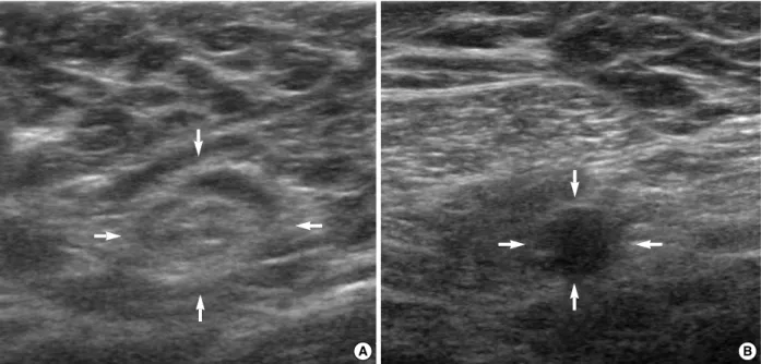
Figure 1. The ultrasonographic findings of axillary lymph node. (A) The reactive lymph node has shown a oval shape, smooth contour, hyperechoic central hilum (arrows). (B) The malignant lymph node has shown a round shape, hypoechoic echogenecity and loss of hyperechoic central hilum (arrows).
A B
술이 102예(59.6%)였고, 유방보존술이 69예(40.4%)였으며, 액 와 수술은 감시 림프절 생검술을 시행 받은 경우는 107예(62.6%), 액와곽청술을 시행 받은 경우는 64예(37.5%)였다. 액와곽청술을 시행 받은 환자에서 감시림프절 생검술에서 양성으로 나와 액와
곽청술을 시행한 경우가 23예(13.5%)였다. 병리학적 T stage는 T1이 104예(60.8%), T2는 65예(38.0%), T3는 2예(1.2%)였다.
조직학적 분화도는 고분화도(well differentiated)를 가진 경우 가 59예(34.5%), 중등도 분화도(moderately differentiated)를 가진 경우가 60예(35.1%), 저분화도(poorly differentiated)인 경우가 52예(30.4%)였다. 호르몬 수용체 상태는 에스트로겐수용 체 양성(IHC >10%)인 경우가 118예(69.0%)였으며, 프로게스테 론수용체 양성(IHC >10%)인 경우는 71예(41.5%)였다. C-erb-
``B2는 37예(21.6%)에서 양성(IHC 3+, FISH+)이었다. 18F-FDG PET/CT에서 평균 SUVmax값은 원발종양 부위는 7.52, 액와림 프절 부위는 3.09였다. 수술 후 시행한 병리학적 검사에서 액와 림프절 전이 양성으로 나온 경우는 52예(30.4%)였다. 이 중에서 미세전이(micrometastasis)로 나온 경우는 2예였다(Table 1).
초음파검사및18F-FDG PET/CT의액와림프절전이평가결과
초음파검사와18F-FDG PET/CT를 통합한 경우에 초음파검사 와18F-FDG PET/CT에서 각각 양성으로 예측한 경우는 30예였 으며 이 중 병리학적으로 액와 림프절 전이 양성인 경우는 28예 (93.3%)였다. 18F-FDG PET/CT에서 양성 또는 초음파검사에서 양성으로 예측한 경우는 41예였으며, 병리학적으로 액와림프절 Table 1. Clinical characteristics (n=171)
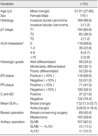
Variables n (%)
Age (yr) Mean (range) 51.01 (27-85)
Sex Female:Male 170:1
Histology Invasive ductal carcinoma 169 (98.8) Invasive lobular carcinoma 2 (1.2)
pT-stage T1 104 (60.8)
T2 65 (38.0)
T3 2 (1.2)
ALN metastasis* 0 119 (69.6)
1-3 39 (22.8)
4-9 8 (4.7)
>10 5 (2.9)
Histologic grade Well differentiated 59 (34.5) Moderately differentiated 60 (35.1) Poorly differentiated 52 (30.4) ER status Positive ( >10% ) 118 (69.0) Negative ( <10% ) 53 (31.0) PR status Positive ( >10% ) 71 (41.5) Negative ( <10% ) 100 (58.5)
C-erb-B2 Positive 37 (21.6)
Negative 134 (78.4)
Mean SUVmax Breast (range) 7.52 (1.5-23.7) Axilla (range) 3.09 (0.4-16.4) Breast operation Breast-conserving surgery 69 (40.4)
Mastectomy 102 (59.6)
Axillary operation SLNB 107 (62.5)
SLNB → ALND 23 (13.5)
ALND 41 (24.0)
ALN=axillary lymph node; ER=estrogen receptor; PR=progesteron receptor; SLNB=sentinel lymph node biopsy; ALND=axillary lymph node dissection.
*Micrometastasis: 2 case.
Table 2. Preoperative staging of axillary lymph node metastasis using 18F-FDG PET/CT and ultrasonography (p=0.000)
Pathologic diagnosis FDG PET/CT
US pN+ pN- Total
US (-), FDG PET/CT (-) 6 94 100
US (+), FDG PET/CT (-) 10 16 26
US (-), FDG PET/CT (+) 8 7 15
US (+), FDG PET/CT (+) 28 2 30
Total 52 119 171
US=ultrasound; pN+=metastasis-positive; pN-=metastasis-negative.
Table 3. Characteristics of patients with false-negative and false-positive axillary lymph node on 18F-FDG PET/CT together with ultra- sonography (n=8)
Case no. Age (yr) Type Tumor size (cm) Tumor grade Tumor SUVmax ALN SUVmax SLNB (+/total) ALND (+/total) FN
1 39 IDC 1.2 Moderately 6.5 1.1 1/2 0/3
2 51 IDC 1.6 Well 4.6 1.1 1/6 0/15
3 63 IDC 1.7 Well 5.9 0.7 1/3 0/14
4 64 IDC 1.4 Moderately 4.0 0.9 1/5 0/7
5 60 IDC 1.2 Well 3.9 0.6 1/2 0/14
6 42 IDC 1.5 Well 5.6 0.7 1/5 0/12
FP
1 60 IDC 0.7 Moderately 2.7 1.4 0/14
2 62 IDC 2.2 Poorly 13.1 1.7 0/7
ALN=axillary lymph node; SLNB=sentinel lymph node biopsy; ALND=axillary lymph node dissection; +=number of metastatic nodes; FN=false negative; FP=false positive; IDC=invasive ductal carcinoma.
전이 양성인 경우는 18예(43.9%)였다. 초음파검사와18F-FDG PET/CT에서 각각 음성으로 예측한 경우는 100예였고, 병리학적 으로 액와림프절 전이 양성인 경우는 6예(6%)였다(p=0.000) (Table 2). 초음파검사와18F-FDG PET/CT를 통합한 경우의 결 과에서 각각 양성으로 예측한 경우의 위양성은 2예(6.66%)였으 며, 각각 음성으로 예측한 경우의 위음성은 6예(6.00%)였다. 위 음성 6예와 위양성 2예에 대한 특성은 Table 3에 요약하였다.
초음파검사에서 액와림프절 전이 양성으로 예측한 경우는 56예 였으며 이 중에서 병리학적으로 액와림프절 전이 양성인 경우는 38예였다. 결과적으로 초음파검사의 액와림프절 전이 평가의 민 감도는 73.07%, 특이도는 84.87%, 양성예측도는 67.85%, 음성 예측도는 87.82%였으며, 정확도는 81.28%였다. 18F-FDG PET/
CT에서 액와림프절 전이 양성으로 예측한 경우는 45예였으며 이 중 병리학적으로 액와림프절 전이 양성인 경우는 36예였다. 18F- FDG PET/CT의 액와림프절 전이 평가의 민감도는 69.23%, 특 이도는 92.43%, 양성예측도는 80.00%, 음성예측도는 87.30%
였으며, 정확도는 85.38%였다(p=0.000) (Table 4). 액와림프절
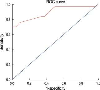
전이에 대한 ROC 곡선을 이용한 SUVmax분석결과 ROC 곡선 하 방의 면적은 0.895±0.040이었다(p=0.000). ROC 곡선을 이용 한 cut-off value는 SUVmax를 1.95로 하였을 때 민감도 70.3%, 특이도 100%로 나타났고, SUVmax를 1.05로 하였을 때 민감도 97.3%, 특이도 50%로 나타났다(Figure 3).
초음파검사와18F-FDG PET/CT를 통합한 경우에 초음파검사 와18F-FDG PET/CT에서 각각 양성으로 예측한 경우와 각각 음성으로 예측한 경우만으로 민감도, 특이도, 양성예측도, 음성 예측도 그리고 정확도를 계산하였을 때 각각 82.35%, 97.91%, 93.33%, 94.00%, 93.84%로 나왔다(p=0.000) (Table 4). 액 와림프절 전이 예측에 대하여18F-FDG PET/CT의 민감도는 초 음파검사에 비하여 3.84% 낮았으나 특이도는 7.71% 높았다. 초 음파 검사와18F-FDG PET/CT를 통합한 경우의 민감도와 특이 도를18F-FDG PET/CT, 초음파 검사 각각의 결과와 비교하면 통합한 경우의 민감도와 특이도가 모두 높았다.
고 찰
최근 유방암에 대한 사회적인 관심과 건강 검진 및 유방암 진단 기기의 발전으로 조기 유방암이 증가하고 있으며 이에 발맞추어 유방 수술에도 많은 변화가 있었다. 유방 전 절제술은 유방보존술 로 액와곽청술은 감시림프절 생검술로 대체되는 환자들이 증가하 고 있다. 특히 감시 림프절 생검술은 액와림프절 전이가 없는 유 방암이 증가함에 따라 액와곽청술의 합병증을 최소화하면서 비 교적 정확한 액와림프절 전이를 평가할 수 있어 현재까지 가장 유 용한 방법으로 알려져 있다. 하지만 감시 림프절 생검술이 액와곽 청술에 비하여 수술 중 동결 절편 조직검사 등의 많은 시간과 노 력, 비용이 필요한 것이 현실이다. 그러므로 수술 전에 액와 림프 절 전이를 정확하게 평가할 수 있다면 불필요한 감시림프절 생검 술을 생략할 수도 있을 것이다. 이를 위하여 수술 전 액와 초음파 검사에서 전이가 의심되는 림프절의 세침흡인세포진검사가 시행 되기도 하며, 최근에는 비침습적인 검사법으로18F-FDG PET/
CT가 연구되고 있다.
초음파검사는 유방암의 진단에 있어 현재 가장 널리 사용되는
Table 4. Accuracy of ultrasonography and 18F-FDG PET/CT and in predicting axillary lymph node status (p=0.000)
TP FP FN TN Sen (%) Sp (%) PPV (%) NPV (%) Acc (%)
Ultrasound 38 18 14 101 73.07 84.87 67.85 87.82 81.28
FDG PET/CT 36 9 16 110 69.23 92.43 80.00 87.30 85.38
Combined* 28 2 6 94 82.35 97.91 93.33 94.00 93.84
TP=true positive; FP=false positive; FN=false negative; TN=true negative; Sen=sensitivity; Sp=specificity; PPV=positive predictive value; NPV=negative predictive value; Acc=accuracy.
*Combination of PET/CT and ultrasound.
Sensitivity
1.0
0.8
0.6
0.4
0.2
0.0
0.0 0.2 0.4 0.6 0.8 1.0
1-specificity ROC curve
Figure 3. Receiver operating characteristics (ROC) curve for semiquantitative SUV analysis and visual interpretation of 18F- FDG PET/CT images for axillary lymph node staging in breast cancer patients (The AUC was 0.895±0.040, p=0.000).
방법이다. 원발 부위인 유방과 액와까지 모두 최소의 노력으로 검 사가 가능하다. 하지만 액와림프절의 전이 평가에 있어 매우 낮은 정확도를 가지고 있다. 몇몇 연구에서 초음파검사에 의한 액와림 프절 전이 평가의 민감도와 특이도를 각각 56-73%, 70-100%로 보고하고 있다.(14-16) 본 연구에서 초음파검사의 액와 림프절 평가의 민감도는 73.07%, 특이도는 84.87%, 정확도는 81.28%
로 이들 연구와 비슷한 결과가 나와 액와림프절 전이를 정확히 예 측하기에는 충분하지 않다. 하지만 Sato 등(7)은 초음파검사에서 액와림프절 음성으로 나온 환자에서는 감시림프절 생검술이 매 우 유용하다고 보고하고 있다.
Noh 등(17)은18F-FDG PET이 유방암에서 액와림프절 전이 의 진단적 특이도가 매우 높다고 보고하였고, Barranger 등(18) 은18F-FDG PET이 유방암에서 액와림프절의 병기결정에 특이 도가 높으므로18F-FDG PET에서 액와림프절 전이 양성으로 예 측한 환자에서는 감시림프절 생검술을 생략하고 액와곽청술을 시행해야 한다고 하였으며, Veronesi 등(19)은 낮은 민감도를 나 타내므로18F-FDG PET에서 액와 림프절 전이 음성으로 예측한 경우에는 감시 림프절 생검술을 시행해야 한다고 하였다. 하지만 Taira 등(20)은18F-FDG PET/CT의 민감도가 48.1%, 특이도 는 92.3%, 양성예측도와 음성예측도가 각각 72.2%, 81.1%로 나 와 낮은 양성예측도로 인하여 감시림프절 생검술의 적응증이 될 수 없다고 보고하였다. 본 연구의 결과에서는18F-FDG PET/CT 에서 민감도는 69.23%, 특이도는 92.43%, 양성예측도가 80.00%, 음성예측도는 87.30% 그리고 정확도는 85.38%로 나와 기존의 연구들과 비슷한 결과를 보여 감시림프절 생검술의 적응증으로 적용하기에는 불충분하다.
Ueda 등(21)은 초음파검사와18F-FDG PET/CT를 통합하여 액와림프절 전이를 평가할 경우 민감도, 특이도, 정확도가 각각 64%, 94%, 85%로 나와 감시림프절 생검술의 적용이 더 적합할 수 있다고 보고하였다. 본 연구에서도 초음파검사와18F-FDG PET/CT를 통합한 경우의 결과에서 초음파검사와18F-FDG PET/
CT에서 각각 음성으로 예측한 경우와 각각 양성으로 예측한 경우 만을 가지고 비교하였을 때 민감도는 82.35%, 특이도는 97.91%, 양성예측도는 93.33%, 음성예측도는 94.00%, 그리고 정확도는 93.84%로 나왔다. 이것은 초음파검사 단독으로 평가한 경우나
18F-FDG PET/CT 단독으로 평가한 경우에 비해 높은 결과를 보 여주어 액와림프절 전이 예측에 대한 진단적 가치를 가진다. 초음 파검사에서 양성 또는18F-FDG PET/CT에서 양성으로 예측한 총 41예에서 18예의 병리학적 액와림프절 전이 확진을 받아43.9%
의 낮은 양성예측도를 보였다. 이 경우에는 감시림프절 생검술을 시행하여 액와림프절의 전이 평가가 필요할 것으로 사료된다. 초 음파검사와18F-FDG PET/CT에서 각각 음성으로 예측한 경우
에 음성예측도는 94.00%, 위음성률은 6예(6.00%)를 보였다. 위 음성을 보인 6예에서 병리학적 진단은 모두 침습성 관암(invasive ductal carcinoma)이었으며, 종양의 크기는 평균 1.43 cm (1.2- 1.7 cm)였다. 모든 경우에서 감시림프절에만 전이가 있었고, 액와 곽청술 후 나온 병리학적 결과에서는 더 이상의 전이된 림프절은 발견되지 않았다. Wahl 등(22)은 전이된 림프절의 크기가 작을 수록, 개수가 적을수록 위음성률이 높다고 하였으며, Fehr 등(23) 은 림프절의 크기는 위음성률에 크게 영향을 미치지 않으며 미세 전이가 있을 경우에 위음성률이 높다고 하였으며, 림프절의 크기 가 1 cm 이상일 경우에는 종양의 특성에 기인한다고 하였다. 본 연구에서도 액와 림프절 전이된 경우에 2예의 미세전이가 있었으 며18F-FDG PET/CT에서는 음성으로 예측되었으나 초음파 검 사에서 양성으로 예측되어 위음성을 보인 6예에서는 제외되었다.
초음파검사와18F-FDG PET/CT에서 각각 양성으로 예측한 경 우에 양성예측도는 93.33%, 위양성률은 2예(6.66%)였다. 위양 성을 보인 2예에서18F-FDG PET/CT의 액와림프절 SUVmax값 은 각각 1.4, 1.7이었으며, 종양의 크기는 각각 0.7, 2.2 cm였다.
액와림프절 전이에 대한 ROC 곡선을 이용한 SUVmax분석결과 cut-off value를 1.95로 하였을 때 민감도 73.3%, 특이도 100%
를 보였으며, 이것을 적용하면 위양성을 보인 2예가 제외되어 양 성예측도가 100%로 불필요한 감시림프절 생검술을 생략하고 액 와곽청술을 시행할 수 있을 것이다. 하지만 민감도가 낮아지기 때 문에 주의 깊게 적용해야 할 것으로 사료된다.
결 론
유방암 환자의 액와 림프절 전이 평가에 있어 초음파검사와18F- FDG PET/CT를 통합한 경우는 초음파검사 단독 또는18F-FDG PET/CT 단독으로 한 경우보다 높은 민감도와 특이도, 양성예측 도 및 음성예측도 그리고 정확도를 보여 수술 전 액와 림프절 전이 평가에 유용함을 알 수 있다. 하지만 6.66%의 위양성률과 6.00%
의 위음성률이 있어 액와 림프절 전이 평가를 정확히 예측할 수는 없지만 신뢰할 수 있는 정확도를 가진다.
초음파 검사는 접근 용이성으로 인하여 유방암의 선별검사에 사용되고 있고, 18F-FDG PET/CT는 한 번에 전신 촬영이 가능하 고 비침습적이라는 장점이 있으며 병변 부위의 해부학적 위치파악 에 용이하다는 장점이 있어 최근에 이용이 증가하고 있기 때문에 향후 유방암 환자의 초기 진단에 있어 초음파 검사 및18F-FDG PET/CT의 통합 검사를 통한 액와 림프절 전이에 대한 더 정확한 예측을 통하여 치료 방향에 도움을 줄 수 있을 것으로 사료된다.
참고문헌
1. McCready DR, Hortobagyi GN, Kau SW, Smith TL, Buzdar AU, Balch CM. The prognostic significance of lymph node metastases after preoperative chemotherapy for locally advanced breast cancer.
Arch Surg 1989;124:21-5.
2. Fisher B, Bauer M, Wickerham DL, Redmond CK, Fisher ER. Re- lation of the number of positive axillary nodes to the prognosis of patients with primary breast cancer. Cancer 1983;52:1551-7.
3. Henderson IC, Canellos GP. Cancer of the breast: the past decade (first of two parts). N Engl J Med 1980;302:17-30.
4. Kwan W, Jackson J, Weir LM, Dingee C, McGregor G, Olivotto IA.
Chronic arm morbidity after curative breast cancer treatment: pre- valence and impact on quality of life. J Clin Oncol 2002;20:4242-8.
5. Aarsvold JN, Alazraki NP. Update on detection of sentinel lymph nodes in patients with breast cancer. Semin Nucl Med 2005;35:116-28.
6. Nori J, Vanzi E, Bazzocchi M, Bufalini FN, Distante V, Branconi F, et al. Role of axillary ultrasound examination in the selection of breast cancer patients for sentinel node biopsy. Am J Surg 2007;193:16-20.
7. Sato K, Tamaki K, Tsuda H, Kosuda S, Kusano S, Hiraide H, et al.
Utility of axillary ultrasound examination to select breast cancer pa- tients suited for optimal sentinel node biopsy. Am J Surg 2004;187:
679-83.
8. Wahl RL, Cody RL, Hutchins GD, Mudgett EE. Primary and meta- static breast carcinoma: Initial clinical evaluation with PET with the radiolabeled glucose analogue 2-[F-18]-fluoro-2-deoxy-D-glucose.
Radiology 1991;179:765-70.
9. Adler LP, Crowe JP, al-Kaisi NK, Sunshine JL. Evaluation of breast masses and axillary lymph nodes with [F-18] 2-deoxy-2-fluoro-D- glucose PET. Radiology 1993;187:743-50.
10. Utech CI, Young CS, Winter PF. Prospective evaluation of fluorine- 18 fluorodeoxyglucose positron emission tomography in breast cancer for staging of the axilla related to surgery and immunocytochemistry.
Eur J Nucl Med 1996;23:1588-93.
11. Avril N, Dose J, Ja_nicke F, Zieqler S, Ro_mer W, Weber W. Assess- ment of axillary lymph node involvement in breast cancer patients with positron emission tomography using radiolabeled 2-(fluorine- 18)-fluoro-2-deoxy-D-glucose. J Natl Cancer Inst 1996;88:1204-9.
12. Adler LP, Faulhaber PF, Schnur KC, Al-Kasi NL, Shenk RR. Axillary lymph node metastases: screening with [F-18]2-deoxy-2-fluoro-D- glucose (FDG) PET. Radiology 1997;203:323-7.
13. Tatsumi M, Cohade C, Mourtzikos KA, Fishman EK, Wahl RL. Initial
experience with FDG-PET/CT in the evaluation of breast cancer. Eur J Nucl Med Mol Imaging 2006;33:254-62.
14. de Kanter AY, van Eijck CH, van Geel AN, Kruijt RH, Henzen SC, Paul MA. Multicentre study of ultrasonographically guided axillary node biopsy in patients with breast cancer. Br J Surg 1999;86:1459- 62.
15. Pamilo M, Soiva M, Lavast EM. Real-time ultrasound, axillary mam- mography, and clinical examination in the detection of axillary lymph node metastases in breast cancer patients. J Ultrasound Med 1989;8:
115-20.
16. Mustonen P, Farin P, Kosunen O. Ultrasonographic detection of meta- static axillary lymph nodes in breast cancer. Ann Chir Gynaecol 1990;
79:15-8.
17. Noh DY, Yun IJ, Kang HS, Kim JS, Lee DS, Chung JK, et al. The diagnostic value of positron emission tomography in detecting the breast cancer. J Korean Breast Cancer Soc 1998;1:6-12.
18. Barranger E, Grahek D, Antoine M, Montravers F, Talbot JN, Uzan S. Evaluation of fluorodeoxyglucose positron emission tomography in the detection of axillary lymph node metastasis in patients with early stage breast cancer. Ann Surg Oncol 2003;10:622-7.
19. Veronesi U, De Cicco C, Galimberti VE, Fernandez JR, Rotmensz N, Viale G, et al. A comparative study in the value of FDG-PET and sentinel node biopsy to identify occult axillary metastases. Ann Oncol 2007;18:473-8.
20. Taira N, Ohsumi S, Takabatake D, Hara F, Takashima S, Aogi K, et al. Determination of indication for sentinel lymph node biopsy in clinical node-negative breast cancer using preoperative 18F-fluoro- deoxyglucose positron emission tomography/computed tomography fusion imaging. Jpn J Clin Oncol 2009;39:16-21.
21. Ueda S, Tsuda H, Asakawa H, Omata J, Fukatsu K, Kondo N, et al.
Utility of 18F-fluoro-deoxyglucose emission tomography/computed tomography fusion imaging (18F-FDG PET/CT) in combination with ultrasonography for axillary staging in primary breast cancer. BMC Cancer 2008;8:165.
22. Wahl RL, Siegel BA, Coleman RE, Gatsonis CG. Prospective mul- ticenter study of axillary nodal staging by positron emission tomo- graphy in breast cancer: a report of the staging breast cancer with PET Study Group. J Clin Oncol 2004;22:277-85.
23. Fehr MK, Hornung R, Varga Z, Burger D, Hess T, Haller U, et al.
Axillary staging using positron emission tomography in breast cancer patients qualifying for sentinel lymph node biopsy. Breast J 2004;10:
89-93.