• 검색 결과가 없습니다.

ββ -Lactamase Producing : A Case Report Acute Osteomyelitis of the Mandible by Extended-Spectrum JOMP

N/A
N/A
Protected

Academic year: 2021

Share "ββ -Lactamase Producing : A Case Report Acute Osteomyelitis of the Mandible by Extended-Spectrum JOMP"

Copied!
5
0
0

로드 중.... (전체 텍스트 보기)

전체 글

(1)

pISSN 2288-9272 eISSN 2383-8493 J Oral Med Pain 2021;46(3):88-92 https://doi.org/10.14476/jomp.2021.46.3.88

Acute Osteomyelitis of the Mandible by Extended-Spectrum ββ-Lactamase Producing Klebsiella Pneumoniae: A Case Report

Gyeo-Woon Jung, Seong-Yong Moon, Ji-Su Oh, Hae-In Choi, Jae-Seek You

Department of Oral and Maxillofacial Surgery, School of Dentistry, Chosun University, Gwangju, Korea

Received June 25, 2021 Revised August 24, 2021 Accepted August 25, 2021

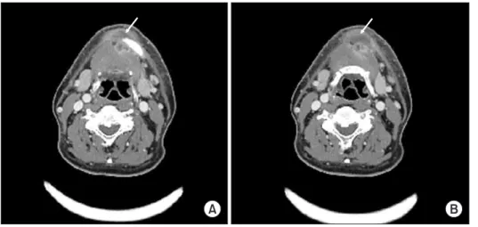
Acute osteomyelitis caused by Klebsiella pneumoniae is rare in the oral and maxillofacial region. Klebsiella pneumoniae is a Gram-negative bacillus and the normal flora of the hu- man body, but it can cause pneumonia, urinary tract infection, meningitis, and osteomy- elitis in patient with compromised immune systems. These infections are mainly caused by nosocomial infection. Microbacterial osteomyelitis was developed by clinical cause such as tooth extraction, fracture, and surgical history, which requires long-term antibiotic ad- ministration and surgical treatment. This report describes that a 56-year-old male patient with acute osteomyelitis caused by Klebsiella pneumoniae infection after implant placement was treated with intravenous administration of ertapenem without open surgery treatment.

Through this case, we report that antibiotic susceptibility test is essential for the treatment of acute osteomyelitis caused by a bacterial infection resistant to empirical antibiotics, and early administration of appropriate antibiotics can reduce the possibility of extensive bone destruction or additional open surgery.

Key Words:

Key Words: Antibiotics, Bacterial infection, Drug resistance, Klebsiella, Osteomyelitis

Correspondence to:

Jae-Seek You

Department of Oral and Maxillofacial Surgery, School of Dentistry, Chosun University, 303 Pilmun-daero, Gwangju 61452, Korea

Tel: +82-62-220-3816 Fax: +82-62-222-3810 E-mail: [email protected] https://orcid.org/0000-0001-7638-9583

JOMP

Journal of Oral Medicine and Pain

Copyright Ⓒ 2021 Korean Academy of Orofacial Pain and Oral Medicine.

CC This is an open-access article distributed under the terms of the Creative Commons Attribution Non-Commercial License (http://creativecommons.org/licenses/by-nc/4.0/), which permits unrestricted non-commercial use, distribution, and reproduction in any medium, provided the original work is properly cited.

INTRODUCTION

Osteomyelitis of the jaw is a relatively rare infectious dis- ease caused by systemic diseases such as hematogenous, bone disease or vascular disease, and odontogenic or non- odontogenic local infection in the oral and maxillofacial region. Osteomyelitis can be classified into two types: acute suppurative osteomyelitis and chronic suppurative osteo- myelitis. Acute suppurative osteomyelitis develops shortly after the predisposition occurs and is accompanied by sys- temic symptoms such as severe pain, tenderness, swell- ing, fever, and malaise. This osteomyelitis is not completely cured even with appropriate treatment and lasts for 1 to 4 months or longer. And it can become fixed as chronic sup- purative osteomyelitis and require the treatments contain- ing sequestrectomy, saucerization, hyperbaric oxygen ther- apy, mandibular resection and reconstruction [1]. Today,

as nutritional status is better and interest in medical care is increasing compared to the past, more patients are vis- iting hospitals early, and osteomyelitis of the jaw, exclud- ing drug-related osteonecrosis, is gradually decreasing [2].

However, it is difficult to diagnose the acute osteomyelitis quickly and to administer susceptible antibiotics because the bone changes do not appear early on the radiograph.

Therefore, acute osteomyelitis is still considered a refractory disease [3].

Enterobacteriaceae producing extended-spectrum β-lac- tamase (ESBL) have recently been reported as nosocomi- al or community infection organism, and ESBL-positive Escherichia coli (E. coli) and Klebsiella pneumoniae are rep- resentative [4].

This case reports that acute osteomyelitis with a rapid

destruction of the mandible caused by ESBL-producing

Klebsiella pneumoniae in a patient with diabetes was

(2)

treated through antibiotics without surgery.

CASE REPORT

Before reporting a case, the conduct with the approv- al of the Institutional Review Board (IRB) of the Chosun University Dental Hospital (CUDHIRB 2102 001 Q) was processed.

A 56-year-old male visited the Department of Oral and Maxillofacial Surgery, Chosun University Dental Hospital and complained of swelling and pain in the right mandibu- lar molar area. The implants of mandibular 2nd premolar, 1st molar, and 2nd molar were placed 1 month before visit, and were removed 3 days before due to consistent pain.

But the pain did not disappear, and mild swelling and fever endured.

The patient was diagnosed with diabetes 2 years before and had taken oral hypoglycemic drugs. The glycemic con- trol was good and there were no other underlying diseases.

There was no history of osteoporosis medication or injec- tion and radiation exposure.

On panoramic radiograph of the first medication exami- nation, irregular bone erosion and bone loss of left mandi- ble, the site of implant removal, were observed. But no ap- parent bone destruction was observed (Fig. 1). There was no distinct symptom excluding nonspecific pain, so anti-biotic, amoxicillin-clavulanic acid (Augmentin; Ilsung Pharma Co., Seoul, Korea), and anti-inflammatory analgesic drug were prescribed.

Two days later, the patient visited the emergency room

due to severe swelling of submental and submandibular area, trismus, fever, and chills. Vital signs were as follows;

blood pressure 130/80 mmHg, pulse rate 80 beats/min, breath rate 20 times/min, body temperature 36 degrees. In peripheral blood examination, the white blood cells (WBCs) were 12,000/mm

3

and the fasting blood glucose was 250 mg/dL. Enhanced neck CT was taken, and abscess was ob- served in the submental space (Fig. 2). Under local anesthe- sia, incision and drainage were performed with pus culture for the antibiotic susceptibility test. Amoxicillin-clavulanic acid (Amocla inj.; Kuhnil Pharma Co., Seoul, Korea) was administered intravenously three times a day at a dose of 1.2 g for a week and dressing on abscess site was done daily with saline irrigation. However, the amount of pus drained was not reduced and severe pain was remained. The symp- toms were not subsided for a week and antibiotic resistance was suspected. ESBL producing Klebsiella pneumoniae was

Fig. 1.

Fig. 1. Panoramic radiographic image (arrow) on first visit was observed irregular bone erosion and bone loss of left mandible, but no apparent bone destruction was observed.

Fig. 2.

Fig. 2. Radiographic images of enhanced neck computed tomography. (A, B) Axial ct image show abscess (arrow) in the submental space.

A B

(3)

identified in the antibiotic susceptibility test and we con- sulted with the Infectious Medicine Department. From the 10th day of admission, ertapenem (Invanz Inj.; Merck Sharp

& Dohme Korea Co., Seoul, Korea) was administered intra- venously once a day at a dose of 1 g. After 18 days of ad- mission, was not discharged on drainage site, the pain and swelling disappeared. Also, WBCs were 6,000/mm

3

in com- plete blood cell test, all symptoms were improved, and the patient was discharged.

Panoramic radiograph and cone beam computer tomog- raphy (CBCT) taken on the day of discharge showed corti- cal bone destruction of mandible border and severe bone erosion (Fig. 3). Under the diagnosis of acute osteomyelitis, ertapenem (Invanz Inj. 1 g) was administered intravenously for 6 weeks and panorama radiograph was taken every 2 weeks to observe the healing pattern. After 4 months of dis- charge, the cortical bone continuity of mandible border was observed on panoramic radiograph and CBCT (Fig. 4).

DISCUSSION

Osteomyelitis is an inflammatory condition of the bones by infection through bone marrow cavity [5]. It is not only mainly caused by bacterial, but also caused by various causes such as dental infection, post-extraction

complications, and trauma [5].

Clinical symptoms including pain, swelling, fever, delayed healing and fistula, and radiological findings including cor- tical bone destruction, a mixed pattern of radiolucent and sclerotic changes, and sequestrum are observed in osteomy- elitis [6,7].

Acute osteomyelitis has also the same infection signs such as pain, malaise, fever, and cellulitis. However, radiographic changes are not observed in the early stages of acute osteo- myelitis, but observed with bone destruction patterns on ra- diographic image after 10 to 12 days [8,9]. Because of this reason, it is difficult to provide adequate treatment in the early stages of acute osteomyelitis.

Even in this case, there was no evidence of osteomyeli- tis on the incipient panoramic radiograph, and the patient’s clinical symptoms were improved by antibiotic treatment, so acute osteomyelitis could not be suspected and diag- nosed. Panoramic radiographs taken at the day of discharge showed partial cortical bone destruction of the mandibu- lar margin, which is to mean the course of healing of acute osteomyelitis.

In the oral and maxillofacial region, Staphylococcus au- reus is a representative microorganism causing osteomyeli- tis, followed by Arachnia, Klebsiella, mycobacterium tuber- culosis, and Eikenella spp. [10]. In addition, osteomyelitis

Fig. 3.

Fig. 3. Radiographic examinations on the day of discharge show cortical bone destruction of mandible border and severe bone erosion (arrow). (A) Panoramic view. (B) Cone beam com- puter tomography-sagittal view.

A B

Fig. 4.

Fig. 4. Radiographic examinations after 4 months of discharge show the cortical bone continuity of mandible border (arrow). (A) Panoramic view. (B) Cone beam computer tomography-sagittal view.

A B

(4)

is caused by fungal organisms such as Aspergillus and Candida parapsilosis, especially in immunocompromised patients [11].

Penicillin is used as the first-choice empiric antibiotic in the oral and maxillofacial region. Intractable microbes can be treated in combination with metronidazole, clindamy- cin, cephalosporins, amoxicillin/clavulanic acid, carbapen- ems, vancomycin, and etc [4]. Acute osteomyelitis should be primarily treated with antibiotics. Also, underlying vul- nerable conditions must be adequately managed [11,12].

Acute osteomyelitis is usually a polymicrobial infection, so antibiotic susceptibility test should be performed and ap- propriate antibiotics should be administered intravenously.

Clindamycin is an antibiotic used for treatment on strepto- cocci and anaerobes found in acute osteomyelitis. Serious or chronic osteomyelitis requires surgical treatment to re- move the infected tissue and necrotic bone [11,12].

Klebsiella pneumoniae identified in this case causes a va- riety of infections, including pneumonia, liver abscess, uri- nary tract infections, and bacteremia. Klebsiella pneumoniae mainly causes infections in immunocompromised patients [13]. ESBL enzyme produced by Klebsiella pneumoniae hy- drates and inactivates β-lactams. Therefore, these strains are resistant to penicillin, cephalosporin, aztreonam, and etc.

used to empirically primary choice [4]. Among the β-lactam family of antibiotics, Carbapenems are the effective primary treatment for ESBL-producing bacteria. However, with the increase in the use of carbapenems enterobacteriaceae resis- tant to carbapenems have emerged, and it make the treat- ment of multi-resistant bacteria difficult [14,15]. Klebsiella pneumoniae carbapenemase producing Klebsiella pneu- moniae requires combination therapy with carbapenem and either colistin-polymyxin B or tigecycline [16].

As reported in this case, when acute osteomyelitis occurs through bacterial infection, antibiotics should be adminis- trated intravenously for a long period of time. And osteo- myelitis can be treated without open surgery by adminis- tering appropriate antibiotics at the exact timing. Although Klebsiella pneumoniae strains are generally resistant to spe- cific antibiotics and carbapenems are effective in many cas- es, it is not proven that they necessarily follow the tenden- cy. Therefore, the antibiotic susceptibility test is necessary in infection patients, and consultation with the Infectious

Medicine Department is very important to administer the appropriate antibiotic. Also, if surgical intervention is re- quired, it should be performed without delay.

CONFLICT OF INTEREST

No potential conflict of interest relevant to this article was reported.

ORCID

Gyeo-Woon Jung

https://orcid.org/0000-0002-1026-0290 Seong-Yong Moon

https://orcid.org/0000-0002-7513-4404 Ji-Su Oh

https://orcid.org/0000-0002-8369-5025 Hae–In Choi

https://orcid.org/0000-0002-2425-9506 Jae-Seek You

https://orcid.org/0000-0001-7638-9583

REFERENCES

1. Schuknecht B, Valavanis A. Osteomyelitis of the mandible. Neu- roimaging Clin N Am 2003;13:605-618.

2. Hudson JW. Osteomyelitis of the jaws: a 50-year perspective. J Oral Maxillofac Surg 1993;51:1294-1301.

3. Tsujiguchi K, Imai K, Mizuno T. Rapidly progressing osteomy- elitis of the mandible: a case report. Jpn J Plast Reconstr Surg 1996;39:641-646.

4. Paterson DL. Recommendation for treatment of severe infections caused by Enterobacteriaceae producing extended-spectrum β-lactamases (ESBLs). Clin Microbiol Infect 2000;6:460-463.

5. Baur DA, Altay MA, Flores-Hidalgo A, Ort Y, Quereshy FA.

Chronic osteomyelitis of the mandible: diagnosis and manage- ment--an institution’s experience over 7 years. J Oral Maxillofac Surg 2015;73:655-665.

6. Haeffs TH, Scott CA, Campbell TH, Chen Y, August M. Acute and chronic suppurative osteomyelitis of the jaws: a 10-year review and assessment of treatment outcome. J Oral Maxillofac Surg 2018;76:2551-2558.

7. Prasad KC, Prasad SC, Mouli N, Agarwal S. Osteomyelitis in the head and neck. Acta Otolaryngol 2007;127:194-205.

8. Andre CV, Khonsari RH, Ernenwein D, Goudot P, Ruhin B. Osteo- myelitis of the jaws: a retrospective series of 40 patients. J Sto- matol Oral Maxillofac Surg 2017;118:261-264.

9. Koorbusch GF, Deatherage JR, Curé JK. How can we diagnose

(5)

and treat osteomyelitis of the jaws as early as possible? Oral Maxillofac Surg Clin North Am 2011;23:557-567, vii.

10. Tuon FF, Russo R, Nicodemo AC. Brain abscess secondary to frontal osteomyelitis. Rev Inst Med Trop Sao Paulo 2006;48:233- 235.

11. Pincus DJ, Armstrong MB, Thaller SR. Osteomyelitis of the cra- niofacial skeleton. Semin Plast Surg 2009;23:73-79.

12. Dym H, Zeidan J. Microbiology of acute and chronic osteomy- elitis and antibiotic treatment. Dent Clin North Am 2017;61:271- 282.

13. Paczosa MK, Mecsas J. Klebsiella pneumoniae: going on the of- fense with a strong defense. Microbiol Mol Biol Rev 2016;80:629-

661.

14. Nathisuwan S, Burgess DS, Lewis JS 2nd. Extended-spectrum β-lactamases: epidemiology, detection, and treatment. Pharmaco- therapy 2001;21:920-928.

15. Rupp ME, Fey PD. Extended spectrum β-lactamase (ESBL)-pro- ducing Enterobacteriaceae: considerations for diagnosis, preven- tion and drug treatment. Drugs 2003;63:353-365.

16. Qureshi ZA, Paterson DL, Potoski BA, et al. Treatment outcome of

bacteremia due to KPC-producing Klebsiella pneumoniae: superi-

ority of combination antimicrobial regimens. Antimicrob Agents

Chemother 2012;56:2108-2113.

수치

Fig. 1. Panoramic radiographic image (arrow) on first visit was  observed irregular bone erosion and bone loss of left mandible, but  no apparent bone destruction was observed.

참조

관련 문서

Case Report of Korean Medical Treatment on Acute Peroneal Nerve Palsy Patient Caused by Prolonged Immobilization.. Min-Soo Kim, K.M.D., Jin-Hee Kim,

Tae-Young Jung: Treatment of osteomyelitis in the rear area of the lingula of the man- dible using sagittal split ramus osteotomy: a case report.. J Korean Assoc Oral Maxillofac

We used oriental treatment for a patient who have the sinsimtong which has been caused by the Acute glomerulonephritis. After treated with oriental treatment,

enterica serotype Enteritidis isolate that harbored the ESBL CTX-M-15, which is the first time this enzyme has been found in Korea.. CTX-M-type β -lactamases are generally much