만성 족관절 외측 불안정성의 관절경적 소견 및 치료
한림대학교 의과대학 정형외과학교실 강동성심병원 이승용⋅김갑래⋅박덕용
Arthroscopic Findings and Treatment of Chronic Lateral Ankle Instability
Seung-Yong Lee, M.D., Gab-Lae Kim, M.D., Deok-Yong Park, M.D.
Department of Orthopaedic Surgery, Kangdong Sacred Heart Hospital, Hallym University College of Medicine, Seoul, Korea
=Abstract=
Purpose: To assess the arthroscopic findings in chronic lateral ankle instability and to evaluate the results of modified Broström operation and arthroscopic procedures.
Materials and Methods: Twenty-nine cases with chronic lateral ankle instability were treated with modified Broström operation and ankle arthroscopy from May 2004 to January 2007. There were 19 male and 10 female with the mean age of 29.7 years. Mean follow up period was 15.8 months. All patients were checked preoperative stress anterior drawer and varus test with X-ray.
Results: Associated injuries were 28 fat impingement projected into the joint between distal tibio-fibular space, 20 anterior impingement of soft tissue, 19 osteochondral defects and 13 loose body. Preoperative AOFAS score of pain, function and alignment were 28.9, 34.1 and 7.9 each other. They were improved into 38.7, 40.8 and 9.8 postoperatively.
Conclusions: Modified Broström operation with ankle arthroscopy for chronic lateral ankle instability is believed to be a reliable option to obtain satisfactory results. Careful attention to the associated injuries such as distal fat impingement, anterior impingement, osteochondral defect and loose body is needed during the arthroscopy.
Key Words: Chronic lateral ankle instability, Distal tibio-fibular fat impingement, Ankle arthroscopy
∙Address for correspondence
Gab-Lae Kim, M.D.
Kangdong Sacred Heart Hospital, 445, Gil-dong, Kangdong-gu, Seoul, 134-701, Korea
Tel: +82-2-2224-2706 Fax: +82-2-489-4391 E-mail: [email protected]
* 본 논문의 요지는 2007년도 대한정형외과학회 추계학술대회에서 poster로 발표되었음.
서 론
족관절은 경골 및 거골과 비골로 이루어지며, 주위의 인
대들에 의해 안정되는 관절로서, 과격한 운동이나 외력의 작용에 의한 내번력에 의해 연부조직 및 인대손상이 발생할 수 있으며 이러한 급성 족관절 염좌는 자주 접하게 되는 질 환이다. 하지만, 족관절 염좌를 과소평가하여 적절한 치료 가 이루어지지 않을 경우 만성 족관절 외측 불안정성으로 이행하게 되며17) 합병증으로 원위부 지방 충돌 및 전방 충 돌 증후군, 골연골 손상, 족관절 내 유리체 등의 문제점들이 함께 동반되는 경우6,15,21,22,24)도 적지 않다. 족관절 염좌는 보존적 치료를 통하여 80%에서 치유되지만9), 20%의 환자 에서는 만성화되어 수술적 치료를 필요로 하게 된다. 수술 적 치료로 여러 가지 방법들이 소개되어 있지만, 특히 변형
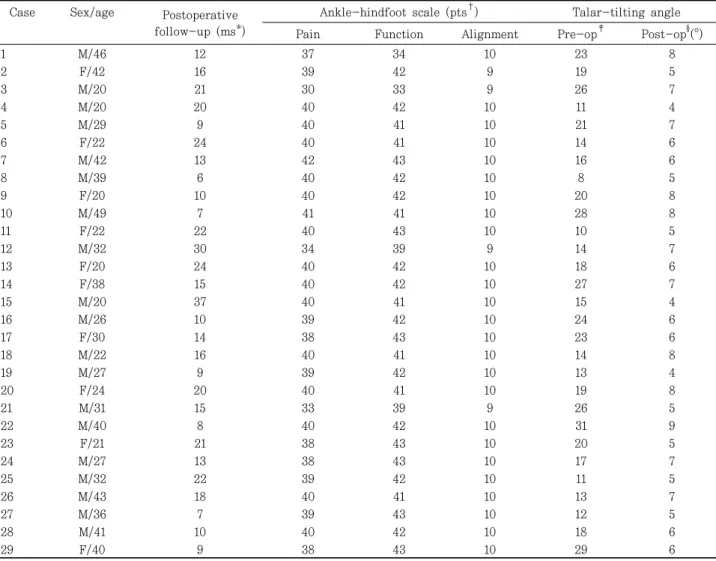
Table 1. Patient Data
Case Sex/age Postoperative follow-up (ms*)
Ankle-hindfoot scale (pts
†) Talar-tilting angle
Pain Function Alignment Pre-op
‡Post-op
§(°)
1 M/46 12 37 34 10 23 8
2 F/42 16 39 42 9 19 5
3 M/20 21 30 33 9 26 7
4 M/20 20 40 42 10 11 4
5 M/29 9 40 41 10 21 7
6 F/22 24 40 41 10 14 6
7 M/42 13 42 43 10 16 6
8 M/39 6 40 42 10 8 5
9 F/20 10 40 42 10 20 8
10 M/49 7 41 41 10 28 8
11 F/22 22 40 43 10 10 5
12 M/32 30 34 39 9 14 7
13 F/20 24 40 42 10 18 6
14 F/38 15 40 42 10 27 7
15 M/20 37 40 41 10 15 4
16 M/26 10 39 42 10 24 6
17 F/30 14 38 43 10 23 6
18 M/22 16 40 41 10 14 8
19 M/27 9 39 42 10 13 4
20 F/24 20 40 41 10 19 8
21 M/31 15 33 39 9 26 5
22 M/40 8 40 42 10 31 9
23 F/21 21 38 43 10 20 5
24 M/27 13 38 43 10 17 7
25 M/32 22 39 42 10 11 5
26 M/43 18 40 41 10 13 7
27 M/36 7 39 43 10 12 5
28 M/41 10 40 42 10 18 6
29 F/40 9 38 43 10 29 6
*ms, months;
†pts, points;
‡Pre-op, Pre-operative;
§Post-op, Post-operative.
Brostrӧm 술식은 비교적 적은 절개로 간단하게 신연된 인대를 해부학적 위치로 복원하는 방법으로서 비골 건의 손 상이나 비복 신경의 손상을 최소화 할 수 있는 장점이 있어 자주 사용되는 방법2,4,12,14,16,18,23)이다. 이에 본원에서는 만성 족관절 불안정성으로 진단된 환자 중 족관절의 후외방 동통 이 있는 환자들을 대상으로 관절경을 통해 관절내 상태를 확인하고 관절내 병변의 치료를 시행하였으며, 그 후 변형 Brostrӧm 술식을 이용하여 해부학적 인대 복원술을 시행 하여 그 결과를 분석하였다.
대상 및 방법
1. 연구대상
2004년 5월부터 2007년 1월까지 신체검진 및 방사선학
적 검사상 만성 족관절 불안정성 중 등급 Ⅲ 이상으로 진단 받고 변형 Brostrӧm 술식을 시행한 환자 25명, 29예를 대상으로 하였다. 환자의 나이는 20세부터 49세까지 평균 29.7세이었다. 남자가 19예, 여자 10예이었으며 술 후 추시 기간은 최단 6개월에서 최장 37개월까지로 평균 15.8개월 이었다. 수상 원인으로는 스포츠 손상 10예, 계단에서 넘어 진 경우 5예, 장시간 보행 후 발생한 경우 3예, 교통사고 3 예, 굽이 높은 구두를 신고 보행 후 발생한 경우 2예, 기타 정확한 원인을 모르는 경우들이 6예이었다. 29예 대부분 에서 급성 외상 후 적절한 치료가 이루어지지 않았다.
29예 모두에서 후외방부의 동통이 동반되어 있었으며, 변형 Brostrӧm 술식을 시행하기 전 족관절 관절경을 시행 하였다.
2. 수술방법
Table 2. Surgical Results in Cases with Associated Injuries
Case Excellent Good Fair Total
DTFFI* 2 2
AI
†1 1
DTFFI + AI 2 2
DTFFI + AI + OCD
‡7 1 8
DTFFI + AI + Loose body 3 3
DTFFI + OCD 3 3
DTFFI + Loose body 2 2
DTFFI + OCD + Loose body 2 2
DTFFI + AI + OCD + Loose body 3 2 1 6
Total 25 3 1 29
*DTFFI, Distal tibio-fibular fat impingement;
†AI, Anterior impingement;
‡OCD, Osteochondral defect.
(A) (B)
Figure 1. Magnetic resonance imaging of 38-year-old female with chronic lateral ankle instability due to sports activity. (A) Axial MR shows anterior tibiofibular ligament tear. (B) Coronal T2WI MR shows tear of anterior tibiofibular ligament, anterior talofibular ligament and calcaneofibular ligament.
(A) (B)
Figure 2. Preoperative ankle stress test, anterior drawer and varus stress test, was done. (A) Anterior drawer test of 46-year- old male patient shows 9.17 mm anterior displacement and 8.16 mm joint widening. (B) Varus stress test of 49-year-old male patient shows 11.92° talar-tilting angle.
수술은 척추마취 하에서 환자를 앙와위로 위치시킨 후 족관절의 전 내외측을 이용하여 관절경을 삽입하였고, 관절 내 병변을 확인하였다. 관절경을 이용한 치료 후, 비복신 경과 비골 건의 주행을 피해 비골 원위부 전하방 앞면을 따 라서 곡선형 피부절개 후 전거비인대와 종비인대, 하신전지 지대를 확인하는 순서로 진행하였다. 변형 Brostrӧm 술식 의 경우 각각의 인대 끝 부위를 일부 제거한 후 비골 전하단 부에서 인대를 봉합하였으며 하신전지지대를 이용, 비골막 에 봉합하였다.
술 후 부목고정을 시행하였고 3일째 석고고정을 하여 족 관절을 중립위로 유지하였다. 약 3주간의 단하지 석고고정 후, 3주간 보조기를 착용하면서 관절운동 및 근육강화 운동 과 동시에 간단한 보행에서부터 재활치료를 병행하였다.
3. 수술 후 평가
수술 후 결과판정은 미국정형외과족부족관절학회 족근
관절 및 후족부 평가표(AOFAS scale)20)를 이용하여 동통 40점, 기능 50점, 정렬 10점으로 총 100점을 기준으로 하 여 91점 이상은 우수, 81점 이상은 양호, 71점 이상은 보통 그리고 70점 이하를 불량으로 분류하였다.
방사선학적 방법으로 술 전과 술 후 족관절 전후방 및 측면 사진, 전방 및 내반 부하검사 단순 방사선 촬영을 하여 거골 경사각을 π view STARPACS system을 이용하여 측정 비교하였다.
결 과
미국정형외과족부족관절학회의 족근 관절 및 후족부 평 가표의 점수를 이용한 임상평가에서 평균 통증 점수는 술 전 28.9점에서 술 후 38.7점(30-40점), 평균 기능 점수는 술 전 34.1점에서 술 후 40.8점(30-43점) 그리고 평균 정렬
(A) (B)
Figure 3. Arthroscopic finding of 22-year-old female patient and treatment of distal tibiofibular fat impingement. (A) There was distal tibiofibular fat impingement via anterior tibiofibular ligament tear at arthroscopic exploration. (B) We treated distal tibiofibular fat impingement with shaving and electrical stimulations.
(A) (B)
Figure 4. Arthroscopic finding of 31-year-old male patient and treatment of distal tibiofibular fat impingement. (A) Distal tibiofibular fat impingement via anterior tibiofibular ligament tear. (B) Arthroscopic treatment with shaving of distal tibiofibular fat impingement.
점수는 술 전 7.9점에서 술 후 9.8점을 보여 우수 25예, 양호 3예, 보통 1예를 보였다. 방사선학적 평가에서는 평균 거골 경사각이 술 전 18.6°에서 술 후 5.9°로 개선되었다(Table 1).
한편 변형 Brostrӧm 술식을 시행하기 전 시행한 관절경 소견에서 원위 경비골간 지방 충돌 소견 28예, 전방 충돌 소견 20예, 골연골 손상 19예, 족관절 내 유리체 13예가 발견되었으며, 2개 이상의 동반 손상을 가진 경우는 27예 이었다. 족관절 내 원위 경비골간 지방 충돌 소견이 발견된 28예와 전방 충돌 소견이 발견된 20예 모두에서 관절경적 지방 제거술 및 변연술(arthroscopic shaving)을 시행하였 으며, 골연골 손상이 발견된 19예 중 14예에서 다발성 미세 골절술(multiple microfracture)을 시행하였고, 유리체가 발견된 13예 모두에서 유리체 제거술을 동시에 시행하였다.
관절경 소견에서 동반 손상을 보였던 29예 중 우수 25예, 양호 3예 그리고 보통 1예의 결과를 보였고, 특히 원위 경비 골간 지방 충돌 소견이 발견된 28예 중 27예에서 우수 및 양호의 결과를 보였으며(Table 2), 술 전에 보이던 후외방 의 통증은 술 후 보이지 않았다.
술 후 진행된 재활치료의 결과 약 2.2개월 후부터 일상생 활로의 복귀가 가능하였다.
고 찰
족관절 염좌는 매우 흔한 족관절 손상 중 하나로서 흔히 보존적 치료로 호전되지만 약 30%에서는 만성 염좌로 갈 수 있고 반복되는 족근 관절의 족저 굴곡 및 내번력에 의해 족관절 외측 측부인대 손상이 발생하게 되고11) 만성적 불안 정성을 보이는 경우 수술적 치료를 요하게 된다2,10).
한편 만성 족관절 불안정성을 보이는 환자에서 초기에 적절한 치료가 시행되지 않을 경우, 전 거비인대를 포함한 외측 측부인대 손상 후 불안정성이 점점 진행하게 되고 반복되는 내반 손상에 따른 원위부 경비골 인대 전방부의 견인이 발생하게 되어 경비골 간격의 미세 움직임이 증가 하게 되고 원위부 경비골 간의 이완이 발생하게 되면서 이 완부를 통한 지방의 과성장과 충돌 현상이 발생하게 된다.
이와 같이 원위부 지방 충돌 증후군을 비롯한 전방 충돌 증후군, 골연골 손상, 관절내 유리체 등의 동반된 관련 손 상이 있는 경우가 많기 때문에 이러한 것들이 족관절에 발생하는 통증의 원인으로 간주되고 있으며, 특히 족관절 후외측의 동통이 있는 환자의 경우에도 원위부 경비골간 지방 충돌 증후군이 존재하는 소견을 보여 술 전 이학적 검사 및 방사선적 진단과 수술 중 관절경 검사를 통한 적극 적인 진단과 치료가 필요하고 동반 손상의 유뮤 및 치료는 전체적인 결과에 많은 영향을 주게 된 것으로 추정된다.
DiGiovanni 등6)은 61명의 환자를 대상으로 한 관절경 소 견에서 전방 충돌 증후군 41예, 골연골 손상 14예, 관절내 유리체 33예, 단 비골 건 파열 15예 등의 동반 손상을 확인 하였다. Kibler19)도 46예의 환자 가운데 38예에서 관절내 동반 손상을 확인하였고 충돌 증후군 12예, 골연골 손상 6 예, 관절내 유리체 6예 및 전방 골극 12예를 보고하였다.
Scranton 등24)은 만성 족관절 외측 불안정성 환자에서 관절경 소견상 57%에서 전방 골극 및 유리체가 있음을 확인 하였고, Cannon과 Hackney4)도 전방 충돌 증후군이 동반 된 경우 골극을 제거하여 좋은 결과를 얻었다고 보고하였다.
Komenda와 Ferkel21)은 술 전 관절경을 시행하여 93%에
서 관절내 손상을 확인하였고, Hintermann 등15)도 관절경 을 통하여 연골 손상을 진단 및 치료하여 좋은 결과를 얻었 다고 보고하였다.
또한 만성 족관절 외측 불안정에 대하여 지금까지 80가 지 이상의 수술방법이 알려져 있으나1), 이들 대부분의 수술 방법들은 단 비골 건과 같은 정상 구조물을 이용해 건을 고정 및 재건하는 비해부학적 방법5,7,8,25)과 신연된 전 거비 인대 와 종비 인대를 단축시켜 봉합하거나2) 여기에 추가하여 신전 지지대의 근위부를 비골 외과 전연에 봉합해 주는12) 해부학적 방법이 있다.
해부학적 방법으로 Brostrӧm과 Sundelin3)은 파열된 족관절 외측 측부인대를 직접 봉합하는 해부학적 술식을 발표하였고, Gould 등12)은 변형 Brostrӧm 술식으로서 족 관절의 내번을 방지할 목적으로 신전지지대의 근위부를 비골에 추가 봉합해주는 방법을 소개하여 좋은 결과를 보고 하였다. Karlsson 등17)과 Liu와 Baker23)도 이 술식이 다른 술식에 비해 전방 거골 전이나 거골 경사각의 정도가 최소화 되어 더 나은 안정성을 제공한다고 보고하였으며, Hennri- kus 등14)도 이 술식을 통해 80% 이상의 환자에서 만족스러 운 안정성을 가져왔다고 하였다. 또한 Hamilton 등13)은 변 형된 Brostrӧm 술식을 시행한 28예에서 합병증 발생없이 93%에서 만족스러운 결과를 얻었다고 발표하였다.
본 연구에서도 수술 전 시행한 자기 공명 영상 검사에서 원위부 경비골 인대의 파열이 확인된 환자에서 관절경 소견 상 파열부위를 통한 관절내로의 지방 충돌 및 전방 충돌과 골연골 손상을 확인할 수 있었으며, 이런 지방 충돌의 제거 및 골연골 손상 부위 미세골절술 시행과 변형 Brostrӧm 술식을 통하여 통증 완화 및 관절 운동의 회복을 얻을 수 있었다.
그러므로 수술 전에 동반 손상의 유무에 대하여 이학적 검사 및 자기 공명영상을 포함한 방사선적 검사를 통하여 충분히 확인을 하고 수술 중 관절경을 통해 병변의 확인 및 적극적인 치료를 하는 것이 중요할 것이라 생각된다.
결 론
만성 측부인대 손상이 있는 환자 중 특히 후외측 부의 동 통이 있는 경우 관절경을 통한 원위부 지방 충돌의 제거술 시행과 변형 Brostrӧm 술식을 통한 해부학적 수술 치료는 불안정성을 해결할 수 있는 좋은 치료 방법이라 생각되며, 초기 인대 손상이 의심되는 환자들의 경우 적극적인 치료가 필요할 것으로 생각되고, 수술 전 관절내 동반 손상에 대한 충분한 검사와 수술 중 관절경을 이용한 진단 및 치료는 전
체적인 결과에 많은 영향을 줄 수 있으므로 족관절의 동통 을 유발시킬 수 있는 병변에 대하여 시행되어져야 한다고 사료된다.
REFERENCES