INTRODUCTION
Neuroendocrine carcinomas are malignant tumors that show histopathological and immunohistochemical evidence of neu- roendocrine differentiation. Neuroendocrine carcinomas can develop at many sites of the body, but primary neuroendocrine carcinoma of the breast is very rare (1). There are different his- tologic subtypes of neuroendocrine carcinomas of the breast that have been reported in the literature (1-4). In 2003, the World Health Organization (WHO) recommended classifi- cation of these tumors into three histologic types: solid, small- cell, and large-cell neuroendocrine carcinoma (5). There are some reports of neuroendocrine carcinoma of the breast in the pathology literature (2, 3), but there are few reports that describe the imaging features (6, 9) and most of them are about small- cell carcinomas. To our knowledge, the radiologic appearance of primary large-cell neuroendocrine carcinoma (LCNC) of the breast has not been described previously. In this case report, we present the radiologic and pathologic findings of prima- ry LCNC of the breast in a 27-yr-old woman.
CASE REPORT
A 27-yr-old woman presented with a palpable mass in her left breast. The mass was first noticed one year previously and
it had grown substantially. She had no remarkable past med- ical history, no family history of breast, colon, or ovarian can- cer, and was not on any medications. On physical examina- tion, a soft, movable mass was palpated in the left breast. The lesion was located at the three o’clock position of the left breast 4 cm from the nipple. There was no axillary lymphadenopa- thy, skin retraction, or abnormal nipple discharge. Mammog- raphy showed a relatively well-demarcated, lobular shaped mass in the mid-outer portion of the left breast (Fig. 1). Breast
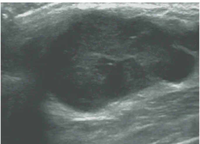
ultrasonography (US) revealed a circumscribed oval, gently lobulated, heterogeneous, low-echoic mass measuring about 3.2×1.3 cm in size in the left breast (Fig. 2). The lesion was thought to be low suggestion of malignancy, BI-RADS cate- gory 4A. An ultrasound-guided 14G core needle biopsy was performed for the mass. On a chest CT scan, the lesion app- eared as a well-defined, highly-enhancing mass in the left breast (Fig. 3). In the right upper lung, there was pulmonary tuberculosis lesion, but we could not find any lesions sug- gestive of primary lung cancer. Abdominal ultrasonography, whole body bone scan and PET CT scan were performed for preoperative metastasis work-up and no distant metastasis was found. She underwent breast-conserving surgery and left axillary lymph node dissection. On microscopic examina- tions (Fig. 4), small nests of large tumor cells with faintly gran- ular cytoplasm were present and separated by dense colla- gen bundles. An immunohistochemical panel (Fig. 5) was
Some breast neoplasms are classified as primary neuroendocrine carcinomas because they are positive for neuroendocrine markers. Although neuroendocrine carcinomas can originate from various organs of the body, primary neuroendocrine carcinomas of the breast are extremely rare. The diagnosis of primary neuroen- docrine carcinoma of the breast can only be made if nonmammary sites are confi- dently excluded or if an in situ component can be found. Here we report a primary large-cell neuroendocrine carcinoma (LCNL) involving the left breast. Breast ultra- sonography revealed a lobulated, heterogeneous, low-echoic mass in the left breast, and the lesion ap-peared as a well-defined, highly-enhancing mass on a chest computed tomography scan. Ultrasound-guided core needle biopsy was performed on the mass, and primary LCNC was confirmed by histopathologic examination.
Key Words : Breast; Ultrasonography; Tomography, X-Ray Computed; Large Cell Neuroendocrine Carcinoma
1118
Jin Woo Kim, Ok Hee Woo, Kyu Ran Cho, Bo Kyung Seo, Hwan Seok Yong, Aeree Kim*, and Eun-Young Kang
Departments of Radiology and Pathology* College of Medicine, Korea University Guro Hospital, Seoul, Korea
Address for correspondence Ok Hee Woo, M.D.
Department of Radiology, Korea University Guro Hospital, 97 Guro-dong, Guro-gu, Seoul 152-703, Korea
Tel : +82.2-2626-1342, Fax : +82.2-2626-1379 E-mail : [email protected]
*This study was supported by Korea University Grant.
J Korean Med Sci 2008; 23: 1118-20 ISSN 1011-8934
DOI: 10.3346/jkms.2008.23.6.1118
Copyright � The Korean Academy of Medical Sciences
Primary Large Cell Neuroendocrine Carcinoma of the Breast:
Radiologic and Pathologic Findings
Received : 12 September 2007 Accepted : 7 January 2008
Primary Large Cell Neuroendocrine Carcinoma of the Breast 1119
performed, and the tumor was found to be positive for neu- roendocrine markers (chromogranins, synaptophysin, and neuron-specific enolase [NSE]). These pathologic findings were compatible with primary LCNC.
She underwent adjuvant chemotherapy and radiation ther- apy. At 18 months post-operation, there was no evidence of local recurrence or distant metastasis.
DISCUSSION
More than 97% of neuroendocrine tumors occur in the gastrointerstinal or respiratory system, and primary neu- roendocrine carcinoma of the breast is very rare. In a series of 1,845 histopathologically-proven breast cancer, the inci- dence was about 0.27% (6).
The diagnosis of primary neuroendocrine carcinoma of the
Fig. 1. Mammography showed a relatively well-demarcated, lob- ular shaped mass in the mid-outer portion of the left breast.
Fig. 2. Breast US revealed an oval shaped, micro-lobulated, het- erogeneous low-echoic mass, measuring about 3.2×1.3 cm in size, in the left breast.
Fig. 3. Chest CT scan showed a well-defined, highly-enhancing mass in the left breast.
Fig. 5. The tumor stained positive for chromogranin A (magnifi- cation ×200) and also expressed neuron-specific enolase and synaptophysin (not shown).
Fig. 4. Photomicrograph of a histopathologic specimen showed small nests of large tumor cells with faintly granular cytoplasm sep- arated by dense collagen bundles (Hematoxylin-eosin, magnifi- cation ×200).
1120 J.W. Kim, O.H. Woo, K.R. Cho, et al.
breast can only be made if nonmammary sites are confident- ly excluded or if an in situ component can be found (9). There are different histologic subtypes of neuroendocrine carcino- mas of the breast reported in the literature (2-4). Papotti et al. divided neuroendocrine carcinomas into seven histologi- cal types (A-G) (2). Sapino et al. described five different his- tological types: solid cohesive, alveolar, small cell, solid pap- illary, and cellular mucinous (3). In 2003, the World Health Organization (WHO) recommended differentiation of these tumors into three histologic types: solid, small-cell, and large- cell neuroendocrine carcinoma (5). There are some features that favor neuroendocrine differentiated carcinoma of the breast and morphologic features should be confirmed immunochem- ically or by positivity for neuroendocrine markers (3). NSE, chromogranin, and synaptophysin are widely accepted as neuroendocrine markers, though NSE is relatively non-spe- cific (4). In our case, the three aforementioned neuroendocrine markers were all positive.
LCNC, one of the subtypes of the 2003 WHO classifica- tion of neuroendocrine tumors, shares common features of the neuroendocrine-differentiated tumor described above and also shows more specific cytologic features: large cell size, polygonal shape, low nuclear-cytoplasmic ratio, finely granu- lar eosinophilic cytoplasm, occasionally prominent nucleoli, peripheral palisading, mitosis, and necrosis (8).
In a clinical setting, neuroendocrine carcinomas usually occur in women around the 7th decade; most patients in the previously reported cases were between 40 and 70 yr old (2, 4-6, 9). Our patient was a 27-yr old woman without risk fac- tors of breast cancer and presented with a painful breast mass that had appeared about one year prior. The mass was soft and movable on physical examination. It is an uncommon presentation of breast cancer, considering the patient’s age and clinical findings. The prognosis of neuroendocrine-dif- ferentiated carcinoma is a subject to debate, probably due to different growth patterns of the subtypes and the low num- ber of cases. In some recent literature, primary neuroendocrine carcinomas of the non-small cell type have shown satisfacto- ry prognosis with adequate surgical excision and adjuvant chemotherapy (5, 10).
Previously noted mammographic findings of neuroendocrine carcionoma included high-density round masses with spicu- lated or lobulated margin (6, 9). Our findings are not signif- icantly different. US findings of our patient were those of gently lobulated, heterogenous, low-echoic mass. In one series of five neuroendocrine carcinomas, most of the lesions demon- strated homogeneous low-echoic masses, though the WHO classification was not used and all of the lesions were of the solid and papillary subtypes (6). US findings in small-cell
carcinoma reported by Mariscal et al. was of a solid, homo- geneous, low-echoic mass with a partially ill-defined margin and mild posterior acoustic enhancement (9). In our case, CT findings are also presented. A chest CT scan was per- formed as part of a metastastic work-up as well as for evalu- ation of pulmonary tuberculosis. The lesion appeared as a well-defined, highly-enhancing mass in the left breast.
In summary, we presented imaging findings of a LCNC of the breast. This is the first report describing imaging features of large-cell type neuroendocrine carcinoma and presenting the CT findings of a neuroendocrine-differentiated carcino- ma in Korea. This is a rare entity, and description of more cases is necessary to determine the radiologic and clinical features of LCNC.
REFERENCES
1. Maluf HM, Koerner FC. Carcinomas of the breast with endocrine dif- ferentiation: a review. Virchows Arch 1994; 425: 449-57.
2. Papotti M, Macri L, Finzi G, Capella C, Eusebi V, Bussolati G. Neu- roendocrine differentiation in carcinomas of the breast: a study of 51 cases. Semin Diagn Pathol 1989; 6: 174-88.
3. Sapino A, Righi L, Cassoni P, Papotti M, Pietribiasi F, Bussolati G.
Expression of the neuroendocrine phenotype in carcinomas of the breast. Semin Diagn Pathol 2000; 17: 127-37.
4. Zekioglu O, Erhan Y, Ciris M, Bayramoglu H. Neuroendocrine dif- ferentiated carcinomas of the breast: a distinct entity. Breast 2003;
12: 251-7.
5. Tsai WC, Yu JC, Lin CK, Hsieh CT. Primary alveolar-type large cell neuroendocrine carcinoma of the breast. Breast J 2005; 11: 487.
6. Gunhan-Bilgen I, Zekioglu O, Ustun EE, Memis A, Erhan Y. Neuroen- docrine differentiated breast carcinoma: imaging features correlat- ed with clinical and histopathological findings. Eur Radiol 2003; 13:
788-93.
7. Miremadi A, Pinder SE, Lee AH, Bell JA, Paish EC, Wencyk P, Elston CW, Nicholson RI, Blamey RW, Robertson JF, Ellis IO. Neuroen- docrine differentiation and prognosis in breast adenocarcinoma.
Histopathology 2002; 40: 215-22.
8. Krivak TC, McBroom JW, Sundborg MJ, Crothers B, Parker MF.
Large cell neuroendocrine cervical carcinoma: a report of two cases and review of the literature. Gynecol Oncol 2001; 82: 187-91.
9. Mariscal A, Balliu E, Diaz R, Casas JD, Gallart AM. Primary oat cell carcinoma of the breast: imaging features. AJR Am J Roentgenol 2004;
183: 1169-71.
10. Berruti A, Saini A, Leonardo E, Cappia S, Borasio P, Dogliotti L. Man- agement of neuroendocrine differentiated breast carcinoma. Breast 2004; 13: 527-9.