INTRODUCTION
Although phototherapy has been used for the treatment of neonatal jaundice for more than 4 decades, the most effi- cacious phototherapy method with the least side effects has not been developed yet (1-3). Its efficacy is dependent on the color (wavelength) and intensity (irradiance) of the light emit- ted during phototherapy, the exposed body surface area, and the duration of exposure (2, 4-6). The currently available pho- totherapy devices such as fluorescent tubes, halogen spotlights and fiberoptic blankets have many disadvantages including high heat production and unstable broad wavelength light output (1, 7, 8). In cases of severe or rapidly increasing neona- tal jaundice, it is important to institute intensive photother- apy to decrease the bilirubin levels as soon as possible to reduce the need for exchange transfusion and the risk of kernicterus (9-11). However, conventionally used phototherapy may be less effective, thereby increasing the risk of bilirubin-induced neurotoxicity (2, 4, 11).
Recently, high intensity gallium nitride light emitting diodes (LEDs) have been developed (12, 13), and studied as possible light sources for the phototherapy of neonatal jaun-
dice (14). Blue LEDs emit a high intensity narrow band of blue light overlapping the peak spectrum of bilirubin break- down (14), resulting in potentially shorter treatment times (15, 16). LEDs are also power efficient, light in weight, pro- duce less heat, and have a longer lifetime (12-14). These uni- que characteristics of LEDs make them very optimal light sources for phototherapy devices. Despite these potential ben- efits and substantial versatility of this device, two recent clini- cal trials of blue gallium nitride LED were not found to be of higher efficacy when applied using relatively low irradi- ances levels (17, 18). These results suggest that besides the color (wavelength), the dose (irradiance) of light is also very important in determining the effectiveness of phototherapy (2, 4-6, 16). As LED devices could generate significantly high- er light irradiance levels compared to all currently available conventional phototherapy units (14), much higher irradi- ance levels are likely to be used clinically in the future. There- fore, in the present study, we developed a prototype blue LED phototherapy unit with high intensity, and compared its effi- cacy to that of commercially used halogen quartz photother- apy device by measuring both in vitro and in vivo bilirubin photodegradation.
Yun Sil Chang, Jong Hee Hwang, Hyuk Nam Kwon*, Chang Won Choi, Sun Young Ko�, Won Soon Park, Son Moon Shin�, Munhyang Lee
Department of Pediatrics, Department of Biomedical Engineering*, Samsung Medical Center, Samsung Cheil Hospital�, Sungkyunkwan University School of Medicine, Seoul, Korea
Address for correspondence Munhyang Lee, M.D.
Department of Pediatrics, Samsung Medical Center, 50 Irwon-dong, Kangnam-gu, Seoul 135-710, Korea Tel : +82.2-3410-3522, Fax : +82.2-3410-0043 E-mail : [email protected]
*This study was supported by a grant of the Korea Health 21 R&D Project, Ministry of Health and Welfare, Republic of Korea (02-PJ1-PG3-21301-0020).
61 J Korean Med Sci 2005; 20: 61-4
ISSN 1011-8934
Copyright � The Korean Academy of Medical Sciences
In vitro and in vivo Efficacy of New Blue Light Emitting Diode Phototherapy Compared to Conventional Halogen Quartz
Phototherapy for Neonatal Jaundice
High intensity light emitting diodes (LEDs) are being studied as possible light sources for the phototherapy of neonatal jaundice, as they can emit high intensity light of narrow wavelength band in the blue region of the visible light spectrum cor- responding to the spectrum of maximal bilirubin absorption. We developed a pro- totype blue gallium nitride LED phototherapy unit with high intensity, and compared its efficacy to commercially used halogen quartz phototherapy device by measur- ing both in vitro and in vivo bilirubin photodegradation. The prototype device with two focused arrays, each with 500 blue LEDs, generated greater irradiance than the conventional device tested. The LED device showed a significantly higher effi- cacy of bilirubin photodegradation than the conventional phototherapy in both in vitro experiment using microhematocrit tubes (44±7% vs. 35±2%) and in vivo experiment using Gunn rats (30±9% vs. 16±8%). We conclude that high intensi- ty blue LED device was much more effective than conventional phototherapy of both in vitro and in vivo bilirubin photodegradation. Further studies will be neces- sary to prove its clinical efficacy.
Key Words : Jaundice, Neonatal; Phototherapy; Rats, Gunn
Received : 13 July 2004 Accepted : 10 August 2004
62 Y.S. Chang, J.H. Hwang, H.N. Kwon, et al.
MATERIALS AND METHODS Phototherapy unit
A prototype of high intensity gallium nitride blue LED phototherapy unit was developed and custom built by the Department of Pediatrics and Biomedical Engineering at the Samsung Medical Center, Sungkyunkwan University School of Medicine. High intensity blue LEDs (HLBB-L55B, Nis- sitronics Korea Inc., Seoul, Korea) had a dominant wavelength at 465-470 nm, and the overhead device had two focused arrays, each array equipped with 500 LEDs (Fig. 1). For con- ventional phototherapy, halogen quartz phototherapy device (Model: 6600-0084-900, Ohmeda Medical Co., MD, U.S.A.) was used. As halogen lamps could incur the risk of burn when positioned closer to the infant, phototherapy was administered 45 cm apart in accordance with the manufacturer’s recom- mendation. At that distance, irradiance was measured with a phototherapy radiometer (BiliBlanket Meter II, Ohmeda Medical Co., MD, U.S.A.), with peak sensitivity at 450 nm, and illuminance was measured with an Illuminance Meter, TL-1 Minolta (Minolta Camera Co., Tokyo, Japan). The in vitro results do not necessarily reflect its in vivo efficacy due to confounding variables including poor skin penetration of blue light spectrum (14-16). We therefore conducted both in vitro and in vivo experiment to determine the bilirubin degra- dation efficacy of LED device in the present study.
In vitro experiment
Bilirubin (Sigma Chemical Co., MO, U.S.A.) was dissolved
in a buffer solution containing 18.5 vol% 0.1 N NaOH, 44.5 vol% human albumin (5%) and 37 vol% 0.055 M Na2HPO4, with the final concentration and pH adjusted to 20 mg/dL and 7.4 respectively. In each group, ten microhematocrit tubes containing 100 L of the bilirubin solution per tube, was placed horizontally on a black non-reflective background, and exposed to each phototherapy light at 45 cm distance for 5 hr at room temperature. In vitro photodegradation of biliru- bin (%) was calculated as the difference between bilirubin concentrations at before and after exposure to the photother- apy divided by the bilirubin concentration at before exposure to the phototherapy times 100. Bilirubin concentration was measured with Bilirubin Tester (Wako Pure Chemical Indus- tries Ltd., Osaka, Japan).
In vivo experiment
For in vivo experiment, Gunn rats were purchased from Harlan Sprague Dawley, Inc. (Indianapolis, IN, U.S.A.). Twen- ty of 8-day old jaundiced (jj) Gunn rats were randomly divid- ed into two groups, and exposed to each phototherapy light at 45 cm distance for 5 hr at room temperature. In vivo biliru- bin degradation (%) was also calculated as the difference bet- ween serum bilirubin concentrations of Gunn rats at before and after exposure to the phototherapy divided by the serum bilirubin concentration at before exposure to the photother- apy times 100.
Statistical analysis
Statistical analysis was performed by Student’s t test. The data are presented as mean±standard deviation. A p-value of <0.05 was considered significant.
Bilirubin degradation (%)
60
50
40
30
20
10
0
in vitro in vivo
LED Conventional
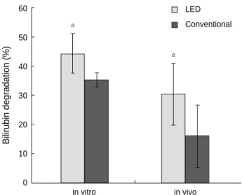
Fig. 2.Comparison of both in vitro and in vivo efficacy of bilirubin photodegradation between the blue LED and conventional pho- totherapy unit. Data are expressed as mean±standard deviation.
a: p<0.05 compared to conventional phototherapy.
a
a
Fig. 1.A prototype phototherapy device made of two focused arrays, each with 500 high intensity blue gallium nitride light emit- ting diodes (LEDs).
RESULTS
Mean irradiance of blue LED and conventional photother- apy unit at 45 cm distance was 75 and 23 W/cm2/nm, and illuminance was 680 and 1999 foot candle, respectively.
Bilirubin photodegradation of blue LED device was sig- nificantly higher than conventional phototherapy in both in vitro (44±7% vs. 35±2%) and in vivo (30±9% vs. 16
±8%) experiment (Fig. 2).
DISCUSSION
In the present study, high intensity gallium nitride blue LED phototherapy was more effective than conventional halo- gen quartz phototherapy in both in vitro and in vivo biliru- bin photodegradation. More rapid breakdown of bilirubin with the use of more efficient LED phototherapy devices would shorten the treatment times of neonatal jaundice (15, 16), and ultimately improve the prognosis of severe neonatal hyper- bilirubinemia by reducing the need for exchange transfusion and minimizing the risk of bilirubin neurotoxicity (2, 4, 11).
The optimum phototherapy method with the least side effects has not been developed yet (1-3). The color (wavelength) and intensity (irradiance) of the light emitted during pho- totherapy are two major factors determining its efficacy (2, 4-6). Blue LEDs used for light sources of phototherapy in the present study would be more effective than day light because they emit a high intensity narrow band of blue light corre- sponding to the peak absorption wavelength at which biliru- bin is broken down (14). Furthermore, as LEDs emit no sig- nificant ultraviolet or infrared light, they would reduce the potential risk of DNA damage (8) or fluid loss (7). However, two recent clinical trials of blue gallium nitride LED device using comparable light intensity were not found to be of high- er efficacy than the conventional phototherapy (17, 18). In contrast, gallium nitride blue LED phototherapy was observed to be more effective than conventional phototherapy in both in vitro and in vivo bilirubin photodegradation in the present study. As the irradiance and illuminance of our LED device was nearly three times higher and three times less than the conventional phototherapy respectively, not the broad light intensity (illuminance) but the specific light intensity match- ing the bilirubin absorption spectrum (irradiance) seems to correlate with the magnitude of bilirubin photodegradation (6, 16). Taken together, these findings suggest that besides the wavelength, the intensity of light (irradiance) emitted during phototherapy is also very important for its best effi- cacy (2, 4-6, 16).
The dose of phototherapy light is the product of irradiance, surface area exposed, and duration of exposure (2, 4-6, 16).
Irradiance depends not only on the power of the light source but also on the distance from the source (2, 4, 5). LEDs can deliver much higher doses of light than any of the conven-
tional devices as they generate greater irradiance (14), and can be placed very close to the skin due to less heat produc- tion in contrast to halogen or fluorescent lights (1, 7). Fur- thermore, such direct contact LEDs in the form of mattress- es, pads or jackets of any size and shape could cover a larger surface area than conventional devices, leading to more effi- cient and convenient phototherapy and abrogating the need for eye patches (2). Therefore, LED devices could markedly improve the efficacy and safety of phototherapy (14).
Tan (5, 19) demonstrated a dose response relationship with increased intensity of phototherapy resulting in greater res- ponses. However, the rate of bilirubin decline progressively decreased with increasing irradiance until a saturation point of 30-40 W/cm2/nm in the 425 to 475 nm range was reach- ed, beyond which an increase in the irradiance produced no added efficacy. At the saturation point, a bilirubin decline of 40 to 50% in 24 hr was achieved (19). However, given that the conversion of bilirubin to excretable photoproducts is partly irreversible and follows first order kinetics (14, 15), it is not certain whether a saturation point suggested by Tan really exists. Our data of more effective bilirubin degradation with blue LEDs well above the saturation point of irradiance also contradict the concept of the saturation point. As LED phototherapy devices can provide significantly higher irra- diance levels compared to all currently available convention- al systems (14), further studies will be necessary to clarify this.
In addition to the features mentioned above, LEDs have several advantages not found in conventional devices such as light weight, compact size, high energy efficiency, use of low voltage battery power, extremely long life, the ability to be focused with a lens or through spatial orientation and no glass parts (2, 12-14). These features make the LED devices ideal light sources for phototherapy during transport or at home.
Furthermore, as LEDs use direct current, the typical flicker- ing glare that may be responsible for the untoward symptoms of headache, nausea and dizziness, reported with the use of conventional blue fluorescent lamps (11), is absent, thereby reducing nursing staff discomfort (17, 18).
In summary, a blue LED phototherapy unit with higher irradiance levels showed higher efficacy of both in vitro and in vivo bilirubin photodegradation compared to that of com- mercially used halogen quartz phototherapy device. As LED devices can provide much higher irradiance, and thus greater efficacy, they can be ideal light sources for the phototherapy of neonatal jaundice. Additional studies will be necessary to prove its clinical efficacy.
REFERENCES
1. McDonagh AF. Phototherapy: from ancient Egypt to the new mil- lennium. J Perinatol 2001; 21 (Suppl 1): S7-12.
2. Maisels MJ. Phototherapy-traditional and nontraditional. J Perina- tol 2001; 21 (Suppl 1): S93-7.
Blue LED Phototherapy 63
64 Y.S. Chang, J.H. Hwang, H.N. Kwon, et al.
3. Hansen TW. Phototherapy for neonatal jaundice- still in need of fine tuning. Acta Paediatr 2000; 89: 770-2.
4. Maisels MJ. Why use homeopathic doses of phototherapy? Pediatrics 1996; 98: 283-7.
5. Tan KL. The nature of the dose-response relationship of photother- apy for neonatal hyperbilirubinemia. J Pediatr 1977; 90: 448-52.
6. Modi N, Keay AJ. Phototherapy for neonatal hyperbilirubinemia:
The importance of does. Arch Dis Child 1983; 58: 406-9.
7. Ente G, Klein SW. Hazards of phototherapy. N Engl J Med 1970;
283: 544-5.
8. Christensen T, Reitan JB, Kinn G. Single-strand breaks in the DNA of human cells exposed to visible light from phototherapy lamps in the presence and absence of bilirubin. J Photochem Photobiol B 1990;
7: 337-46.
9. Kang JH, Shankaran S. Double phototherapy with high irradiance compared with single phototherapy in neonates with hyperbilirubine- mia. Am J Perinatol 1995; 12: 178-80.
10. Hansen TW. Acute management of extreme neonatal jaundice-the potential benefits of intensified phototherapy and interruption of entero- hepatic circulation. Acta Paediatr 1997; 86: 843-6.
11. Tan KL, Lim GC, Boey KW. Efficacy of high intensity blue light and
standard daylight phototherapy for non-haemolytic hyperbilirubine- mia. Acta Paediatr 1992; 81: 870-4.
12. Nakamura S, Fasol G. InGaN single-quantum-well LEDs. In: The Blue Laser Diode. Berlin: Springer-Verlag, 1997; 201-21.
13. Fasol G. Longer life for the blue laser. Science 1997; 278: 1902-3.
14. Vreman HJ, Wong RJ, Stevenson DK, Route RK, Reader SD, Fejer MM, Gale R, Seidman DS. Light-emitting diodes: A novel light source for phototherapy. Pediatr Res 1998; 44: 804-9.
15. Ennever JF, McDonagh AF, Speck WT. Phototherapy for neonatal jaundice: Optimal wavelengths of light. J Pediatr 1983; 103: 295-9.
16. Ennever J. Blue light, green light, white light, more light: Treatment of neonatal jaundice. Clin Perinatol 1990; 17: 467-81.
17. Seidman DS, Moise J, Ergaz Z, Laor A, Vreman HJ, Stevenson DK, Gale R. A prospective randomized controlled study of phototherapy using blue and blue-green light-emitting devices, and conventional halogen-quartz phototherapy. J Perinatol 2003; 23: 123-7.
18. Seidman DS, Moise J, Ergaz Z, Laor A, Vreman HJ, Stevenson DK, Gale R. A new blue light-emitting phototherapy device: a prospective randomized controlled study. J Pediatr 2000; 136: 771-4.
19. Tan KL. The pattern of bilirubin response to phototherapy for neona- tal hyperbilirubinaemia. Pediatr Res 1982; 16: 670-4.