ISSN 2288-0356(Online) http://dx.doi.org/10.14518/crals.2013.31.4.003 Original Article
Reinforcing Efficiencies of Two Different Cellulose Nanocrystals in Polyvinyl Alcohol-Based Nanocomposites
Byung-Dae Park1,*, Valerio Causin2
1Department of Wood Science and Technology, Kyungpook National University Daegu, 702-701, Republic of Korea
2Department of Chemical Sciences, University of Padova, via Marzolo 1, 35131 Padova, Italy
Abstract
As a renewable nanomaterial, cellulose nanocrystal (CNC) isolated from wood grants excellent mechanical properties in developing high performance nanocomposites. This study was undertaken to compare the reinforcing efficiency of two different CNCs, i.e., cellulose nanowhiskers (CNWs) and cellulose nanofibrils (CNFs) from hardwood bleached kraft pulp (HW-BKP) as reinforcing agent in polyvinyl alcohol (PVA)-based nanocomposite.
The CNWs were isolated by sulfuric acid hydrolysis while the CNFs were isolated by 2,2,6,6-tetramethylpiperidine-1-oxyl radical (TEMPO)-mediated oxidation. Based on measurements using transmission electron microscopy, the individual CNWs were about 6.96±0.87 nm wide and 178±55 nm long, while CNFs were 7.07±0.99 nm wide. The incorporation of CNWs and CNFs into the PVA matrix at 5% and 1% levels, respectively, resulted in the maximum tensile strength, indicating different efficiencies of these CNCs in the nanocomposites. Therefore, these results suggest a relationship between the reinforcing potential of CNCs and their physical characteristics, such as their morphology, dimensions, and aspect ratio.
Keywords:cellulose nanocrystal, reinforcing efficiency, tensile properties, aspect ratio
Introduction1)
In recent years, the use of natural fibers as a reinforcement in composites has attracted much attention due to environmental concerns. Among such natural fibers, cellulose is the most abundant, renewable and biodegradable natural polymer in the world. Cellulose fibrils consist of different hierarchical microstructures, and are commonly known as nano-sized microfibrils.
Their cellulose chains are stabilized laterally by intra- and inter-molecular hydrogen bonds between hydroxyl groups, resulting in high structural strength and stiffness. These nano-sized fibrils are composed of a crystalline and amorphous part. Nano-sized cellulose isolated from the crystalline region is called cellulose nanocrystals (CNCs) (Sain and Oksman, 2005). In theory, the elastic modulus of native cellulose Ι crystal has been reported to be 167.5 GPa (Tashiro and Kobayashi, 1991) and that of tunicin whiskers is 143 GPa (Sturcová et al. 2005). Thus due to their excellent properties, CNCs have generated a great deal of interest as nanometer size reinforcement agent for a range of nanocomposites.
CNCs can be isolated by various methods, including acid hydrolysis, mechanical grinding, enzyme hydrolysis, and cryo-grinding, and have different names, such as nanocellulose (NC), cellulose nanowhiskers (CNWs), and microfibrillated
cellulose (MFC). In general, CNCs isolated by the acid hydrolysis of woody cellulose are called CNWs due to their whisker shape. In contrast, chemical oxidation or mechanical treatment produces nano-sized fibrils, called cellulose nanofibril (CNFs).
Acid hydrolysis removes the amorphous region of cellulose to isolate CNWs, and is stopped before the complete depolymerization of the non-accessible cellulose (i.e. crystalline component) (Renneckar et al. 2006). The hydrolysis of cellulose is affected by the acid concentration, hydrolysis time and hydrolysis temperature. Bondeson et al. (2006) studied the optimum condition for cellulose hydrolysis using sulfuric acid and commercially available microcrystalline cellulose (MCC).
Bai and his co-workers (2009) reported that the CNW had narrow width and short length with increasing RPM In contrast, CNFs is produced by using 2,2,6,6-tetramethylpiperidine- 1-oxyradical (TEMPO), a water-soluble, commercially available and stable nitroxyl radical, which has paved the way for a new field of conversion chemistry that allows the efficient and selective conversion of alcoholic hydroxyl groups to aldehydes, ketones and carboxyl groups under mild conditions (de Nooy et al. 1995; de Nooy et al. 1996). TEMPO-mediated oxidation has been employed for isolating cellulose nanofibrils from Received: December 11 2013 / Revised: December 20 2013 / Accept: December 31 2013
*Corresponding Author: Byung-Dae Park, Tel. 82-53-950-9597, Fax. 82-53-950-6751, Email. [email protected]
©2012 College of Agricultural and Life Science, Kyungpook National University
several wood pulps and cotton linters (Fukuzumi et al. 2009;
Saito et al. 2006). Many related studies on wood cellulose have been extensively carried out in the last few years (Saito et al.
2006;Saito et al. 2007), and have been reviewed in detail (Isogai et al. 2011). The CNFs obtained by the TEMPO-mediated oxidation have a high crystallinity, mostly uniform widths (3-4 nm), and large aspect ratios (>50) when compared with other nanocelluloses (Saito et al. 2006; Saito et al. 2007; Shinoda et al. 2012). Moreover, these CNFs can be completely dispersed at the individual nanofibril level in water by electrostatic repulsion and/or osmotic effects due to anionically charged sodium carboxylate groups densely present on the surfaces (Saito and Isogai, 2004).
The utilization of various types of CNCs has attracted much attention for preparing high performance nanocomposites owing to advances in nanotechnology. These cellulose nanomaterials are also environmentally compatible, and endowed with several other unique and desirable features. Thus, cellulose-based nanomaterials are being increasingly employed for fabricating a wide variety of polymer-based nanocomposites, and such developments are also benefitting from rapid advances in nanoscience and nanotechnologies (Samir et al. 2005; Eichhorn et al. 2010). CNC-based nanocomposites have been extensively investigated in anticipation of many desirable property contributions from CNCs, such as high strength, high stiffness, biodegradability and light weight. Favier et al. (1995) first observed significant reinforcing effect of the cellulose whiskers.
In fact, tunicin cellulose whisker with 10-20 nm width and several micrometers length revealed significant reinforcing effect in the copolymer of styrene and butylacrylate. Moreover, polyvinyl alcohol (PVA) is a water soluble synthetic polymer and has excellent film forming and emulsifying properties. It also has a high tensile strength and flexibility. Many papers were published on PVA-based nanocomposites. For example, sugar beet cellulose (Leitner et al. 2007), sulfite pulp cellulose nanofiber (Zimmermann et al. 2004), cellulose whiskers isolated from microcrystalline cellulose (MCC) (Kvien and Oksman, 2007), and microfibrillated cellulose (MFC) (Lu et al. 2008) were used for PVA based nanocomposite.
However, despite numerous studies, there is a great variation in the degree of their reinforcement effect. Thus, this study compared the reinforcing efficiency of two different CNCs, i.e., CNWs isolated by sulfuric acid hydrolysis and CNFs isolated by TEMPO-mediated oxidation in PVA-based nanocomposites.
Materials and Methods
Two different molecular weights of PVA, i.e., 22,000 g/mol
and 66,000 g/mol (Duksan Pure Chemical Co., Ltd., Korea) as the matrix polymer were used as received. A sample of hardwood bleached kraft pulp (HW-BKP) was obtained from Moorim Paper Co. Ltd., Korea and stored in a humidity chamber at 25℃ before use. Sulfuric acid (95%, DC chemical Co., Ltd., Korea) was used for the acid hydrolysis of the HW-BKP. The TEMPO, sodium bromide (NaBr), sodium hypochlorite (NaOCl) solution and other chemicals were all purchased from Sigma-Aldrich. 2)
Isolation of CNWs and CNFs
The CNWs were isolated according to the following procedure.
About 10 g of the kraft pulp was mixed with 100 mL of deionized water. The water/pulp suspension was then put in an ice bath with mechanical stirring, and 100 mL sulfuric acid was added drop by drop. After the adding sulfuric acid, the suspension was hydrolyzed at 44℃ for 130 min. The suspensions were then washed with deionized water for 20 min. at 5000 rpm, using repeated centrifugation (Hanil Centrifuge Co., Ltd, Korea).
The supernatant was removed from the sediment replaced with new deionized water and mixed. The centrifugation step continued until the pH value of the supernatant became 1. The last wash was conducted using dialysis with deionized water to remove the last residue of the sulfuric acid until the wash water maintained a constant pH and became pH 4. The ultrasonication (Sonosmasher, Jeio Tech, Korea) was carried out for 20 min. at a power output of 30% in an ice bath to avoid overheating, which can cause desulfonation of the sulfate groups on the cellulose. Then CNC suspension solutions were then kept in a refrigerator until use.
The CNFs were isolated from the cellulose pulp using a modified procedure of the TEMPO-mediated oxidation method (Saito et al. 2006). In brief, the kraft pulp sample (2 g of cellulose content) was suspended in water containing TEMPO and sodium bromide. TEMPO-mediated oxidation of the cellulose slurry was initiated by adding 5 mmol of 13% NaClO per gram of cellulose and carried out at room temperature under gentle agitation. The pH was maintained at 10.5 by adding 0.5 M NaOH. When no further decrease in the pH was observed, the reaction was finished and the pH was adjusted to 7 by adding 0.5 M HCl. The TEMPO-oxidized product was thoroughly washed with water by filtration and physically fibrillated by ultra-sonication using a sonicator (Sonosmasher, Jeio Tech, Korea) for 20 min. The suspension was then centrifuged at 5000 rpm two to three times for 30 min each. The supernatant was decanted and collected as the CNFs suspension. The yield of the CNFs was calculated as a percentage of the initial weight
of CNFs suspension after drying in a drying oven at 105℃.
The final concentration of CNFs was adjusted to 0.1% by weight/volume.
3)
Characterization of CNWs and CNFs
The morphology of the two different CNCs was characterized by transmission electron microscopy (TEM) (H-7600, Hitachi, Japan), operating at an acceleration voltage of 100 kV. The samples were diluted to a concentration of 0.01 % w/v. To examine the morphology of the two CNCs, three droplets of each suspension were placed on Cu-grids coated with a thin carbon film and then allowed to dry at 30℃ for 10 min. To enhance the contrast in the TEM, the grids were floated on drops of 3% solution of uranyl acetate for 3 min and then dried at 70℃ for 10 min. The CNWs and CNFs were then examined by TEM and their dimensions measured using TEM micrographs and image analysis software. The particle dimensions were obtained by measuring 30 individual CNWs or CNFs on TEM micrographs.
A wet particle size analysis was also conducted using a particle analyzer (N5/LS-13320. Beckman Coulter, USA) to approximately measure the size of the CNWs and CNFs. The CNW and CNF suspensions were exposed for 200 s using a 25mV Helium-Neon Laser Light source.
Preparation of CNC-reinforced PVA nanocomposites The PVA solution was first prepared by dissolving 10 g of the PVA powder in 90 g of deionized water at 80℃ with stirring for 6 hours. The different CNC suspensions were added at 1,3, 5, and 7 wt% loadings. The PVA/CNC suspensions were then stirred mechanically for another 2 hours and sonicated for 10min before being cast onto a petri dish coated with Teflon.
(a) (b)
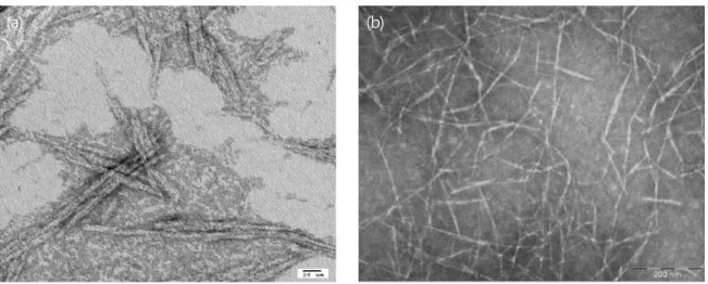
Figure 1. TEM images of two types of CNCs: (a) CNWs, and (b) CNFs
The resulting mixtures were placed in a 50℃ vacuum oven (281A Fisher Scientific, U.S.A) to evaporate the water. The resulting films were kept in a desiccator for 24 hr to remove any remaining water.
Measurement of mechanical properties of nocomposites The mechanical tests were performed using an Instron testing machine (model 3343, load cell 1 kN). The cross head speed was 5 mm/min. The specimens were cut into stripes with a width of 15 mm and a length of 50 mm. The thickness of the samples was calculated before the test. The gauge between the top and bottom clamp was 25 mm.
Results and Discussion
High magnification TEM images of the CNWs prepared by sulfuric acid hydrolysis and CNFs separated by the TEMPO-mediated oxidation method are displayed in Figure 1.
As expected, CNWs wih a rod-like whisker form were clearly identified in the suspension prepared by the acid hydrolysis, and there were also a few aggregated CNWs. This agglomeration could be due to the water used for the sample preparation (Bondeson et al. 2006). An image analysis of the TEM images revealed that the CNWs isolated from the pulp had a width (d) of 6.96±0.87 nm and length (L) of 178±55 nm, giving an aspect ratio (L/d) of about 25. These measurement results are well with the previously published results (Bondeson et al. 2006; Dong et al.1996). For example, Dong et al. (1996) reported that the isolated cellulose crystallites were 70~170 nm long and 7 nm wide.
In contrast, the morphology of the CNFs produced by the TEMPO-mediated oxidation was clearly in a fibrillated form with an average diameter of about 7.07±0.99 nm, which also matched well with other measurements (Shinoda et al. 2012).
Although the length of the CNFs could not be measured in the present study, a published report has recorded a length of about 1 m (Shinoda et al. 2012). This gives an aspect ratio of about 141, which is much greater than that of the CNWs.
It was expected that the fibrillated CNFs would expect to provide more efficient reinforcement than the CNWs in the PVA-based nanocomposites.4)
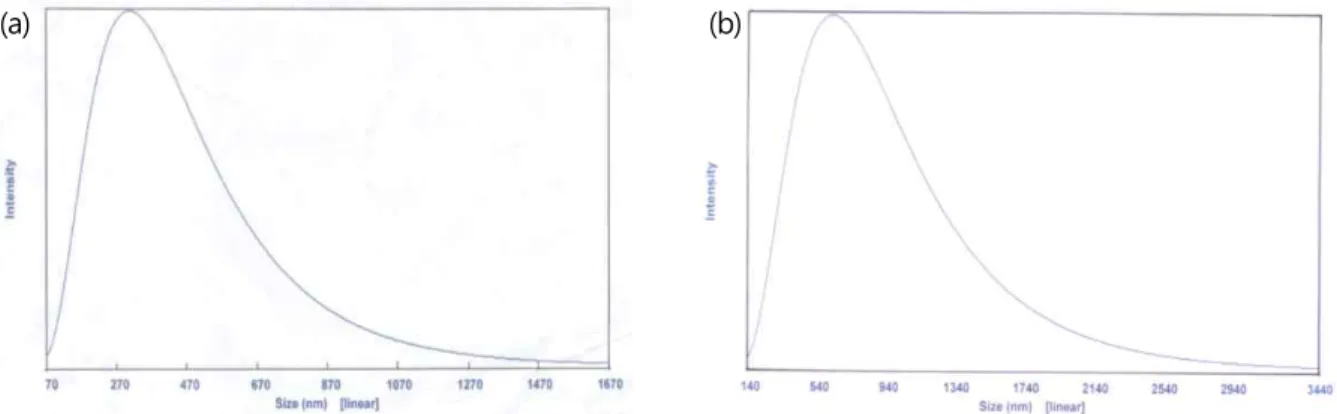
The particle size distribution of the two CNCs in suspension was also measured using a wet size analysis method, and the results are presented in Figure 2. The size distribution of the CNWs ranged from 70 nm to 1670 nm, which gives an average of 340.6 nm, based on an analysis of more than 1.27 million particles (Figure 2, a). This method measured both the width and the length of the CNWs using light scattering. This average value was quite different from the dimensions measured by TEM. Meanwhile, the average size of the CNFs was 703.9 nm with a range from 140 nm to 3440 nm, which was much larger than that of the CNWs (Figure 2b). This could have been due to much greater length of the CNFs in the suspension.
(a) (b)
Figure 2. Wet size analysis results of two types of CNCs: (a) CNWs, and (b) CNFs Figures 3 and 4 show the tensile modulus and strength values
of the two types of CNC-reinforced PVA nanocomposite, depending on the molecular weight (MW) of the PVA. The pure PVA film showed a greater tensile modulus with a higher MW PVA than with low MW PVA, which was quite reasonable.
When the PVA MW 66,000 was reinforced by adding 1%
CNWs, the tensile modulus of the resultant nanocomposites reached a maximum at 5% CNW level and thereafter decreased.
However, the CNF-reinforced nanocomposite showed a maximum tensile modulus at a 1% level for a PVA MW of
22,000. Thus, these results indicated that the incorporation of the CNWs or CNFs into the polymer matrix resulted in different reinforcing efficiencies with the same polymer matrix, most likely due to differences in the morphology, dimension, or aspect ratio between the CNWs and CNFs. The main contributing factor was probably the greater aspect ratio of the CNFs when compared to that of the CNWs.
CNC content (wt%)
0 1 3 5 7
Tensile modulus (GPa)
0 1 2 3 4 8 9 10
CNW-66000 CNF-66000 CNF-22000
Figure 3. Tensile modulus of CNC/PVA nanocomposites as a function of CNC content and MW of PVA
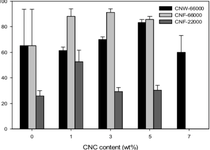
The tensile strengths of the nanocomposites are shown in Figure 4. When the PVA matrix with a MW of 66,000 was reinforced by adding the CNWs, the tensile strength of the nanocomposites gradually increased up to a 5% CNW content and thereafter decreased. Plus, the tensile strength values were smaller than those for the CNF reinforced nanocomposites. Meanwhile, the tensile strength of the nanocomposites reinforced by the CNFs increased only slightly up to a 3% CNW content and thereafter decreased. The tensile strength of the CNF reinforced nanocomposites with a PVA MW of 22,000 showed a maximum
at a 1% level, yet all the tensile strength values for the CNF reinforced nanocomposites were smaller than those for the CNW reinforced nanocomposites. Therefore, the results indicate that the reinforcing efficiency of the CNFs was greater than that of the CNWs. This may also have been due to the greater aspect ratio of the CNFs, which provided a better stress transfer from the matrix to the reinforcing agent, i.e., the CNWs or CNFs in this work.5)
In summary, when comparing the reinforcing efficiency of the two different CNCs, the CNFs were more efficient than the CNWs on the basis of the tensile modulus and strength. These different reinforcing efficiencies could be ascribed to the differences in their morphology, size, aspect ratio, or the MW of the matrix.
CNC content (wt%)
0 1 3 5 7
Tensile strength (MPa)
0 20 40 60 80 100
CNW-66000 CNF-66000 CNF-22000
Figure 4. Tensile strength of CNC/PVA nanocomposites as a function of CNC content and MW of PVA
Conclusions
This study compared the reinforcing efficiency of two different CNCs, i.e., cellulose nanowhiskers (CNWs) and cellulose nanofibrils (CNFs) from hardwood bleached kraft pulp (HW-BKP), as a reinforcing agent in polyvinyl alcohol (PVA)-based nanocomposites. The morphology, dimension, and size distribution of the CNWs and CNFs were characterized by TEM and a wet size analysis. The tensile modulus and strength of the resultant nanocomposites were determined to compare the reinforcing efficiency. The following conclusions were drawn from the results:
1. According to the TEM-based measurements, the individual CNWs were about 6.96±0.87 nm wide and 178±55 nm long, while the CNFs were 7.07±0.99 nm wide, which provided aspect ratios of about 25 and 141, respectively.
2. The incorporation of the CNWs and CNFs into the PVA
matrix at 5% and 1% levels, respectively, resulted in the maximum tensile strength, indicating that the nanocrystals had different efficiencies in reinforcing the nanocomposites.
3. The present results suggest a relationship between the reinforcing potential of CNCs and their physical characteristics, such as their morphology, dimensions, and aspect ratio.
Acknowledgement
The authors acknowledge the contribution of both the National Research Foundation (NRF) of Korea funded by the Ministry of Education, Science and Technology, and the Ministerodegli Affari Esteri, Direzione Generale per la Promozione del Sistema Paese of Italy.
References
Bai W, Holbery J, Li K (2009) Technique for production of nanocrystalline cellulose with a narrow size distribution.
Cellulose 16:455-465.
Bondeson D, Kvien I, Oksman K (2006) Strategies for preparation of cellulose whiskers from microcrystalline cellulose as reinforcement in nanocomposites. Oksman K., Sain M. Ed., pp. 10-25, ACS Symposium Series 938, Washington, DC.
de Nooy AEJ, Besemer AC, van Bekkum H (1995) Highly selective nitroxyl radical-mediated oxidation of primary alcohol groups in water-soluble glucans.Carbohyd Res269:
89-98.
de Nooy AEJ, Besemer AC, van Bekkum H (1996) On the use of stable organic nitroxyl radicals for the oxidation of primary and secondary alcohols. Synth 10:1153-1176.
Dong XM, Revol JF, Gray DG (1998) Effect of microcrystallite preparation conditions on the formation of colloid crystals of cellulose. Cellulose 5:19-32.
Eichhorn SJ, Dufresne A, Aranguren M., Marcovich NE, Capadona JR, Rowan SJ, Weder C, Thielemans W, Roman M, Renneckar S, Gindl W, Veigel S, Keckes J, Yano H, Abe K, Nogi M, Nakagaito AN, Mangalam A, Simonsen J, Benight AS, Bismarck A, Berglund LA, Peijs T (2010) Review: current international research into cellulose nanofibres and nanocomposites. J Mater Sci 45:1-33.
Favier V, Canova GR, Cavaille JY, Chanzy H, Dufresne A, Gauthier C. (1995) Nanocomposite materials from latex and cellulose whiskers.Polym Advan Technol 6:351-355.
Fukuzumi H, Saito T, Iwata T, Kumamoto Y, Isogai A (2009) Transparent and high gas barrier films of cellulose nanofibers prepared by TEMPO-mediated oxidation. Biomacromol
10:162-165.
Isogai A, Saito T, Fukuzumi H (2011) TEMPO-oxidized cellulose nanofibers. Nanoscale 3:71-85.6)
Kvien I, Tanem BS, Oksman K (2005) Characterization of cellulose whiskers and their nanocomposites by atomic force and electron microscopy. Biomacromol 6:3160-3165.
Leitner J, Hinterstoisser B, Wastyn M, Keckes J, Gindle W (2007) Sugar beet cellulose nanofibril-reinforced composites.
Cellulose 14:419-425
Lu J, Wang T, Drzal LT (2008) Preparation and properties of microfibrillated cellulose polyvinyl alcohol composite materials.Compos. Part A: Appl Sci Manufact39:738-746.
Renneckar S, Zink-Sharp A, Esker AR, Johnson RK, Glasser WG (2006) Novel methods for interfacial modification of cellulose-reinforced composites. Cellulose Nanocomposites:
processing, characterization and properties, Oksman K., Sain M. Ed., pp. 78-96, ACS Symposium Series 938, Washington, DC.
Sain M, Oksman K (2006) Cellulose Nanocomposites: Processing, Characterization, and Properties, ACS Symposium Series 938, 2006, ACS, Washington DC, USA
Saito T, Isogai A (2004) TEMPO-mediated oxidation of native cellulose. The effect of oxidation conditions on chemical and crystal structures of the water-insoluble fractions.
Biomacromol 5:1983-1989.
Saito T, Nishiyama Y, Putaux JL, Vignon M, Isogai A (2006) Homogeneous suspensions of individualized microfibrils from TEMPO-catalyzed oxidation of native cellulose.
Biomacromol 7:1687-1691.
Saito T, Kimura S, Nishiyama Y, andIsogai A (2007) Cellulose nanofibers prepared by TEMPO-mediated oxidation of native cellulose. Biomacromol 8:2485-2491.
Samir MASA, Alloin F, Dufresne A (2005) Review of recent research into cellulosic whiskers, their properties and their application in nanocomposite field. Biomacromol 6:
612-626.
Shinoda R, Saito T, Okita Y, Isogai A (2012) Relationship between length and degree of polymerization of TEMPO- oxidized cellulose nanofibrils. Biomacromol 13:842-849.
Sturcová A, Davies GR, Eichhorn SJ (2005) Elastic modulus and stress transfer properties of tunicate cellulose whiskers.
Biomacromol 6:1055-1061.
Tashiro K, Kobayashi M (1991) Theoretical evaluation of three dimensional elastic constants of native and regenerated cellulose: role of hydrogen bonds. Polym32:1516-1526.