• 검색 결과가 없습니다.

Development of Single-layer-structured Glucose Biosensor

N/A
N/A
Protected

Academic year: 2021

Share "Development of Single-layer-structured Glucose Biosensor"

Copied!
5
0
0

로드 중.... (전체 텍스트 보기)

전체 글

(1)

http://dx.doi.org/10.5369/JSST.2015.24.2.83 pISSN 1225-5475/eISSN 2093-7563

Development of Single-layer-structured Glucose Biosensor

Young-Tae Lee

1,+

and Min Su Kwon

2

Abstract

In this paper, we fabricated a low-cost glucose sensor with a simpler structure and fabrication process than the existing glucose sensor.

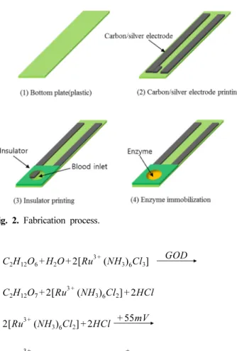
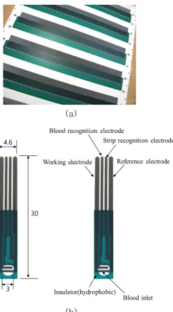
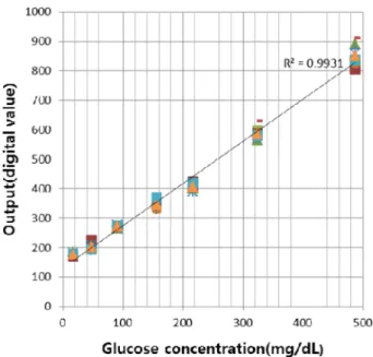
The currently used glucose sensor has a three-layer structure with upper, middle, and bottom plates; here, we fabricated a single-layer glucose sensor using only a printing and dispensing process. We successfully fabricated the glucose sensor using a simple method involving the formation of an electrode and insulator layer through a 2- or 3-step printing process on plastic or paper film, followed by the dispensing of glucose oxidase solution on the electrode. Cyclic voltammetry (CV) and cyclic amperometry (CA) measurements were used to evaluate the characteristics of the fabricated single-layer glucose sensor. Also, its sensitivity was analyzed through glucose-con- trolled blood measurements. Hence, a low-cost single-layer glucose sensor was fabricated with evaluation of its characteristics dem- onstrating that it has useful application in medicine.

Keywords: Glucose, Single-layer, Electrochemical, Biosensor, Screen printing

1. INTRODUCTION

Recently, due to the rapidly aging global population and other lifestyle- and environment-related factors, the prevalence of diabetes has increased. According to the International Diabetes Federation (IDF) [1], the number of diabetics in Korea in 2013 came to 3.32 million, ranking Korea 20

th

in the world as regards sufferer numbers. It is anticipated that, in 2035, the global number of diabetics will increase to 592 million, i.e., 1 in 10 people, the majority of whom (over 80%) will be from low or middle-income countries and aged under sixty. In south-east Asia, as it is not possible for half of diabetics to be diagnosed correctly, concerns have been raised regarding increasing death rates due to complications, along with the cost of treatment. In 2013, $548 billion was spent on diabetes treatment and the scale is predicted to grow rapidly every year. Thus, each country is actively framing

policies to reduce the cost of diabetes-related treatment. To monitor the blood glucose values of the majority of a nation’s population, including those in the low-income bracket, personal glucose measurement devices can be distributed; however, the device price must first be reduced significantly. This would also contribute to the exportation of glucose sensors and decrease the global social cost of health care, by making it possible to monitor glucose levels in south-east Asia and Africa.

In this paper, we have fabricated a glucose sensor with a single- layer structure. At present, this device is fabricated using three- layer plastic film and electrochemical methods, so the cost of the glucose sensor is reduced [2-5]. The currently used glucose sensor has a three-layer structure comprised of upper, middle, and bottom plates, but the single-layer glucose sensor suggested in this paper is fabricated using a 3-step printing process (silver/carbon/

insulator) only, using screen printing technology [6] on plastic [7]

or paper film [8]. Thus, the device price can be decreased significantly by simplifying the sensor structure and fabrication process. For the existing three-layer glucose sensor, the upper, middle, and bottom plates are each fabricated individually and film is then bonded on each layer through double lamination [7].

However, in the case of the single-layer glucose sensor, the equipment is simple and the material cost is reduced, because only 2- or 3-step screen printing is required. So, we estimate that the price of the sensor can be decreased significantly.

1

Department of Electronic Engineering Education, College of Education, Andong National University

1375 Gyeongdong-ro, Andong, Gyeongsangbuk-do 760-749, Korea

2

Center for Integrated Smart Sensors

309 ITC Building(N1), Kaist, 290 Daehak-ro, Yuseong-gu, Daejeon 305-701, Korea

+

Corresponding author: [email protected]

(Received : Jan. 7, 2015, Revised : Mar. 24, 2015, Accepted : Mar. 25, 2015)

This is an Open Access article distributed under the terms of the Creative Commons Attribution Non-Commercial License(http://creativecommons.org/

licenses/bync/3.0) which permits unrestricted non-commercial use, distribution,

and reproduction in any medium, provided the original work is properly cited.

(2)

2. SENSOR STRUCTURE AND SENSING PRINCIPLES

2.1 Glucose Sensor Structure

Fig. 1 shows the structure of the single-layer glucose sensor.

The working and reference electrodes (made from silver and carbon ink) are placed on the plastic film, and an insulator is formed on all parts apart from the blood inlet to protect the electrodes. Glucose oxidase (GOD) liquid is immobilized on the blood inlet. The single-layer glucose sensor is fabricated using a 3-step silver, carbon, and insulator printing process. So, its structure and fabrication process are simple and the production cost is very low compared to the existing sensors. The sensor is 30-mm long, 4.6-mm wide, and approximately 0.2-mm thick. The blood inlet diameter is 3 mm ψ, with a depth of 12 μm, and a volume of approximately 0.085 μl.

2.2 Sensing Principle

As the single-layer glucose sensor mechanism is electrochemical, the enzyme layer formed on the electrodes is made by mixing GOD, ruthenium ([Ru(NH

3

)

6

]Cl

3

), stabilizer, and surfactants in the appropriate ratio [3]. As shown in formulae (1) and (2) below, the ruthenium functions as an electron mediator, receiving electrons during the course of the glucose oxidization caused by the GOD and delivering them to the carbon electrode to form an electric current.

(1)

(2) By measuring this current, we can determine the glucose concentration. This measurement does not require oxygen since it uses an electron mediator, and its sensitivity and linearity are excellent.

3. FABRICATION PROCESS

Fig. 2 shows the single-layer glucose sensor fabrication process.

This design produces low-cost glucose sensors, as it has a very simple fabrication technique involving a 2- or 3-step printing process at a very low unit cost. As shown in Fig. 2, we formed carbon electrodes on plastic (polyethylene terephthalate, PET) or paper substrate using screen printing [6] (Fig. 2(b)). Then, we formed the blood inlet through screen printing using hydrophobic insulation ink (Fig. 2(c)). Next, we dispensed 1.25-μl enzyme liquid onto the blood inlet using a dispenser (Fig. 2(d)). At that point, the enzyme liquid was dispensed onto the blood inlet only, because the hydrophobic insulation layer had already been formed. Finally, we dried the enzyme liquid in a drying oven at 50

o

C for 15 min to complete the glucose sensor.

Fig. 3 is a photograph of the fabricated single-layer glucose sensor. Fig. 3(a) shows the printed sheet before it was cut into

C

2

H

12

O

6

+ H

2

O 2 Ru + [

3 +

( NH

3

)

6

Cl

3

] GOD

C

2

H

12

O

7

+ 2 Ru [

3 +

( NH

3

)

6

Cl

2

] 2HCl +

2 Ru [

3 +

( NH

3

)

6

Cl

2

] 2HCl + 55mV +

2 Ru [

3 +

( NH

3

)

6

Cl

3

] 2e +

+ 2H

+

Fig. 1. Single-layer glucose sensor.

Fig. 2. Fabrication process.

(3)

individual sensors. For a substrate size of 400 × 270 mm

2

, it is possible to fabricate a total of 312 sensors at a time. Fig. 3(b) shows a cut individual sensor, with dimensions of 30 × 4.6 mm

2

. The blood inlet diameter is 3 mm and the blood injection volume is estimated to be approximately 0.085 μl. The glucose sensor and its blood inlet can be downsized to allow a small-size glucose sensor to be fabricated. So, we estimate that we can reduce the glucose sensor unit-cost by increasing the number of fabricated glucose sensors per substrate.

4. RESULTS AND DISCUSSION

We used whole blood samples to evaluate the characteristics of the fabricated glucose sensor. A Yellow Spring Instruments (YSI) 2300 STAT PLUS and a Won-a-Tech WPG Potentiostat/

Galvanostat, which are standard devices for glucose measurement, were used for the characteristic evaluation [8]. A cyclic voltammetry (CV) measurement method [8] was used to evaluate the oxidation and reduction of the fabricated single-layer glucose sensor. Fig. 4 shows the measured results. For the CV measurement, blood samples with glucose concentrations of 43, 89, 147, 207, 327, and 495 mg/dL were used, with a voltage in the

range of -0.7 V-+0.7 V. From Fig. 4, we determined that the oxidation and reduction peak occurred at approximately 55-mV applied voltage. We found that, as the glucose concentration increased, the current value also rose. Sufficient resolution was exhibited for each concentration.

Fig. 5 shows the time response of the fabricated single-layer glucose sensor measured using cyclic amperometry (CA) [8]. For the CA measurement, blood samples with fixed glucose concentrations of 43, 89, 147, 207, 327, and 495 mg/dL were used. The applied voltage was 55 mV, as previously, and the measurement time was fixed to 10 s. We found that the output current was saturated 3 s after the measurement began, following the blood injection and voltage application.

Fig. 6 shows the glucose concentration-current characteristics of the single-layer glucose sensor. Blood samples with glucose concentrations of 16.4, 48, 90.5, 156, 216, 324, and 487 mg/dL were used for the measurement and the applied voltage was again fixed to 55 mV. We measured the current value 3 s after blood injection for each glucose concentration, 10 times per concentration. The output characteristic shown in Fig. 6 is the 10- bit digital value, which corresponds to the output current value.

The measured results exhibited good output linearity, as R

2

= Fig. 3. Photograph of single-layer glucose sensor.

Fig. 4. Cyclic voltammetry waveform.

Fig. 5. Cyclic amperometry waveform.

(4)

0.9931, and the sensing reproducibility was also found to be excellent. The measured result given above (3-s measurement time, over 0.085-μl blood volume) led us to conclude that the single-layer glucose sensor is fully applicable for personal glucose-level monitoring.

Fig. 7 is a photograph of water making contact with the blood inlet of the single-layer glucose sensor. We found that the water made direct contact with the blood inlet only (which included electrodes). This phenomenon occurred because the printed carbon electrode was hydrophilic, while the insulation layer was hydrophobic.

Compared to the existing glucose sensor, the single-layer glucose sensor requires a somewhat large volume of blood for measurement, but the amount of blood collected by a typical lancet may be sufficient. The existing three-layer glucose sensor utilizes a channel structure (which is the reasoning behind the three-layer structure with upper, middle, and bottom plates), to allow a small or fixed amount of blood to be used for

measurement. Its fabrication process is relatively complex: a channel and air vent are formed on the upper and middle plates, and a lamination process is used to bond each layer. Further, if the channel is fabricated incorrectly, measurement errors can occur.

High-accuracy and high-cost facilities are utilized to reduce such measurement errors, which makes price competitiveness difficult.

Therefore, the glucose sensor structure must be simplified in order to reduce fabrication costs and a newly structured glucose sensor with reduced potential for measurement errors must be developed.

The single-layer glucose sensor suggested in this paper has a very simple fabrication process compared to the existing three- layer glucose sensor, as it is fabricated through a 2- or 3-step printing process only. Because of its simple structure, we also estimate that the sensing reproducibility can be improved through reduction of the measurement errors. In the case of the existing three-layer glucose sensor, capillary action through a small-size channel is used when blood is collected. However, the part in contact with the blood is so small that old or sick individuals cannot use this device easily. Also, the extremely small amount of blood that is drawn and the blood drawing method itself are considered problematic by the medical community. In contrast, the single-layer glucose sensor proposed in this paper can be used by the old or infirm relatively easily, because blood is injected onto the blood inlet on the glucose sensor. In addition, the single- layer glucose sensor draws blood evenly as opposed to partly.

Therefore, we estimate that problems occurring as a result of the small amount of drawn blood can be somewhat improved with this device.

5. CONCLUSIONS

In summary, we developed a low-cost single-layer glucose sensor with a simpler structure and fabrication process than that of the three-layer structure (upper, middle, and bottom plate) glucose sensor. The single-layer glucose sensor can be fabricated using a 2- or 3-step printing process only, on PET or paper film, followed by enzyme liquid dispensation. So, the materials as well as the fabrication process can be simplified; hence, the device cost can be lowered. In this paper, we have fabricated a single-layer glucose sensor by forming electrodes and an insulation layer through three-step screen printing of silver/carbon/insulator layers and then dispensing enzyme liquid onto the electrodes. We have also analyzed the characteristics of the developed single-layer glucose sensor and examined its potential for use as a personal glucose monitor. We measured the CV, CA, and glucose Fig. 6. Output characteristics of single-layer glucose sensor.

Fig. 7. Wetting behavior of water droplet on glucose sensor inlet.

(5)

concentration vs output current characteristics. In particular, we used blood with various glucose concentrations of 16.4-487 mg/

dL to measure the glucose concentration vs output current characteristics. A total of 10 measurements were taken for each glucose concentration and an excellent output linearity of R

2

= 0.9931 was obtained. Consequently, we conclude that the single- layer glucose sensor developed in this paper can be used for medical purposes.

REFERENCES [1] http://www.idf.org/.

[2] L. C. Clark, and C. Lyons, “Electorde system for continuous monitoring in cardiovacular”, Annals New York Academy of Sciences, Vol. 102, pp. 29-45, 1962.

[3] Joseph Wang, “Electrochemical glucose biosensors”, Chem-

ical Reviews, Vol. 108, No. 2, pp. 814-825, 2008.

[4] J. R. Mor, and R. Guarnaccia, “Assay of glucose using an electrochemical enzymatic sensor”, Analytical Biochemis- try, Vol. 79, pp. 319-328, 1977.

[5] J. Anzai, Y. Kobayashi, Y. Suzuki, H. Takeshita, Q. Chen, T. Osa, T. Hoshi, and X. Du, “Enzyme sensors prepared by layer-by-layer deposition of enzyme on the platinum elec- trode through avidin-biotin interaction”, Sensors and Actu- ators B-Chem., Vol. 52, Issues 1-2, pp. 3-9, 1998.

[6] N. G. Patel, S. Meier, K. Cammann, and G. C. Chemntius,

“Screen-printed biosensors using defferent alcohol oxi- dase”, Sensors and Actuators B-Chem., Vol. 75, Issues 1-2, pp. 101-110, 2001.

[7] Y. T. Lee, and S. R. Lee, “Development of the disposable glucose sensor using Cu/Ni/Au electrode”, Journal. of the Korea Sensors Society, Vol. 15, No. 5, pp.352-356, 2006.

[8] A. Y. Kim, and Y. T. Lee, “Development of eco-friendly

paper glucose bio-sensor”, Journal of Sensor Science and

Technology, Vol. 22, No. 3, pp.202-206, 2013.

수치

Fig. 1 shows the structure of the single-layer glucose sensor.
Fig. 6 shows the glucose concentration-current characteristics of the single-layer glucose sensor
Fig. 7 is a photograph of water making contact with the blood inlet of the single-layer glucose sensor

참조

관련 문서