Introduction
Cervical radiculopathy (CR) is a neurologic con- dition characterized by either dysfunction of a cer- vical spinal nerve, the roots of the nerve, or both (Bogduk, 2011). It usually presents with the neck and unilateral arm pain, a combination of loss of the sensory and motor function, or change of reflex in the affected nerve-root distribution (Bogduk, 2011).
In the incidence of CR, a lesion of the nerve root is most frequent, secondary to cervical disc herniation and spondylosis (Wainner and Gill, 2000).
Particularly, monoradiculopathy involving C7 nerve root is the most frequent, followed by C6 (Radhakrishan et al, 1994).
Previous studies demonstrated that people with chronic neck pain has several problems; limited cer- vical range of motion (ROM), impaired motor func- tion, and abnormal control of neck posture with cer- vical spine (Bogduk, 2011; Falla et al, 2007; Jull et al, 2007). Dysfunctions of deep neck flexor muscle (Falla et al, 2004; Jull et al, 2007; Mayoux-Benhamou et al, 1994), and inappropriate length of muscles con- nected with neck region (Sahrmann, 2010) are asso- ciated with chronic neck pain and disability.
The treatments for reducing symptoms for patients with cervical problems included stretching the neck and shoulder muscles (Sahrmann, 2010), increasing cervical ROM (Martínez-Segura et al, 2006), and im- proving neuromuscular control of the cervical spine Corresponding author: Oh-yun Kwon [email protected]
Effect of Cervical Corrective Exercises on Pain, Neck Posture, and Intersegmental Motion of Cervical Spine in a Patient With
Cervical Radiculopathy: A Case Report
Sung-joon Yun1,2, MSc, PT, Moon-hwan Kim1, PhD, PT, Jong-hyuck Weon3, PhD, PT, Oh-yun Kwon4,5, PhD, PT
1Dept. of Rehabilitation Medicine, Wonju Severance Christian Hospital
2Dept. of Physical Therapy, The Graduate School, Yonsei University
3Dept. of Physical Therapy, College of Tourism & Health Science, Joongbu University
4Dept. of Physical Therapy, College of Health Science, Yonsei University
5Dept. of Ergonomic Therapy, The Graduate School of Health and Environment, Yonsei University
Abstract
1)This case report describes the effectiveness of cervical corrective exercises in a patient with cervical radiculopathy (CR) who experienced radicular pain, upper limb paresis, and limited functional activity. A 39-year-old male with cervical radiculopathy performed the cervical corrective exercises for reducing pain.
Pain intensity, cervical posture, and active range of motion of cervical intersegmental spine motion were measured baseline, after 4 weeks, and after 8 weeks with self-reported questionnaire and radiographs.
After 8 weeks of intervention, the patient demonstrated alleviated radicular symptoms, improved neck posture and active range of flexion and extension of the cervical intersegmental spine. Especially in the angle between the cervical vertebra 6 and 7, the angle was changed from -4.69° to 3.30° during resting position after intervention. The present case indicates that the cervical corrective exercises might be a possible treatment to effectively reduce radicular symptoms, improve neck posture, and active cervical intersegmental motion for patient with CR.
Key Words: Cervical exercise; Cervical spine; Intersegmental spine motion; Radiculopathy.
한국전문물리치료학회지 2015년 22권 4호 1-7 Phys Ther Korea 2015;22(4):1-7
and the shoulder girdle (Falla et al, 2004). Although many literatures recommended cervical exercise and correcting alignment of cervical spine for reducing cervical pain (Cassidy et al, 1992; Falla et al, 2004;
Falla et al, 2007; Martínez-Segura et al, 2006;
Sahrmann, 2010), there has not been a study to find the effects of cervical exercises on the change of the posture and active flexion and extension range of motion of cervical intersegmental spine in patients with CR. The purpose of this case study was to find the effectiveness of exercise regimes, with consist of deep neck flexor strengthening and cervicoscapular muscle stretching, on pain, neck posture, and active ROM of the cervical intersegmental spine in a pa- tient with CR.
Case Report
A 39-year-old male (weight 60 ㎏; height 174 ㎝), a radiological technician, and a graduate student par- ticipated in this study. The patient’s work involved performance of visual display tasks (VDTs) and lift- ing patients during office hours. He had a history of left-sided neurological cervicobrachial pain [Numeric Pain Rating Scale (NPRS)=8.2/10; Neck Disability Index (NDI)=25/50] in the C5/C8 dermatome for 6 months prior to the study (Figure 1). His symptoms were exacerbated by prolonged VDT activity. The
patient was diagnosed with CR at the C6/C7 level of disc herniation by magnetic resonance imaging (MRI) and referred to us by a neurosurgeon. The patient underwent 3 months of conventional physical therapy (hot packing, interferencial current therapy, and cer- vical traction) for reducing the pain, but his symp- toms did not improve. Although the patient’s work station was also evaluated and modified monitor height and mouse position, but there was little change in symptoms. Thus patient participated in the experiment for reducing the symptoms. He provided informed consent to anonymously publish his case in a medical journal. In addition, this study followed principles in the Declaration of Helsinki (World Medical Association General Assembly, 2004).
While looking straight ahead, the patient had poor neck posture in both the sitting and standing posi- tions; he exhibited a flexed cervical spine, forward head posture, and protracted shoulders. The left shoulder level was higher than the right. His head was laterally flexed toward the left.
The patient’s neck posture and active flexion and extension ROM of cervical intersegmental spine were evaluated on a lateral X-ray following previously published guidelines (Barrey et al, 2012). The cer- vical spine X-ray findings showed a flexed posture with C6/C7 intersegmental spine during the resting period (Figure 2) and limited active ROM of cervical intersegmental spine according to the Harrison pos- terior tangent method. Cervical MRI revealed thecal sac impression at C6/C7, slightly to the left of the midline (Figure 3). Radiologic imaging reading was achieved by a radiologist with more than 10 years of experience.
The therapist used a universal goniometer to measure the cervical rotation ROM and shoulder ROM (Norkin and White, 1995). The device was marked in 2-degree increments. On physical exami- nation, the cervical spine and shoulder exhibited re- stricted ROM on neck rotation (right 30°; left 15°), left shoulder abduction (150°), and internal rotation (50°) due to pain. Decreased strength was noted in Figure 1. Body chart illustrating pain
presentation.
upper limb muscles on the left; shoulder internal ro- tators (3/5), biceps brachii (4/5), and deltoid (4/5);
however, the C5, C6, and C7 reflexes were normal.
There were positive results in nerve tension tests, the upper limb tension test (ULTT) 1 and 2 (median nerve), and the ULTT 3 (ulnar nerve) (Butler and Jones, 1991).
Treatment plan
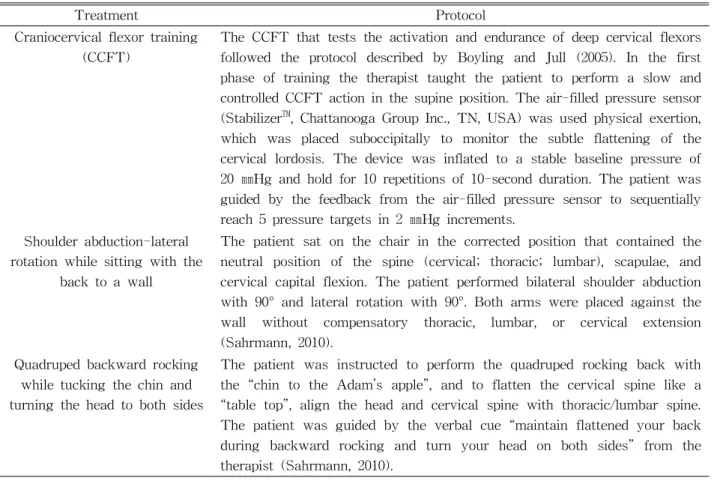
The cervical exercises included craniocervical flex- or training (CCFT) (Boyling and Jull, 2005), shoulder
abduction-lateral rotation while sitting with the back to a wall, and quadruped backward rocking while tucking the chin and turning the head to both sides (Sahrmann, 2010) (Table 1). The cervical exercise protocol comprised 24 sessions of exercises, 3 times per week for 8 weeks.
Outcome measures were collected at baseline and at weeks 4 and 8 using the NPRS, NDI, shoulder ROM and imaging findings (X-ray and MRI). The deep neck flexors strength was assessed by the air-filled pressure sensor. The NPRS and NDI were
A B
Figure 2. X-ray finding comparison for change in upright posture (A: baseline, B: after 8 weeks exercise).
Figure 3. Axial MRI at the C6/C7 level and sagittal MRI of cervical spine.
한국전문물리치료학회지 2015년 22권 4호 1-7 Phys Ther Korea 2015;22(4):1-7
used to measure the intensity of the pain during sustained VDT activity and the perceived disability (Cleland et al, 2008). The NDI and NPRS exhibited moderate test-retest reliability using an intraclass correlation coefficient (ICC) [NDI ICC=.50; 95% con- fidence interval (CI): .25∼.67; NPRS ICC=.76; 95%
CI: .51∼.87) (Cleland et al, 2008). The NDI and NPRS had moderately high correleations (.69∼.70) (Vernon and Mior 1991) and .79 to .95 convergent validity (Good et al, 2001). The imaging findings were used to determine the neck posture and active ROM of the cervical intersegmental spine.
Results
Figure 4 illustrates the change in the NPRS score and NDI. After 8 weeks of cervical corrective ex- ercises, the NPRS score and NDI had decreased from
8.2 to 1.2 and from 25 to 10, respectively.
Through the CCFT, the patient had been run from a baseline of 20 ㎜Hg to the final level of 30 ㎜Hg.
The patient was able to reach task of shoulder ab- duction-lateral rotation from a baseline of 90° to the final level of 150° after intervention.
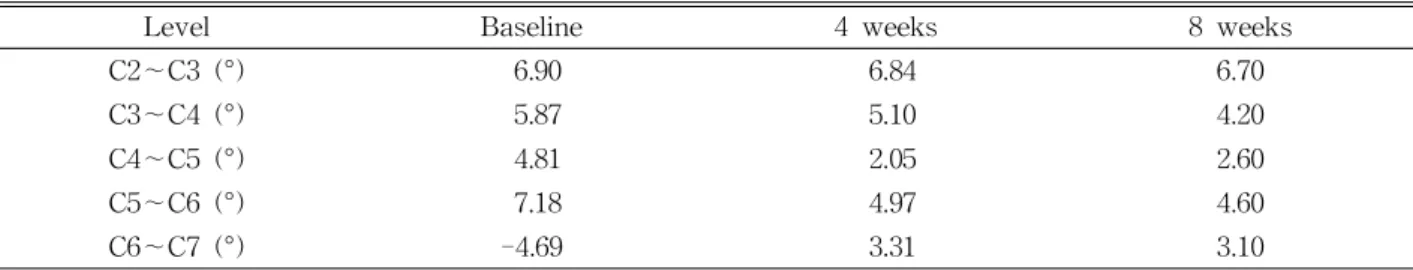
Table 2 and 3 show the results of each assess- ment: baseline, week 4, and week 8. Especially in the cervical posture, the C6/C7 intersegmental angle in- creased from -4.69° to 3.30° (Figure 2). Active ROM of cervical intersegmental spine increased when the baseline and after 8 weeks exercise were compared, were compared with normal range of intersegmental spine (C3∼C7 level) (Dvorak et al, 1987). Pain free ROM on neck rotation (from right 30°; left 15° to right 34°; left 25°), left shoulder abduction (from 150°
to 170°), and left shoulder internal rotation (from 50°
to 55°) increased, however, no change of muscle strength after 8 weeks exercise. In addition, the
Treatment Protocol
Craniocervical flexor training (CCFT)
The CCFT that tests the activation and endurance of deep cervical flexors followed the protocol described by Boyling and Jull (2005). In the first phase of training the therapist taught the patient to perform a slow and controlled CCFT action in the supine position. The air-filled pressure sensor (Stabilizer™, Chattanooga Group Inc., TN, USA) was used physical exertion, which was placed suboccipitally to monitor the subtle flattening of the cervical lordosis. The device was inflated to a stable baseline pressure of 20 ㎜Hg and hold for 10 repetitions of 10-second duration. The patient was guided by the feedback from the air-filled pressure sensor to sequentially reach 5 pressure targets in 2 ㎜Hg increments.
Shoulder abduction-lateral rotation while sitting with the
back to a wall
The patient sat on the chair in the corrected position that contained the neutral position of the spine (cervical; thoracic; lumbar), scapulae, and cervical capital flexion. The patient performed bilateral shoulder abduction with 90° and lateral rotation with 90°. Both arms were placed against the wall without compensatory thoracic, lumbar, or cervical extension (Sahrmann, 2010).
Quadruped backward rocking while tucking the chin and turning the head to both sides
The patient was instructed to perform the quadruped rocking back with the “chin to the Adam’s apple”, and to flatten the cervical spine like a
“table top”, align the head and cervical spine with thoracic/lumbar spine.
The patient was guided by the verbal cue “maintain flattened your back during backward rocking and turn your head on both sides” from the therapist (Sahrmann, 2010).
Table 1. Interventions of cervical corrective exercise
finding of the MRI revealed no changes in thecal sac size between baseline and after 8 weeks cervical exercises.
Discussion
Patients with CR have poor control of their up- right cervical posture because of various elements,
such as dysfunction of sensory input from the deep neck muscles, which have the highest muscle spindle density (Treleaven et al, 2006), and abnormal length and motor control of the shoulder girdle musculature (Sahrmann, 2010). The present case study was per- formed to determine the effects of cervical exercises involving deep neck flexor strengthening and cervi- coscapular muscle stretching on radicular symptoms in a patient with CR. The exercises resulted in de- Figure 4. Numeric Pain Rating Scale and Neck Disability Index score over the
sessions (NPRS: Numeric Pain Rating Scale, NDI: Neck Disability Index).
Level Baseline 4 weeks 8 weeks
C2∼C3 (°) 6.90 6.84 6.70
C3∼C4 (°) 5.87 5.10 4.20
C4∼C5 (°) 4.81 2.05 2.60
C5∼C6 (°) 7.18 4.97 4.60
C6∼C7 (°) -4.69 3.31 3.10
Table 2. The neutral posture of the cervical intersegmental angle
Level Baseline 4 weeks 8 weeks Normal range
C2∼C3 (°) 8.38 5.22 6.00 10 (5∼15)
C3∼C4 (°) 9.67 11.48 12.90 15 (7∼23)
C4∼C5 (°) 10.79 14.62 15.60 19 (13∼26)
C5∼C6 (°) 12.18 10.35 18.40 20 (13∼28)
C6∼C7 (°) 11.27 14.20 13.60 19 (11∼26)
Normal active ranges of cervical intersegmental motion by Dvorak et al (1987).
Table 3. The active ranges of cervical intersegmental flexion and extension motion
한국전문물리치료학회지 2015년 22권 4호 1-7 Phys Ther Korea 2015;22(4):1-7
creased pain and perceived disability, an improved neck posture, and increased active ROM of the cer- vical intersegmental spine.
The CCFT reportedly has a functional role in sup- porting posture (Boyling and Jull, 2005). The im- provement in the performance of these muscles counteracts the cervical lordosis increment caused by the weight of the head and increases the interverte- bral foramen space (Mayoux-Benhamou et al, 1994).
However, the MRI findings in the present study re- vealed no changes in the thecal sac size between the baseline and final assessment.
A noticeable reduction was present in the NPRS score and NDI. Sustained forward flexion of the spine has been associated with increased cervical compressive loading (Kolehmainen et al, 1989) and impaired activation of the deep cervical flexor mus- cles (Falla et al, 2004). According to previous stud- ies, deep cervical flexor training reduces neck pain (Falla et al, 2007; Falla et al, 2013) and neck dis- ability (Falla et al, 2007; Falla et al, 2013; Jull et al, 2007) and enhances proprioception of the head posi- tion (Falla et al, 2013). Furthermore, Cassidy et al (1992) reported that increased ROM of cervical rota- tion was associated with decreased pain. The pain reduction was associated with a lessening of the in- terference with transmission of afferent input to the dorsal horn or a reduction of the abnormal com- pressive loading of the head through an improvement in the ability to maintain the cervical spine in an upright posture (Harms-Ringdahl et al, 1986).
Abnormal cervical intersegmental motion in flexion and extension means a sign of instability of cervical spine (Dvorak et al, 1991). Following 8 weeks of cervical corrective exercises, a noticeable improve- ment occurred in the alignment of the neck posture and active ROM of the cervical intersegmental spine on X-ray examination. In particular, the C6/C7 inter- segmental angle markedly increased after the intervention. These improvements may have occurred secondary to improvement in the strength and en- durance of the deep cervical flexors and the im-
proved flexibility of the cervicoscapular muscles (i.e., the levator scapular, the pectoralis minor). The cer- vical corrective exercise used in this study may be important factor in the prevention of pain and mala- lignment of cervical spine in patient with CR.
Further studies regarding the duration of the effect of the cervical corrective exercise, and developing a step-by-step treatment protocol for patient with CR are necessary.
Conclusion
This case study investigated the effectiveness of cervical corrective exercises with an emphasis on changes in the cervical alignment and intersegmental motion in a patient with CR. The results showed that 8 weeks of cervical corrective exercises had considerable effects on reduction of pain and per- ceived disability, and improved the upright posture with the cervical spine and active ROM in the cer- vical intersegmental spine.
References
Barrey C, Champain S, Campana S, et al. Sagittal alignment and kinematics at instrumented and adjacent levels after total disc replacement in the cervical spine. Eur Spine J. 2012;21(8):
1648-1659.
Bogduk N. The anatomy and pathophysiology of neck pain. Phys Med Rehabil Clin N Am. 2011;
22(3):367-382.
Boyling JD, Jull GA. Grieve’s Modern Manual Therapy: The vertebral column. 3rd ed. Edinburgh, Churchill Livingstone, 2005:451-470.
Butler DS, Jones MA. Mobilisation of the Nervous System. 1st ed. Melbourne, Churchill Livingstone, 1991:148-154, 157-159.
Cassidy JD, Lopes AA, Yong-Hing K. The immedi- ate effect of manipulation versus mobilization on
pain and range of motion in the cervical spine:
A randomized controlled trial. J Manipulative Physiol Ther. 1992;15(9):570-575.
Cleland JA, Childs JD, Whitman JM. Psychometric properties of the neck disability index and nu- meric pain rating scale in patients with me- chanical neck pain. Arch Phys Med Rehabil.
2008;89(1):69-74.
Dvorak J, Hayek J, Zehnder R. CT-functional diag- nostics of the rotatory instability of the upper cervical spine. Part 2. An evaluation on healthy adults and patients with suspected instability.
Spine (Phila Pa 1976). 1987;12(8):726-731.
Dvorak J, Panjabi MM, Novotny JE, et al. In vivo flexion/extension of the normal cervical spine. J Orthop Res. 1991;9(6):828-834.
Falla D, Lindstrøm R, Rechter L, et al. Effectiveness of an 8-week exercise programme on pain and specificity of neck muscle activity in patients with chronic neck pain: A randomized controlled study. Eur J Pain. 2013;17(10):1517-1528.
Falla D, O’Leary S, Fagan A, et al. Recruitment of the deep cervical flexor muscles during a pos- tural-correction exercise performed in sitting.
Man Ther. 2007;12(2):139-143.
Falla DL, Jull GA, Hodges PW. Patients with neck pain demonstrate reduced electromyographic ac- tivity of the deep cervical flexor muscles during performance of the craniocervical flexion test.
Spine (Phila Pa 1976). 2004;29(19):2108-2114.
Good M, Stiller C, Zauszniewski JA, et al. Sensation and distress of pain scales: Reliability, validity, and sensitivity. J Nurs Meas. 2001;9(3):219-238.
Harms-Ringdahl K, Ekholm J, Schüldt K, et al. Load moments and myoelectric activity when the cer- vical spine is held in full flexion and extension.
Ergonomics. 1986;29(12):1539-1552.
Jull G, Falla D, Treleaven J, et al. Retraining cer- vical joint position sense: The effect of two ex- ercise regimes. J Orthop Res. 2007;25(3):404-412.
Kolehmainen I, Harms-Ringdahl K, Lanshammart H.
Cervical spine positions and load moments dur-
ing bicycling with different handlebar positions.
Clin Biomech (Bristol Avon). 1989;4(2):105-110.
Martínez-Segura R, Fernández-de-las-Peñas C, Ruiz-Sáez M, et al. Immediate effects on neck pain and active range of motion after a single cervical high-velocity low-amplitude manipu- lation in subjects presenting with mechanical neck pain: A randomized controlled trial. J Manipulative Physiol Ther. 2006;29(7):511-517.
Mayoux-Benhamou MA, Revel M, Vallée C, et al.
Longus colli has a postural function on cervical curvature. Surg Radiol Anat. 1994;16(4):367-371.
Norkin CC, White DJ. Measurement of Joint Motion:
A guide to goniometry. 2nd ed. Philadelphia, PA, F.A. Davis Co., 1995:54-62, 196-197.
Rahdakrishnan K, Litchy WJ, O’Fallon WM, et al.
Epidemiology of cervical radiculopathy. A pop- ulation-based study from rochester, minnesota, 1976 through 1990. Brain. 1994;117(Pt 2):325-335.
Sahrmann S. Movement System Impairment Syndromes of the Extremities, Cervical and Thoracic Spines. St Louis, Mosby, 2010:51-86.
Treleaven J, Jull G, LowChoy N. The relationship of cervical joint position error to balance and eye movement disturbances in persistent whiplash.
Man Ther. 2006;11(2):99-106.
Vernon H, Mior S. The neck disability index: A study of reliability and validity. J Manipulative Physiol Ther. 1991;14(7):409-415.
Wainner RS, Gill H. Diagnosis and nonoperative management of cervical radiculopathy. J Orthop Sports Phys Ther. 2000;30(12):728-744.
World Medical Association General Assembly. World medical association declaration of helsinki:
Ethical principles for medical research involving human subjects. J Int Bioethique. 2004;15(1):
124-129.
This article was received October 7, 2015, was reviewed October 7, 2015, and was accepted November 7, 2015.