ABSTRACT
Obesity is associated with increased risk and aggressiveness of many types of cancer. Women with obesity and breast cancer are more likely to be diagnosed with larger and higher-grade tumors and have higher incidence of metastases than lean individuals. Increasing evidence indicates that obesity includes systemic, chronic low-grade inflammation, and that adipose tissue can act as an important endocrine site, secreting a variety of substances that may regulate inflammation, immune response, and cancer predisposition. Obesity-associated inflammation appears to be initially mediated by macrophage infiltration into adipose tissue.
Macrophages can surround damaged or necrotic adipocytes, forming “crown-like” structures (CLS). CLS are increased in breast adipose tissue from breast cancer patients and are more abundant in patients with obesity conditions. Moreover, the CLS index-ratio from individuals with obesity seems to influence breast cancer recurrence rates and survival. In this review, we discuss the most recent cellular and molecular mechanisms involved in CLS establishment in the white adipose tissue of women with obesity and their implications for breast cancer biology. We also explain how CLS influence the tumor microenvironment and affect breast cancer behavior. Targeting breast adipose tissue CLS can be a crucial therapeutic tool in cancer treatment, especially in patients with obesity.
Keywords:
Adipocytes; Adipose tissue; Breast neoplasms; Macrophages; Obesity
INTRODUCTION
Overweight and obesity are major risk factors for cancer and chronic diseases such as diabetes, insulin resistance, and cardiovascular disease [1]. These conditions are currently on the rise in low- and middle-income countries. Obesity is characterized by an excessive accumulation of adipose tissue accompanied by systemic chronic inflammation, and it is associated with several types of cancer: breast, ovarian, liver, and pancreatic cancers, among others [2,3]. Overweight and obesity are defined by body mass index (BMI), the ratio between an individual's weight (in kilograms) and the square of their height (in meters). A person with
Review Article
Received: May 30, 2019 Accepted: Apr 15, 2020 Correspondence to Kelly Grace Magalhães
Laboratory of Immunology and Inflammation, Department of Cell Biology, University of Brasilia, DF, Brasilia 70910-900, Brazil.
E-mail: [email protected]
© 2020 Korean Breast Cancer Society This is an Open Access article distributed under the terms of the Creative Commons Attribution Non-Commercial License (https://
creativecommons.org/licenses/by-nc/4.0/) which permits unrestricted non-commercial use, distribution, and reproduction in any medium, provided the original work is properly cited.
ORCID iDs Sara Socorro Faria
https://orcid.org/0000-0003-2508-8160 Luís Henrique Corrêa
https://orcid.org/0000-0003-2825-0127 Gabriella Simões Heyn
https://orcid.org/0000-0001-7120-1086 Lívia Pimentel de Sant'Ana
https://orcid.org/0000-0003-2448-3927 Raquel das Neves Almeida
https://orcid.org/0000-0003-1829-8143 Kelly Grace Magalhães
https://orcid.org/0000-0002-7435-5272 Conflict of Interest
The authors declare that they have no competing interests.
Sara Socorro Faria , Luís Henrique Corrêa , Gabriella Simões Heyn , Lívia Pimentel de Sant'Ana , Raquel das Neves Almeida ,
Kelly Grace Magalhães
Laboratory of Immunology and Inflammation, Department of Cell Biology, University of Brasilia, Brasilia, Brazil
Obesity and Breast Cancer: The Role
of Crown-Like Structures in Breast
Adipose Tissue in Tumor Progression,
Prognosis, and Therapy
Author Contributions
Conceptualization: Magalhães KG; Data curation: Faria SS, Heyn GS, Almeida RN;
Formal analysis: Faria SS, Heyn GS, Almeida RN; Supervision: Magalhães KG; Writing - original draft: Faria SS, Corrêa LH, Heyn GS, Sant'Ana LP, Almeida RN; Writing - review &
editing: Magalhães KG.
a BMI of 25 or more is considered overweight, and a BMI of 30 or more is considered obese, according to the World Health Organization (WHO) classification [4]. However, BMI is an imprecise tool to determine obesity status, since it does not provide a quantitative analysis of the adiposity directly. Instead, it correlates with a few direct measures of body fat content [5]. In contrast, computed tomography is capable of providing an estimate of the amount of fat stored in different adipose tissue compartments. However, this technique is not routinely used in the diagnosis of obesity [6].
Adipose tissue is an endocrine and immune organ [7,8] composed of an intricate network of heterogeneous cell types, including infiltrating immune cells, such as lymphocytes (T and B cells), mast cells, and antigen-presenting leukocytes (macrophages and dendritic cells).
Granulocytes, fibroblasts, endothelial cells, extracellular matrix (ECM), and other stromal components are also present [9,10]. This complex network may have a dramatic impact on carcinogenesis and tumor promotion [8,9,11,12]. Obesity has been associated with a higher risk of developing breast cancer, particularly in postmenopausal women, as well as with a worse disease outcome for women of all ages [13].
Breast cancer is the most common cancer in women worldwide [14-16]. Several risk factors for breast cancer are well-established by epidemiologic studies and include exogenous hormones, family history of cancer, genetic traits, race, ethnicity, and physical inactivity [16]. There are three surface receptors commonly used to characterize breast cancer: the estrogen receptor (ER), the progesterone receptor (PR), and the human epidermal growth factor receptor 2 (HER2). According to their presence or absence, breast cancer can be classified into ER+/−, PR+/−, and HER2+/−. Triple negative breast cancer (TNBC) is characterized by the absence of ER-/PR-/HER2- receptors [17]. TNBC is considered more aggressive with a poorer prognosis than other types of breast cancer, mainly because there are fewer targeted medicines that treat triple-negative breast cancer [18]. Systemic effects of adiposity are believed to be implicated in breast cancer development and aggressiveness, and may involve breast fat [19].
Adipocytes secrete many paracrine and endocrine hormones, as well as adipokines, and are classified into four types: white, brown, beige, and pink [8,20]. These regulate local and systemic metabolism and inflammation [8,9,11,21]. In mammary tumors, white adipocytes are the major component of the stromal microenvironment, and they can accelerate cancer progression by releasing free fatty acids and producing several inflammatory cytokines [22], inducing tumor proliferation and angiogenesis [8,9,11,23].
Expansion of white adipose tissue, characterized by white adipocyte hyperplasia (an increase in adipocyte number) and/or hypertrophy (an increase in adipocyte size), promotes inflammation mediated by macrophage infiltration, activation, and polarization [24]. Adipose tissue
macrophages (ATMs) are highly inflammatory and can secrete proinflammatory cytokines such as tumor necrosis factor α (TNF-α). ATMs likely contribute to propagation of the recruitment of additional macrophages by releasing chemokines such as monocyte chemoattractant protein-1 and chemokine (C-C motif ) ligand 2 (MCP1/CCL2). An increase of ATMs can result in the formation of crown-like structures (CLS) that surround dead adipocytes [25-27]. During obesity, ATM infiltration positively correlates with adipocyte size and CLS density [27,28].
CLS formation in the breast adipose tissue is related to the production of several immunomodulatory molecules which may favor breast cancer cell proliferation and
progression. Here, we provide an overview of the mechanisms involved in CLS formation and
their correlation with breast cancer. Moreover, we discuss the role of CLS in breast cancer prognosis, clinical-pathological parameters, and therapeutic approaches.
ADIPOSE TISSUE-ASSOCIATED MACROPHAGES AND CROWN-LIKE STRUCTURES
Adipose tissue is heterogeneous and contains a diversified pattern of immune cells and adipocytes depending on the anatomical localization. During weight gain, adipose deposits expand beyond the tissue capacity, resulting in white adipose tissue dysfunction and secretion of a greater number of inflammatory cytokines [29]. This moves the tissue toward a phenotype with more pro-inflammatory immune cells inducing a higher inflammatory response [30].
Adipose tissue can increase in size (hypertrophy) and in number of adipocytes (hyperplasia), accompanied by structural and cellular changes in the tissue in response to excess energy.
These changes include fibrosis, local inflammation, infiltration of immune cells, polarization of macrophages from an anti-inflammatory to a pro-inflammatory phenotype, adipocyte death, hypoxia, and mechanical stress in the ECM [31,32].
Excessively hypertrophied adipocytes can induce secretion of chemotactic factors and promote recruitment of immune cells to the adipose tissue. Elevated expression of monocyte chemoattractant protein-1 (MCP-1) and chemokine (C-C motif ) ligand 2 (CCL2) in the white adipose tissue (WAT) of subjects with obesity can prompt an increased influx of monocytes that will differentiate into macrophages to migrate and infiltrate into WAT, setting up a feed- forward inflammatory process [33].
The newly recruited monocytes then become polarized, turning into either proinflammatory M1 macrophages (or classically activated macrophages) or into anti-inflammatory M2 macrophages (alternatively activated macrophages) [34]. Macrophage populations can be distinguished based on the expression of surface markers and their location; for example, CD206, CD163, arginase-1, Mg1l, and IL-10 characterize the M2 macrophage population, which is involved in adipocyte tissue remodeling. M1 macrophages can be distinguished based on iNOS, CD86, and CD80 markers, while also expressing genes such as interleukin-6 (IL-6) and TNF-α [35]. Adipose tissue from lean individuals presents more M2 than M1 macrophages, while adipose tissue from obese individuals present more abundant M1 compared to M2 macrophages [36]. Macrophages in lean adipose tissue play an important role in maintaining the function and homeostasis of the tissue through phagocytosis of dead adipocytes, secretion of anti-inflammatory cytokines, and regulation of iron flux, which plays an important role in adipogenesis [37,38].
In conditions of obesity and overweight, M1 macrophages can accumulate and upregulate
CD11c and F4/80 [39], producing proinflammatory factors that potentiate inflammation
and insulin resistance [40]. M1 macrophages can deregulate adipocyte signaling processes,
increase the production of reactive oxygen species, and secrete proinflammatory cytokines
associated with oxidative stress and tissue destruction [40]. M1 macrophages in adipose
tissue can form a characteristic “crown-like structure (CLS)” around the necrotic,
hypertrophied, and dying adipocytes that need to be resorbed [27,28,41,42]. Electron
microscopic analysis of human and mouse adipose tissue in conditions of obesity showed
ruptured adipocyte membranes and the presence of cellular debris, dilated endoplasmic reticulum, and cytoplasmic lipid droplets, suggesting necrosis [43]. CLS is consider a hallmark of the proinflammatory process in adipose tissue, characterized by adipocyte cell death and intense release of free fatty acids, and infiltration of other immune cells such as lymphocytes, neutrophils, and mast cells, promoting and maintaining the exacerbated inflammatory state [44]. Moreover, adipose tissue macrophages are an important source of the proinflammatory cytokines TNF-α and IL-6, which can block the insulin receptors, leading to insulin resistance [45].
However, a deeper characterization is needed of the adipocyte cell death type that occurs during obesity; how this influences the polarization of macrophages is not yet clear. Even though CLS is a proinflammatory microenvironment, debate about the M1 or M2 profiles of macrophages in obesity is ongoing. Recent work indicates that a complex mixture of M1 and M2 macrophage phenotypes can be observed in white adipose tissue during obesity [46], indicating that macrophages cannot be classified using the simple dual M1/M2 model. Importantly, excess accumulation of adipose tissue produces a proinflammatory
“metabolically activated” macrophage (MMe) phenotype, mechanistically distinct from M1 or M2 activation [47,48]. It was demonstrated that MMe accumulation in mammary adipose tissue promotes the establishment of TNBC during obesity, indicating that the metabolic status of these macrophages under conditions of weight gain may be crucial to cancer progression [49].
CROWN-LIKE STRUCTURES AND BREAST CANCER:
CLINICAL-PATHOLOGICAL PARAMETERS AND PROGNOSIS
CLS are related to free fatty acid release in adipose tissue, NF-κB activation, and generation of a pro-inflammatory microenvironment [50]. For these reasons, CLS are often used as a biomarker of adipose tissue inflammation [51]. Adipose tissue inflammation associated with metabolic syndrome may favor breast cancer development and progression [52]. Given the important biological function of CLS, methods have been developed to measure these structures by light microscopy, and an index has been created to quantify CLS severity on a scale ranging from 0 to 1.0 [30]. CLS were found to be related to several stages of cancer formation and progression in different types of cancer, including breast cancer in women [53] and men [54], squamous cell carcinoma [55], endometrial cancer [56], prostate cancer [57], and hepatocellular cancer [58].
Consistent with clinical and experimental observations, the prognostic value of CLS in breast cancer may vary according to race/ethnicity [59], menopausal status [60,61], tumor subtype [62], presence of fibrosis [63], increased mammary tumor vascular density [64], resistance to therapy [65], and treatment responsiveness [66]. The breast cancer microenvironment is highly heterogeneous, composed of several immune cell types and molecules that are reprogrammed to sustain tumor growth and spread. The most abundant immune cells are macrophages, and more than 50% of macrophages are tumor-associated macrophages (TAMs) [67].
During carcinogenesis, circulating monocytes are recruited by tumor-derived
chemoattractants including CCL2 (MCP-1) and CSF-1, and also differentiate into TAM. An
enhanced understanding of how obesity modulates M1 and M2 macrophage density and function in human breast tissue is important, since most of TAM are composed of M2 macrophages [68] and could play a role in tumorigenesis by suppressing anti-tumor immune responses [69,70]. TAM are found along the tumor-stroma border, an area characterized by improved fibrotic ECM remodeling [71], allowing macrophages with an M1 profile to polarize to M2 through contact with molecules that mimic biochemical and biophysical alterations of ECM [72], thus contributing to worse cancer clinical outcomes [73].
The TAM with an M1 profile exhibit antitumor properties that identify and destroy cancer cells via phagocytosis and cytotoxicity. However, experimental data suggest that during tumor initiation, proinflammatory macrophages might promote malignant transformation through mutagenic reactive species of oxygen and nitrogen [74]. Additionally, the presence of inflammatory configurations is related to NF-κB activation and enhanced levels of proinflammatory mediators, including TNF-α, IL-6, and cyclooxygenase-2 (COX-2)-derived prostaglandin E
2(PGE
2) [30]. These mediators can act to upregulate the transcription of the CYP19 gene encoding aromatase, leading to estrogen production [75] and worse breast cancer prognosis.
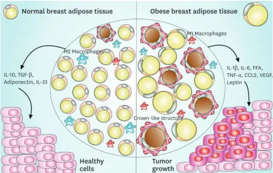
The presence of more CD68 and CD163 macrophage markers in breast adipose tissue is correlated with increased numbers of cancer-associated adipocytes in the breast cancer microenvironment [76,77], and these events are related to decreased survival in breast cancer patients [62,76,78]. These findings indicate that CLS can affect cancer development and impact breast cancer patients' overall survival (Figure 1).
Crown-like structure
M1 Macrophages
M2 Macrophages
Obese breast adipose tissue Normal breast adipose tissue
Healthy
cells Tumor
growth
IL-10, TGF-β, Adiponectin, IL-33
IL-1β, IL-6, FFA, TNF-α, CCL2, VEGF, Leptin
Figure 1. CLS-mediated signaling pathway in normal and obese adipose tissue and impact on breast cancer. Normal breast adipose tissue is characterized by the presence of smaller and less numerous white adipocytes, fewer crown-like structures, a prevalent M2 macrophages subset, with the secretion of anti- inflammatory cytokines such as IL-10, TGF-β, IL-33, and adiponectin. Obese breast adipose tissue is characterized by larger size and more abundant white adipocytes, with the secretion of pro-inflammatory cytokines such as IL-1β, IL-6, FFA, TNF-α, macrophages-chemoattractant CCL-2 chemokine, angiogenesis inducers such as VEGF, a prevalent M1 macrophages subset and an increased production of leptin, a negative feedback signal in the regulation of energy balance. This obese breast adipose tissue provides a favorable microenvironment to cancer establishment and progression.
CLS = crown-like structures; IL = interleukin; TGF = transforming growth factor; FFA = free fatty acid; TNF = tumor necrosis factor; CCL = chemokine (C-C motif) ligand; VEGF = vascular endothelial growth factor.
CLS are also correlated with race with regard to breast cancer prognosis. Iyengar et al. [59]
reported that menopausal Taiwanese women had pathologically enlarged adipocytes and increased presence of CLS in breast tissue, despite having a lower BMI than Caucasian women in the United States. In contrast, in Hispanic/Latin breast cancer survivors who underwent mastectomy, CLS were absent in 35% of patients with grade II and III obesity [79], indicating that a subset of patients with normal BMI and breast cancer can also present signs of inflammation and metabolic abnormalities (Table 1).
This healthier phenotype presented by some individuals with obesity is called metabolically healthy obesity, characterized by a lower degree of systemic inflammation, favorable
inflammatory and hormonal profiles, normal adipokine secretion patterns, and reduced levels of ectopic and visceral fat storage [80,81]. A subgroup of normal-weight individuals with abnormal metabolic parameters (those exhibiting metabolically unhealthy non-obesity or metabolically obese normal weight) has also been described [82]. However, metabolically healthy obese people may be more prone to the risk of obesity-associated cancer mortality [83,84].
CROWN-LIKE STRUCTURES AND BREAST CANCER TREATMENT
Since the presence of CLS in breast adipose tissue involves the modulation of several components such as proinflammatory immunological cells, proinflammatory cytokines,
Table 1. Clinical and experimental studies in breast cancer and crown-like structuresReference Model Target Results
Morris et al., 2011 [53] Human Aromatase activity; adipocyte size; serum
inflammation marker; CLS-B index CLS-B correlated with BMI and adipocyte size Subbaramaiah et al., 2011 [91] Animals NF-kB Activation; TNF-α, IL-1β, and COX-2
levels; aromatase activity; CLS Obesity-inflammation-aromatase axis in the mammary gland and WAT, and its association with CLS, increased risk of ER breast cancer Subbaramaiah et al., 2012 [75] Human Aromatase activity; CLS-B index CLS-B index as an improved correlation marker of breast cancer and
obesity Iyengar et al., 2015 [60] Human CLS-B presence; Circulating inflammatory
mediators CLS-B is associated with metabolic syndrome and worse breast cancer prognosis
Cowen et al., 2015 [64] Animal Effect of a high-fat/high-calorie diet on
mammary carcinogenesis Breast adipose tissue inflammation induced by diet increases tumor vascular density
Seo et al., 2015 [63] Animal; in vitro
and human Role of obesity in interstitial fibrosis in
mammary fat α-SMA levels correlated with CLS-B contribute to increased levels of matrix extracellular remodeling in obesity
Koru-Sengul et al., 2016 [62] Human Association between the number of TAM
and/or CLS and ethnicities Race is associated with the numbers of TAM and CLS in breast cancer
Mullooly et al., 2017 [89] Human CLS-B and sex steroid hormones in breast
adipose tissue CLS were not related to hormone levels or tumor characteristics, but were associated with hormone ratios
Vaysse et al., 2017 [90] Human CLS-B associated with mammary adipocyte size, body composition, and serum biomarkers
CLS was associated with systemic markers
Brown et al., 2017 [61] Human Effect of menopause on breast aromatase expression in relation to BMI, CLS and systemic markers of metabolic dysfunction
Postmenopausal women had higher BMI and presence of CLS than did premenopausal women
Cha et al., 2018 [76] Human Macrophage infiltration and identification of
CLS status CD68+ and/or CD163+ macrophages and CLS are present in adipose tissue near the breast cancer lesion
Iyengar et al., 2018 [59] Human CLS compared in Taiwanese vs Caucasian
women Compared with Caucasians, Taiwanese women had larger
breast adipocytes despite lower BMI after adjusting for BMI and menopausal status
Greenlee et al., 2019 [79] Human Inflammation and BMI in Hispanic/Latina
breast cancer patients Prevalence of CLS-B was associated with Hispanic/Latina patients Springer et al., 2019 [73] Human and
mice Extracellular matrix affects macrophage
phenotype Fibrotic extracellular matrix remodeling promotes a TAM phenotype of macrophages in CLS
CLS-B = crown-like structures in breast; BMI = body mass index; TNF-α = tumor necrosis factor α; IL = interleukin; COX-2 = cyclooxygenase-2; WAT = white adipose tissue; ER = estrogen receptor; α-SMA = α-smooth muscle actin; TAM = tumor-associated macrophage.
adipocyte cell death, release of free fatty acids, and different hormones, it is important to note that any anti-cancer therapy targeting CLS can be potentially mediated by all these parameters. Endocrine therapy is one treatment tool for breast cancer, as it blocks estrogenic activity and suppress adipose tissue aromatization of androgens to estrogens [85]. Although aromatase inhibitors reduce circulating estrogens in most cases, conditions of overweight and obesity in patients with breast cancer (premenopausal and postmenopausal) make patients more prone to a higher risk of recurrence and resistance to therapy [86]. Estrogen replacement therapy in ovariectomized mice with obesity revealed that treatment with 17β-estradiol is enough to attenuate weight gain, reduce production of inflammatory markers and number of CLS [87], and reduce the expression of genes related to inflammation (Cd68,
Mcp1, and Tnf) [88].The presence of CLS and inflammatory mediators in breast adipose tissue in women with both breast cancer and obesity is associated with changes in intracellular signaling and significant changes in cell dysfunction [30]. The CLS are implicated not only in inflammation, rather than in obesity alone, but also as drivers of aromatase activity in the breast via a complex and dynamic system of paracrine interactions between macrophages and other cells [53,75], a process also associated with increased estrogen-to-androgen ratios [89].
These findings underscore the role of CLS as a potential booster of estrogenic signaling and may be crucial for endocrine therapy selection during breast cancer treatment [90]. In vitro experiments further defined the pathways mediating this effect, showing that macrophage COX-2 expression and PGE
2production promote estrogen receptor (ERα) target gene expression [91]. Based on these results, post-menopausal patients with obesity and breast cancer may benefit from an aromatase inhibitor/COX-2 inhibitor combination treatment during breast cancer [92].
Supplementation of breast cancer patients with omega-3 fatty acids can also modulate CLS in breast cancer tissue and serve as an important adjuvant in the treatment of this cancer. There are multiple preclinical and epidemiologic studies suggesting that eicosapentaenoic acid (EPA) and docosahexaenoic acid (DHA) attenuate inflammation and reduce risks for breast cancer [93,94]. In vitro studies also corroborate the anti-tumor effects of omega-3 against human breast cancer cells [95,96]. In a study using rodents, omega-3 supplementation significantly decreased the number and size of CLS and F4/80+ macrophages and decreased expression of proinflammatory mediators including Ptgs2, IL-6, CCL2, TNF-α, NF-κB, and interferon-γ proteins in the mammary fat pad [97].
CLS can be also modulated by other compounds such as polyphenols (resveratrol) decreasing inflammation in breast tissue and potentially affecting breast cancer. Supplementation with resveratrol in the diet of mice with obesity and breast cancer resulted in lower numbers of CLS and decreased proinflammatory cytokine gene expression [66].
Since CLS can be also be identified in a significant proportion of normal-BMI women undergoing mastectomy for breast cancer risk reduction or therapy, CLS may be also important in breast cancer treatment in normal weight women [98], not only in obese breast cancer patients.
To date, at least three clinical trials have advanced in the investigation of adiposity and
inflammation to evaluate the risk factors associated with metabolic profile and tumor
growth, which influence disease-free survival, overall mortality, and breast-cancer-specific
mortality (NCT02240836; NCT03091842; NCT02598557). The forthcoming results of these studies should elucidate the relationship of CLS function to prognosis in different subtypes of breast cancer, providing new therapeutic approaches.
CONCLUSION
CLS are increased in breast adipose tissue from breast cancer patients and are particularly abundant in patients with conditions of obesity. Breast cancer associated with chronic obesity is most common in postmenopausal women. In this review, we discussed how CLS are related to the inflammation status of breast adipose tissue, and how these events can be decisive for breast cancer development and treatment, especially in obese women.
To improve interventions to prevent and treat breast cancer, a better understanding of the physiological and molecular mechanisms involved in breast tissue inflammation is crucial. It is important to understand that breast adipose tissue CLS are important keys to breast tissue inflammation and, consequently, may directly influence the development and treatment of breast cancer.
Targeting breast adipose tissue CLS can be a prominent therapeutic tool during cancer treatment, especially in patients with obesity. Therefore, tests based on body adipose tissue composition and inflammation in daily medical practice could be very effective to better stratify patients and direct combinatorial approaches in clinically relevant ways toward breast cancer treatment.
REFERENCES
1. Langin D. In and out: adipose tissue lipid turnover in obesity and dyslipidemia. Cell Metab 2011;14:569-70.
PUBMED | CROSSREF
2. Kyrgiou M, Kalliala I, Markozannes G, Gunter MJ, Paraskevaidis E, Gabra H, et al. Adiposity and cancer at major anatomical sites: umbrella review of the literature. BMJ 2017;356:j477.
PUBMED | CROSSREF
3. Jankovic N, Geelen A, Winkels RM, Mwungura B, Fedirko V, Jenab M, et al. Adherence to the WCRF/AICR dietary recommendations for cancer prevention and risk of cancer in elderly from Europe and the United States: a meta-analysis within the CHANCES project. Cancer Epidemiol Biomarkers Prev 2017;26:136-44.
PUBMED | CROSSREF
4. Choi B, Steiss D, Garcia-Rivas J, Kojaku S, Schnall P, Dobson M, et al. Comparison of body mass index with waist circumference and skinfold-based percent body fat in firefighters: adiposity classification and associations with cardiovascular disease risk factors. Int Arch Occup Environ Health 2016;89:435-48.
PUBMED | CROSSREF
5. González-Muniesa P, Mártinez-González MA, Hu FB, Després JP, Matsuzawa Y, Loos RJ, et al. Obesity.
Nat Rev Dis Primers 2017;3:17034.
PUBMED | CROSSREF
6. Parikh AM, Coletta AM, Yu ZH, Rauch GM, Cheung JP, Court LE, et al. Development and validation of a rapid and robust method to determine visceral adipose tissue volume using computed tomography images. PLoS One 2017;12:e0183515.
PUBMED | CROSSREF
7. Cinti S. The adipose organ. Prostaglandins Leukot Essent Fatty Acids 2005;73:9-15.
PUBMED | CROSSREF
8. Corrêa LH, Heyn GS, Magalhaes KGCorrêa. The impact of the adipose organ plasticity on inflammation and cancer progression. Cells 2019;8:E662.
PUBMED | CROSSREF
9. Corrêa LH, Corrêa R, Farinasso CM, de Sant'Ana Dourado LP, Magalhães KG. Adipocytes and macrophages interplay in the orchestration of tumor microenvironment: new implications in cancer progression. Front Immunol 2017;8:1129.
PUBMED | CROSSREF
10. Cinti S. The adipose organ: morphological perspectives of adipose tissues. Proc Nutr Soc 2001;60:319-28.
PUBMED | CROSSREF
11. Quail DF, Dannenberg AJ. The obese adipose tissue microenvironment in cancer development and progression. Nat Rev Endocrinol 2019;15:139-54.
PUBMED | CROSSREF
12. Goodwin PJ, Stambolic V. Impact of the obesity epidemic on cancer. Annu Rev Med 2015;66:281-96.
PUBMED | CROSSREF
13. Picon-Ruiz M, Morata-Tarifa C, Valle-Goffin JJ, Friedman ER, Slingerland JM. Obesity and adverse breast cancer risk and outcome: mechanistic insights and strategies for intervention. CA Cancer J Clin 2017;67:378-97.
PUBMED | CROSSREF
14. Ferlay J, Soerjomataram I, Dikshit R, Eser S, Mathers C, Rebelo M, et al. Cancer incidence and mortality worldwide: sources, methods and major patterns in GLOBOCAN 2012. Int J Cancer 2015;136:E359-86.
PUBMED | CROSSREF
15. Bray F, Ferlay J, Soerjomataram I, Siegel RL, Torre LA, Jemal A. Global cancer statistics 2018: GLOBOCAN estimates of incidence and mortality worldwide for 36 cancers in 185 countries. CA Cancer J Clin 2018;68:394-424.
PUBMED | CROSSREF
16. Coughlin SS. Epidemiology of breast cancer in women. 2019. http://link.springer.com/10.1007/978-3-030- 20301-6_2.
17. Onitilo AA, Engel JM, Greenlee RT, Mukesh BN. Breast cancer subtypes based on ER/PR and Her2 expression: comparison of clinicopathologic features and survival. Clin Med Res 2009;7:4-13.
PUBMED | CROSSREF
18. Geyer FC, Pareja F, Weigelt B, Rakha E, Ellis IO, Schnitt SJ, et al. The spectrum of triple-negative breast disease. Am J Pathol 2017;187:2139-51.
PUBMED | CROSSREF
19. Soguel L, Durocher F, Tchernof A, Diorio C. Adiposity, breast density, and breast cancer risk:
epidemiological and biological considerations. Eur J Cancer Prev 2017;26:511-20.
PUBMED | CROSSREF
20. Cinti S. Adipose organ development and remodeling. Compr Physiol 2018;8:1357-431.
PUBMED | CROSSREF
21. Scherer PE. The multifaceted roles of adipose tissue-therapeutic targets for diabetes and beyond: the 2015 banting lecture. Diabetes 2016;65:1452-61.
PUBMED | CROSSREF
22. Nieman KM, Romero IL, Van Houten B, Lengyel E. Adipose tissue and adipocytes support tumorigenesis and metastasis. Biochim Biophys Acta 2013;1831:1533-41.
PUBMED | CROSSREF
23. Blücher C, Stadler SC. Obesity and breast cancer: current insights on the role of fatty acids and lipid metabolism in promoting breast cancer growth and progression. Front Endocrinol (Lausanne) 2017;8:293.
PUBMED | CROSSREF
24. Xu H, Barnes GT, Yang Q, Tan G, Yang D, Chou CJ, et al. Chronic inflammation in fat plays a crucial role in the development of obesity-related insulin resistance. J Clin Invest 2003;112:1821-30.
PUBMED | CROSSREF
25. Surmi BK, Hasty AH. Macrophage infiltration into adipose tissue: initiation, propagation and remodeling. Future Lipidol 2008;3:545-56.
PUBMED | CROSSREF
26. Cinti S, Mitchell G, Barbatelli G, Murano I, Ceresi E, Faloia E, et al. Adipocyte death defines macrophage localization and function in adipose tissue of obese mice and humans. J Lipid Res 2005;46:2347-55.
PUBMED | CROSSREF
27. Murano I, Barbatelli G, Parisani V, Latini C, Muzzonigro G, Castellucci M, et al. Dead adipocytes, detected as crown-like structures, are prevalent in visceral fat depots of genetically obese mice. J Lipid Res 2008;49:1562-8.
PUBMED | CROSSREF
28. Weisberg SP, McCann D, Desai M, Rosenbaum M, Leibel RL, Ferrante AW Jr. Obesity is associated with macrophage accumulation in adipose tissue. J Clin Invest 2003;112:1796-808.
PUBMED | CROSSREF
29. Crewe C, An YA, Scherer PE. The ominous triad of adipose tissue dysfunction: inflammation, fibrosis, and impaired angiogenesis. J Clin Invest 2017;127:74-82.
PUBMED | CROSSREF
30. Iyengar NM, Hudis CA, Dannenberg AJ. Obesity and inflammation: new insights into breast cancer development and progression. Am Soc Clin Oncol Educ Book 2013;33:46-51.
PUBMED | CROSSREF
31. Reilly SM, Saltiel AR. Adapting to obesity with adipose tissue inflammation. Nat Rev Endocrinol 2017;13:633-43.
PUBMED | CROSSREF
32. Saltiel AR, Olefsky JM, Saltiel AR, Olefsky JM. Inflammatory mechanisms linking obesity and metabolic disease. J Clin Invest 2017;127:1-4.
PUBMED | CROSSREF
33. Henninger AM, Eliasson B, Jenndahl LE, Hammarstedt A. Adipocyte hypertrophy, inflammation and fibrosis characterize subcutaneous adipose tissue of healthy, non-obese subjects predisposed to type 2 diabetes. PLoS One 2014;9:e105262.
PUBMED | CROSSREF
34. Itoh M, Suganami T, Hachiya R, Ogawa Y. Adipose tissue remodeling as homeostatic inflammation. Int J Inflamm 2011;2011:720926.
PUBMED | CROSSREF
35. Shapouri-Moghaddam A, Mohammadian S, Vazini H, Taghadosi M, Esmaeili SA, Mardani F, et al.
Macrophage plasticity, polarization, and function in health and disease. J Cell Physiol 2018;233:6425-40.
PUBMED | CROSSREF
36. Castoldi A, Naffah de Souza C, Câmara NO, Moraes-Vieira PM. The macrophage switch in obesity development. Front Immunol 2016;6:637.
PUBMED | CROSSREF
37. Mantovani A, Sica A. Macrophages, innate immunity and cancer: balance, tolerance, and diversity. Curr Opin Immunol 2010;22:231-7.
PUBMED | CROSSREF
38. Hubler MJ, Peterson KR, Hasty AH. Iron homeostasis: a new job for macrophages in adipose tissue?
Trends Endocrinol Metab 2015;26:101-9.
PUBMED | CROSSREF
39. Lumeng CN, DelProposto JB, Westcott DJ, Saltiel AR. Phenotypic switching of adipose tissue macrophages with obesity is generated by spatiotemporal differences in macrophage subtypes. Diabetes 2008;57:3239-46.
PUBMED | CROSSREF
40. Lumeng CN, Bodzin JL, Saltiel AR. Obesity induces a phenotypic switch in adipose tissue macrophage polarization. J Clin Invest 2007;117:175-84.
PUBMED | CROSSREF
41. Kang YE, Kim JM, Joung KH, Lee JH, You BR, Choi MJ, et al. The roles of adipokines, proinflammatory cytokines, and adipose tissue macrophages in obesity-associated insulin resistance in modest obesity and early metabolic dysfunction. PLoS One 2016;11:e0154003.
PUBMED | CROSSREF
42. Murano I, Rutkowski JM, Wang QA, Cho YR, Scherer PE, Cinti S. Time course of histomorphological changes in adipose tissue upon acute lipoatrophy. Nutr Metab Cardiovasc Dis 2013;23:723-31.
PUBMED | CROSSREF
43. Giordano A, Murano I, Mondini E, Perugini J, Smorlesi A, Severi I, et al. Obese adipocytes show ultrastructural features of stressed cells and die of pyroptosis. J Lipid Res 2013;54:2423-36.
PUBMED | CROSSREF
44. Cildir G, Akıncılar SC, Tergaonkar V. Chronic adipose tissue inflammation: all immune cells on the stage.
Trends Mol Med 2013;19:487-500.
PUBMED | CROSSREF
45. Hotamisligil GS, Murray DL, Choy LN, Spiegelman BM. Tumor necrosis factor α inhibits signaling from the insulin receptor. Proc Natl Acad Sci U S A 1994;91:4854-8.
PUBMED | CROSSREF
46. Shaul ME, Bennett G, Strissel KJ, Greenberg AS, Obin MS. Dynamic, M2-like remodeling phenotypes of CD11c+ adipose tissue macrophages during high-fat diet--induced obesity in mice. Diabetes 2010;59:1171-81.
PUBMED | CROSSREF
47. Kratz M, Coats BR, Hisert KB, Hagman D, Mutskov V, Peris E, et al. Metabolic dysfunction drives a mechanistically distinct proinflammatory phenotype in adipose tissue macrophages. Cell Metab 2014;20:614-25.
PUBMED | CROSSREF
48. Xu X, Grijalva A, Skowronski A, van Eijk M, Serlie MJ, Ferrante AW Jr. Obesity activates a program of lysosomal-dependent lipid metabolism in adipose tissue macrophages independently of classic activation. Cell Metab 2013;18:816-30.
PUBMED | CROSSREF
49. Tiwari P, Blank A, Cui C, Schoenfelt KQ, Zhou G, Xu Y, et al. Metabolically activated adipose tissue macrophages link obesity to triple-negative breast cancer. J Exp Med 2019;216:1345-58.
PUBMED | CROSSREF
50. Jung UJ, Choi MS. Obesity and its metabolic complications: the role of adipokines and the relationship between obesity, inflammation, insulin resistance, dyslipidemia and nonalcoholic fatty liver disease. Int J Mol Sci 2014;15:6184-223.
PUBMED | CROSSREF
51. Iyengar NM, Gucalp A, Dannenberg AJ, Hudis CA. Obesity and cancer mechanisms: tumor microenvironment and inflammation. J Clin Oncol 2016;34:4270-6.
PUBMED | CROSSREF
52. Howe LR, Subbaramaiah K, Hudis CA, Dannenberg AJ. Molecular pathways: adipose inflammation as a mediator of obesity-associated cancer. Clin Cancer Res 2013;19:6074-83.
PUBMED | CROSSREF
53. Morris PG, Hudis CA, Giri D, Morrow M, Falcone DJ, Zhou XK, et al. Inflammation and increased aromatase expression occur in the breast tissue of obese women with breast cancer. Cancer Prev Res (Phila) 2011;4:1021-9.
PUBMED | CROSSREF
54. Lees T, Cullinane A, Condon A, Shabaan AM, Humphries MP, Speirs V. Characterising the adipose- inflammatory microenvironment in male breast cancer. Endocr Relat Cancer 2018;25:773-81.
PUBMED | CROSSREF
55. Khan T, Muise ES, Iyengar P, Wang ZV, Chandalia M, Abate N, et al. Metabolic dysregulation and adipose tissue fibrosis: role of collagen VI. Mol Cell Biol 2009;29:1575-91.
PUBMED | CROSSREF
56. Berstein LM, Iyevleva AG, Mukhina MS, Vasilyev DA, Poroshina TE. Features of omental adipose tissue in endometrial cancer patients with ‘standard’ or ‘metabolically healthy’ obesity: associations with tumor process characteristics. Springerplus 2016;5:1900.
PUBMED | CROSSREF
57. Miyazawa M, Subbaramaiah K, Bhardwaj P, Zhou XK, Wang H, Falcone DJ, et al. Pioglitazone inhibits periprostatic white adipose tissue inflammation in obese mice. Cancer Prev Res (Phila) 2018;11:215-26.
PUBMED | CROSSREF
58. Lade A, Noon LA, Friedman SL. Contributions of metabolic dysregulation and inflammation to nonalcoholic steatohepatitis, hepatic fibrosis, and cancer. Curr Opin Oncol 2014;26:100-7.
PUBMED | CROSSREF
59. Iyengar NM, Chen IC, Zhou XK, Giri DD, Falcone DJ, Winston LA, et al. Adiposity, inflammation, and breast cancer pathogenesis in Asian women. Cancer Prev Res (Phila) 2018;11:227-36.
PUBMED | CROSSREF
60. Iyengar NM, Morris PG, Zhou XK, Gucalp A, Giri D, Harbus MD, et al. Menopause is a determinant of breast adipose inflammation. Cancer Prev Res (Phila) 2015;8:349-58.
PUBMED | CROSSREF
61. Brown KA, Iyengar NM, Zhou XK, Gucalp A, Subbaramaiah K, Wang H, et al. Menopause is a determinant of breast aromatase expression and its associations with BMI, inflammation, and systemic markers. J Clin Endocrinol Metab 2017;102:1692-701.
PUBMED | CROSSREF
62. Koru-Sengul T, Santander AM, Miao F, Sanchez LG, Jorda M, Glück S, et al. Breast cancers from black women exhibit higher numbers of immunosuppressive macrophages with proliferative activity and of crown-like structures associated with lower survival compared to non-black Latinas and Caucasians.
Breast Cancer Res Treat 2016;158:113-26.
PUBMED | CROSSREF
63. Seo BR, Bhardwaj P, Choi S, Gonzalez J, Andresen Eguiluz RC, Wang K, et al. Obesity-dependent changes in interstitial ECM mechanics promote breast tumorigenesis. Sci Transl Med 2015;7:301ra130.
PUBMED | CROSSREF
64. Cowen S, McLaughlin SL, Hobbs G, Coad J, Martin KH, Olfert IM, et al. High-fat, high-calorie diet enhances mammary carcinogenesis and local inflammation in MMTV-PyMT mouse model of breast cancer. Cancers (Basel) 2015;7:1125-42.
PUBMED | CROSSREF
65. Giles ED, Wellberg EA, Astling DP, Anderson SM, Thor AD, Jindal S, et al. Obesity and overfeeding affecting both tumor and systemic metabolism activates the progesterone receptor to contribute to postmenopausal breast cancer. Cancer Res 2012;72:6490-501.
PUBMED | CROSSREF
66. Rossi EL, Khatib SA, Doerstling SS, Bowers LW, Pruski M, Ford NA, et al. Resveratrol inhibits obesity- associated adipose tissue dysfunction and tumor growth in a mouse model of postmenopausal claudin- low breast cancer. Mol Carcinog 2018;57:393-407.
PUBMED | CROSSREF
67. Lewis CE, Pollard JW. Distinct role of macrophages in different tumor microenvironments. Cancer Res 2006;66:605-12.
PUBMED | CROSSREF
68. Medrek C, Pontén F, Jirström K, Leandersson K. The presence of tumor associated macrophages in tumor stroma as a prognostic marker for breast cancer patients. BMC Cancer 2012;12:306.
PUBMED | CROSSREF
69. Sousa S, Brion R, Lintunen M, Kronqvist P, Sandholm J, Mönkkönen J, et al. Human breast cancer cells educate macrophages toward the M2 activation status. Breast Cancer Res 2015;17:101.
PUBMED | CROSSREF
70. Noy R, Pollard JW. Tumor-associated macrophages: from mechanisms to therapy. Immunity 2014;41:49-61.
PUBMED | CROSSREF
71. Arwert EN, Harney AS, Entenberg D, Wang Y, Sahai E, Pollard JW, et al. A unidirectional transition from migratory to perivascular macrophage is required for tumor cell intravasation. Cell Rep 2018;23:1239-48.
PUBMED | CROSSREF
72. McWhorter FY, Wang T, Nguyen P, Chung T, Liu WF. Modulation of macrophage phenotype by cell shape.
Proc Natl Acad Sci U S A 2013;110:17253-8.
PUBMED | CROSSREF
73. Springer NL, Iyengar NM, Bareja R, Verma A, Jochelson MS, Giri DD, et al. Obesity-associated extracellular matrix remodeling promotes a macrophage phenotype similar to tumor-associated macrophages. Am J Pathol 2019;189:2019-35.
PUBMED | CROSSREF
74. Hotamisligil GS. Inflammation, metaflammation and immunometabolic disorders. Nature 2017;542:177-85.
PUBMED | CROSSREF
75. Subbaramaiah K, Morris PG, Zhou XK, Morrow M, Du B, Giri D, et al. Increased levels of COX-2 and prostaglandin E2 contribute to elevated aromatase expression in inflamed breast tissue of obese women.
Cancer Discov 2012;2:356-65.
PUBMED | CROSSREF
76. Cha YJ, Kim ES, Koo JS. Tumor-associated macrophages and crown-like structures in adipose tissue in breast cancer. Breast Cancer Res Treat 2018;170:15-25.
PUBMED | CROSSREF
77. Cha YJ, Koo JS. Expression of autotaxin-lysophosphatidate signaling-related proteins in breast cancer with adipose stroma. Int J Mol Sci 2019;20:20.
CROSSREF
78. Quail DF, Olson OC, Bhardwaj P, Walsh LA, Akkari L, Quick ML, et al. Obesity alters the lung myeloid cell landscape to enhance breast cancer metastasis through IL5 and GM-CSF. Nat Cell Biol 2017;19:974-87.
PUBMED | CROSSREF
79. Greenlee H, Shi Z, Hibshoosh H, Giri DD, Ahmed A, Williams S, et al. Obesity-associated breast inflammation among Hispanic/Latina breast cancer patients. Cancer Prev Res (Phila) 2019;12:21-30.
PUBMED | CROSSREF
80. Phillips CM. Metabolically healthy obesity: definitions, determinants and clinical implications. Rev Endocr Metab Disord 2013;14:219-27.
PUBMED | CROSSREF
81. Stefan N, Häring HU, Hu FB, Schulze MB. Metabolically healthy obesity: epidemiology, mechanisms, and clinical implications. Lancet Diabetes Endocrinol 2013;1:152-62.
PUBMED | CROSSREF
82. Samocha-Bonet D, Dixit VD, Kahn CR, Leibel RL, Lin X, Nieuwdorp M, et al. Metabolically healthy and unhealthy obese--the 2013 Stock Conference report. Obes Rev 2014;15:697-708.
PUBMED | CROSSREF
83. Oh CM, Jun JK, Suh M. Risk of cancer mortality according to the metabolic health status and degree of obesity. Asian Pac J Cancer Prev 2014;15:10027-31.
PUBMED | CROSSREF
84. Blüher M. Are metabolically healthy obese individuals really healthy? Eur J Endocrinol 2014;171:R209-19.
PUBMED | CROSSREF
85. Cleary MP, Grossmann ME. Minireview: obesity and breast cancer: the estrogen connection.
Endocrinology 2009;150:2537-42.
PUBMED | CROSSREF
86. Sini V, Lunardi G, Cirillo M, Turazza M, Bighin C, Giraudi S, et al. Body mass index and circulating oestrone sulphate in women treated with adjuvant letrozole. Br J Cancer 2014;110:1133-8.
PUBMED | CROSSREF
87. Bhardwaj P, Du B, Zhou XK, Sue E, Giri D, Harbus MD, et al. Estrogen protects against obesity-induced mammary gland inflammation in mice. Cancer Prev Res (Phila) 2015;8:751-9.
PUBMED | CROSSREF
88. Bhardwaj P, Ikeda T, Zhou XK, Wang H, Zheng XE, Giri DD, et al. Supplemental estrogen and caloric restriction reduce obesity-induced periprostatic white adipose inflammation in mice. Carcinogenesis 2019;40:914-23.
PUBMED | CROSSREF
89. Mullooly M, Yang HP, Falk RT, Nyante SJ, Cora R, Pfeiffer RM, et al. Relationship between crown-like structures and sex-steroid hormones in breast adipose tissue and serum among postmenopausal breast cancer patients. Breast Cancer Res 2017;19:8.
PUBMED | CROSSREF
90. Vaysse C, Lømo J, Garred Ø, Fjeldheim F, Lofteroed T, Schlichting E, et al. Inflammation of mammary adipose tissue occurs in overweight and obese patients exhibiting early-stage breast cancer. NPJ Breast Cancer 2017;3:19.
PUBMED | CROSSREF
91. Subbaramaiah K, Howe LR, Bhardwaj P, Du B, Gravaghi C, Yantiss RK, et al. Obesity is associated with inflammation and elevated aromatase expression in the mouse mammary gland. Cancer Prev Res (Phila) 2011;4:329-46.
PUBMED | CROSSREF
92. Bowers LW, Brenner AJ, Hursting SD, Tekmal RR, deGraffenried LA. Obesity-associated systemic interleukin-6 promotes pre-adipocyte aromatase expression via increased breast cancer cell prostaglandin E2 production. Breast Cancer Res Treat 2015;149:49-57.
PUBMED | CROSSREF
93. Paixão EM, Oliveira AC, Pizato N, Muniz-Junqueira MI, Magalhães KG, Nakano EY, et al. The effects of EPA and DHA enriched fish oil on nutritional and immunological markers of treatment naïve breast cancer patients: a randomized double-blind controlled trial. Nutr J 2017;16:71.
PUBMED | CROSSREF
94. Fabian CJ, Kimler BF, Phillips TA, Box JA, Kreutzjans AL, Carlson SE, et al. Modulation of breast cancer risk biomarkers by high-dose omega-3 fatty acids: phase II pilot study in premenopausal women. Cancer Prev Res (Phila) 2015;8:912-21.
PUBMED | CROSSREF
95. Pizato N, Luzete BC, Kiffer LF, Corrêa LH, de Oliveira Santos I, Assumpção JA, et al. Omega-3 docosahexaenoic acid induces pyroptosis cell death in triple-negative breast cancer cells. Sci Rep 2018;8:1952.
PUBMED | CROSSREF
96. Pizato N, Kiffer LF, Luzete BC, Assumpção JA, Correa LH, Melo HA, et al. Omega 3-DHA and Delta- Tocotrienol modulate lipid droplet biogenesis and lipophagy in breast cancer cells: the impact in cancer aggressiveness. Nutrients 2019;11:1199.
PUBMED | CROSSREF
97. Khadge S, Thiele GM, Sharp JG, McGuire TR, Klassen LW, Black PN, et al. Long-chain omega-3
polyunsaturated fatty acids modulate mammary gland composition and inflammation. J Mammary Gland Biol Neoplasia 2018;23:43-58.
PUBMED | CROSSREF
98. Iyengar NM, Brown KA, Zhou XK, Gucalp A, Subbaramaiah K, Giri DD, et al. Metabolic obesity, adipose inflammation and elevated breast aromatase in women with normal body mass index. Cancer Prev Res (Phila) 2017;10:235-43.
PUBMED | CROSSREF