RESEARCH ARTICLE
Received: April 24, 2019, Revised: May 20, 2019, Accepted: May 22, 2019 eISSN 2233-7679
†Correspondence to: Soon-Jeong Jeong, https://orcid.org/0000-0002-8959-4663
Department of Dental Hygiene, College of Health Science, Youngsan University, 288 Junam-ro, Yangsan 50510, Korea Tel: +82-55-380-9453, Fax: +82-55-380-9305, E-mail: [email protected]
Copyright © The Korean Society of Dental Hygiene Science.
Inflammatory Effect of Light-Emitting Diodes Curing Light Irradiation on Raw264.7 Macrophage
Moon-Jin Jeong
1, Ki-Sung Kil
1, Myoung-Hwa Lee
1, Seung-Yeon Lee
1, Hye-Jin Lee
2, Do-Seon Lim
3, and Soon-Jeong Jeong
4,†1
Department of Oral Histology and Developmental Biology, School of Dentistry, Chosun University, Gwangju 61452,
2
Department of Dental Hygiene, Dong-Pusan College, Busan 48000,
3
Department of Dental Hygiene, Graduate School of Public Health Science, Eulji University, Seongnam 13135,
4
Department of Dental Hygiene, College of Health Science, Youngsan University, Yangsan 50510, Korea
Background: The light-emitting diode (LED) curing light used is presumed to be safe. However, the scientific basis for this is unclear, and the safety
of LED curing light is still controversial. The purpose of this study was to investigate the effect of LED curing light irradiation according to the conditions applied for the polymerization of composite resins in dental clinic on the cell viability and inflammatory response in Raw264.7 macrophages and to confirm the stability of LED curing light.
Methods: Cell viability and cell morphology of Raw264.7 macrophages treated with 100 ng/ml of lipopolysaccharide (LPS) or/and LED curing light
with a wavelength of 440∼490 nm for 20 seconds were confirmed by methylthiazolydiphenyl-tetrazolium bromide assay and microscopic observation. The production of nitric oxide (NO) and prostaglandin E
2(PGE
2) was confirmed by NO assay and PGE
2enzyme-linked immunosorbent assay kit. Expression of interleukin (IL)-1 and tumor necrosis factor (TNF)- in total RNA and protein was confirmed by reverse transcription polymerase chain reaction and Western blot analysis.
Results:
The LED curing light did not affect the viability and morphology of normal Raw264.7 cells but affected the cell viability and induced cytotoxicity in the inflammation-induced Raw264.7 cells by LPS. The irradiation of the LED curing light did not progress to the inflammatory state in the inflammation-induced Raw264.7 macrophage. However, LED curing light irradiation in normal Raw264.7 cells induced an increase in NO and PGE
2production and mRNA and protein expression of IL-1 and TNF-, indicating that it is possible to induce the inflammatory state.
Conclusion:
The irradiation of LED curing light in RAW264.7 macrophage may induce an excessive inflammatory reaction and damage oral tissues.
Therefore, it is necessary to limit the long-term irradiation which is inappropriate when applying LED curing light in a dental clinic.
Key Words: Inflammatory effect, Light-emitting diode, Light curing light, Macrophage, Raw264.7 cell
Introduction
The light-emitting diode (LED), introduced in the 1990s, consists of a combination of two semiconductors which effectively convert electricity into light
1). The LED has also been developed as light sources for light curing units for the polymerization of composite resins used for tooth restoration
1,2). LED light is a blue light with a wavelength of 420∼600 nm with a semi-permanent life span of 10,000 hours or more
3,4)and is widely used in
dental clinic because of its low cost and strong resistance
to vibration and impact
5,6). Although the LED curing light
used for polymerization of composite resins is presumed
to be safe, the scientific basis for this is insufficient and
unclear
7,8). In addition, there is some evidence that it directly
or indirectly affects oral tissues (including non-dental and
dental), and the safety of LED curing light is still
controversial
7,8). It has also been reported that the effect of
the LED curing light differs depending on the time and
intensity of light irradiation and the cells and tissues
Table 1. Light Curing Unit
Light source Wave length Exposure time Dose of light Company Model no.
LED 440∼490 nm 20 s 20 J/cm
2Dematec Co., Ltd., Busan, Korea Skylight
LED: light-emitting diode.
examined
7-9). It is therefore pertinent to explore the effects of LED curing light on non-dental and dental tissue.
Macrophages overexpress pro-inflammatory mediators such as reactive oxygen species, nitric oxide (NO) and prostaglandin E
2(PGE
2) and pro-inflammatory cytokines such as interleukin (IL)-1, IL-6, IL-10, and tumor necrosis factor (TNF)- when exposed to toxic substances
10,11). Over-expressed pro-inflammatory mediators and pro-inflammatory cytokines play an important role in the induction of inflammatory diseases
10-12). Periodontal disease is also an inflammatory disease caused by the interaction of leukocytes such as macrophages and certain pathogenic microorganisms living in the oral cavity
13-15). Therefore, the Raw264.7 macrophage is an ideal cell line to study the effect of LED curing light in the oral cavity.
The purpose of this study was to investigate the effect of LED curing light irradiation according to the conditions applied to polymerize of composite resins in dental clinics on the cell viability and inflammatory response in Raw264.7 macrophages and to confirm the stability of LED curing light.
Materials and Methods
1. Cell culture
The RAW264.7 macrophage were purchased from the Korean Cell Line Bank (KCLB, Seoul, Korea). The cells were stabilized and cultured in Dulbecco’s modified Eagle’s medium (DMEM; Gibco-BRL, Seoul, Korea) in a 37
oC 5% CO
2incubator. A 10% fetal bovine serum (Gibco-BRL) and 1% antibiotic-antimycotic solution (Gibco-BRL) were added to the medium. Cells were divided into control, lipopolysaccharide (LPS) only treated LPS+ group, LED only irradiated LED+ group, and LPS+/LED+ group treated with LPS treatment and LED irradiation.
2. Lipopolysaccharide treatment and light-emitting diode curing light irradiation
The prepared cells were replaced with fresh medium, treated with 100 ng/ml of LPS or/and irradiated with LED curing light for 20 seconds according to the conditions applied for the polymerization of composite resins in dental clinics and then replaced with fresh medium. The light curing unit used in this experiment has a wavelength of 440∼490 nm and the LED is the right source (Table 1).
The treated cells were incubated for 0, 24, and 48 hours and then used for experiments.
3. Methylthiazolydiphenyl-tetrazolium bromide assay The effect of LED curing light irradiation on cell viability was evaluated by methylthiazolydiphenyl- tetrazolium bromide (MTT) assay. Prepared cells were treated with 0.5 mg/ml of MTT (Sigma-Aldrich Chemical Co., St. Louis, MO, USA) and reacted in a dark incubator for 2 hours. After removal of MTT from the cell, the cells were treated with dimethyl sulfoxide (DMSO; Sigma- Aldrich Chemical Co.), and 200 l of the DMSO-treated solution was transferred to a 96-well plate, and the absorbance was read at 540 nm using an enzyme-linked immunosorbent assay (ELISA) reader (Molecular Devices, Sunnyvale, CA, USA) was measured.
4. Microscopic examination
The morphological changes of cells by LED curing light irradiation were observed with an inverted microscope (Olympus, Tokyo, Japan). Prepared cells were fixed with 2.5% glutaraldehyde (MERCK, Frankfurter, Germany) in phosphate buffered saline (PBS) (pH 7.4), washed three times with PBS, and the morphology of cell was observed.
5. Nitric oxide assay
The changes in a pro-inflammatory mediator, NO by
LED curing light irradiation were measured using NO
assay kit (R&D Systems, Minneapolis, MN, USA). The
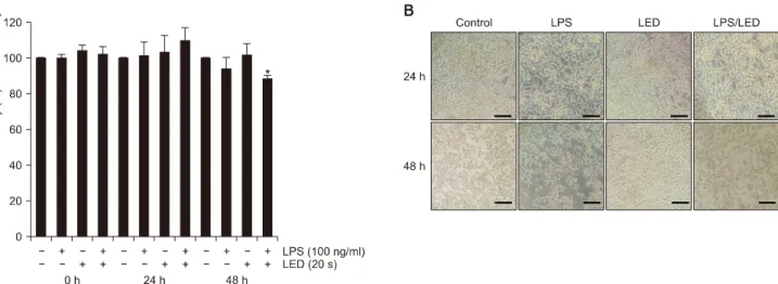
Fig. 1. Cell viability of Raw264.7 macrophages irradiated with LED curing light. (A) Cell viability of Raw264.7 macrophages treated with 100 ng/ml of LPS or/and LED curing light irradiation with wavelength of 440∼490 nm for 20 seconds were confirmed by MTT assay. The experiment was repeated three times and performed independently, and the results were expressed as mean and standard deviation. (B) Morphological changes of prepared Raw264.7 cells were observed by inverted microscope. All scale bars indicate 60
m. LED: light-emitting diode, LPS: lipopolysaccharide, MTT: methylthiazolydiphenyl-tetrazolium bromide. *p<0.05.
supernatants of the cells treated with the reagent according to the manufacturer’s method were transferred to a 96-well plate and measured at 540 nm absorbance using the ELISA reader.
6. Prostaglandin E
2enzyme-linked immunosorbent assay
The concentration of PGE
2, which is a pro-inflammatory mediator that plays an important role in the initiation of inflammatory responses, was measured using a PGE
2ELISA kit (R&D Systems). The supernatants of the prepared cells were treated according to the manu- facturer’s method and the absorbance at 490 nm was measured using an ELISA reader to determine the PGE
2concentration.
7. RNA extraction and reverse transcription polymerase chain reaction analysis
Total RNA was isolated using TRIzol reagent (Molecular Research Center Inc., Cincinnati, OH, USA) according to the manufacturer's instructions. Isolated total RNA was reverse transcribed into complementary DNA (cDNA). Specific primers (Bioneer Co., Ltd., Daejeon, Korea) were used for reverse transcription polymerase chain reaction (RT-PCR). The desired genes were amplified according to the Jeong et al.
16)and were then
electrophoresed on 1.5% agarose gel. The bands were visualized by Gel-Doc System (BioRad Laboratories Inc., Hercules, CA, USA) and measured by Science Lab Image Guage (Fujifilm, Tokyo, Japan).
8. Protein extraction and Western blot analysis According to the method of Jeong et al.
16), total protein was separated, the concentration of the protein was measured, prepared total protein was electrophoresed on 10% SDS-polyacrylamide gel and then western blot and immunoprecipitation were performed on specific protein.
The intensity of the band detected with ECL solution (Merck Millipore, Burlington, MA, USA) was measured by Science Lab Image Guage (Fujifilm).
9. Statistical analysis
Using Excel 2010 statistical software (Microsoft, Redmond, WA, USA), the mean, standard deviation, p-values (*p<0.05, **p<0.01, ***p<0.001) of the results from three replica experiments were calculated.
Significant differences were analyzed using the Student’s
t-test.
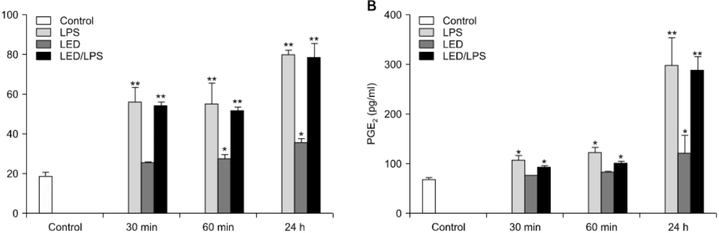
Fig. 2. NO and PGE2 production of Raw264.7 macrophages irradiated with LED curing light. Raw264.7 macrophage were irradiated with LED curing light for 20 seconds or/and treated with 100 ng/ml LPS. The production of NO (A) and PGE2 (B) in the prepared Raw264.7 macrophage was confirmed by NO assay and PGE2 ELISA kit. All experiments were carried out three times independently and their values were expressed as mean and standard deviation. NO: nitric oxide, PGE2: prostaglandin E2, LED: light-emitting diode, LPS: lipopolysaccharide, ELISA: enzyme-linked immunosorbent assay. *p<0.05 and **p<0.01 compared with the control group.
Results
1. Cell viability of Raw264.7 macrophages irradiated with light-emitting diode curing light
Cell viability and cytotoxic effect of Raw264.7 macrophages treated with 100 ng/ml of LPS or/and LED curing light with a wavelength of 440∼490 nm for 20 seconds of general applying the composite resin for polymerization in dental clinics were confirmed by MTT assay. There was no significant change in cell viability of all groups, except for 48 hours LPS+/LED+ group, a significant decrease was seen compared to the control (p<
0.05) (Fig. 1A). This suggests that although the LED curing light did not affect the cell viability and cytotoxicity of normal Raw264.7 macrophages, LPS- induced inflammation in Raw264.7 cells may decrease cell viability and may have a cytotoxic effect. Microscopic observations of Raw264.7 cells revealed that the LPS+
and LPS+/LED+ group at 24 hours and 48 hours had an extended cytoplasmic process resulting from LPS stimulation, but no change in cell morphology was observed in the LED+ group (Fig. 1B). Therefore, the microscopic observation of the Raw264.7 cell morphology showed no effect of the LED curing light.
2. Nitric oxide and prostaglandin E
2production of Raw264.7 macrophages irradiated with light- emitting diode curing light
The production of NO and PGE
2in the prepared Raw264.7 macrophage was confirmed by NO assay and PGE
2ELISA kit (Fig. 2). Significant increase in NO was observed in the LPS+ and LPS+/LED+ group with increasing time (p<0.01) and the amount of NO production in both groups was similar (Fig. 2A). The increase in NO production in both groups was thought to be due to LPS treatment and the effect of LED irradiation was not confirmed. In the case of NO of LED+ group, NO production increased with increasing time, and at 60 minutes and 24 hours, the increase was significant compared with the control group (Fig. 2A) (p<0.05).
These results imply that LED curing light irradiation can affect the production of NO in normal Raw264.7 macrophage. PGE
2also increased significantly in LPS+
and LPS+/LED+ groups with time compared to control.
The amount of PGE
2production was similar in both groups, but the value of LPS+/LED+ group was lower than that of LPS+ group (p<0.05, p<0.01) (Fig. 1B).
PGE
2production in both groups was also considered to be
due to LPS, and the effect of LED curing light irradiation
was not confirmed. A significant increase in PGE
2was
also observed in the LED+ group after 24 hours, which
suggests that LED irradiation could affect the production
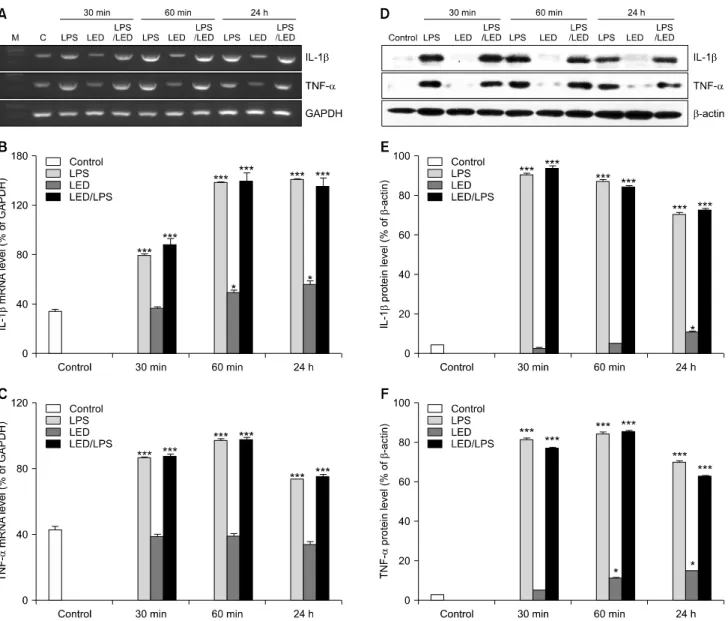
Fig. 3. IL-1 and TNF- expression of Raw264.7 macrophages irradiated with LED curing light. Total RNA and protein were extracted from Raw264.7 macrophages treated with LPS (100 ng/ml) or/and irradiated with LED curing light for 20 seconds and RT-PCR (A∼C) and Western blot analysis (D∼F) of IL-1 and TNF- were performed. Values are represented the mean±standard deviation of results obtained in three independent experiments. IL: interleukin, TNF: tumor necrosis factor, LED: light-emitting diode, LPS: lip- opolysaccharide, RT-PCR: reverse transcription polymerase chain reaction, GAPDH: glyceraldehyde 3-phosphate dehydrogenase.
*p<0.05 and ***p<0.001 compared with the control group.