• 검색 결과가 없습니다.

The Effect of Topiramate on Status Epilepticus- Induced Neurotoxicity in Immature Mouse Brain

N/A
N/A
Protected

Academic year: 2021

Share "The Effect of Topiramate on Status Epilepticus- Induced Neurotoxicity in Immature Mouse Brain"

Copied!
14
0
0

로드 중.... (전체 텍스트 보기)

전체 글

(1)

□ 원 저 □ Vol. 14, No. 2, November, 2006

1)

책임저자 : 황규근, 동아대학교 의과대학 소아과학교실 Tel : 051)240-2958, Fax : 051)242-2765 E-mail : [email protected]

Introduction

Status epilepticus (SE) is a medical emer- gency and more common in children than in

The Effect of Topiramate on Status Epilepticus- Induced Neurotoxicity in Immature Mouse Brain

Sang Soo Park, M.D., Hae Rahn Bae, M.D.*

and Kyu Geun Hwang, M.D.

Department of Pediatrics and Physiology

*

, Dong-A University Medical School, Busan, Korea

= 국문 요약 =

미성숙 쥐의 뇌에서 간질중첩증에 의한 신경세포 손상에 미치는 Topiramate의 효과

동아대학교 의과대학 소아과학교실, 생리학교실

*

박상수·배혜란

*

·황규근

목 적 : 미성숙 뇌는 경련에 대한 감수성, 경련의 특징 및 항간질약제의 반응성 등에서 성 숙 뇌와 다르다. 출생 초기의 간질 경련과 항간질약제의 사용이 이 후 뇌 성숙 과정에 어떠 한 영향을 주는지가 소아신경학 분야의 주요 관심사이다. 이에 본 연구자는 미성숙 쥐에서 간질중첩증을 유발시킨 후 신경세포의 손상 및 신경 흥분성 정도를 성숙 뇌와 비교하고 항 간질약제인 topiramate가 신경세포 손상 및 뇌 성숙 과정에 미치는 효과를 알아보고자 하였 다.

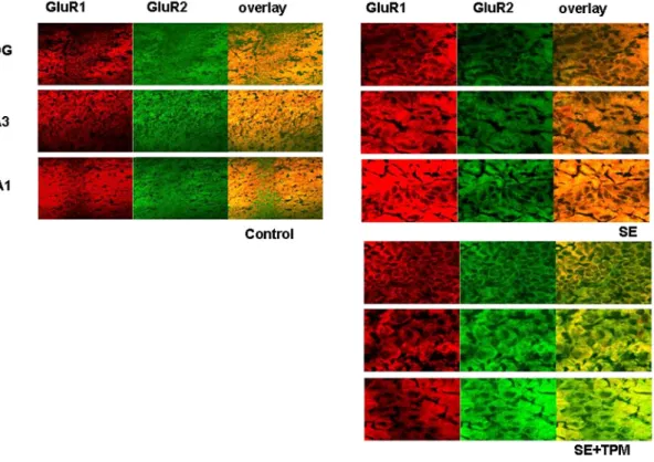
방 법 : 생후 14일된 미성숙 쥐에 kainate를 주입하여 간질중첩증을 유발시킨 뒤 실험군은 1주 혹은 1달 동안 topiramate를 투여한 뒤 뇌를 적출하여, 간질중첩증으로 인한 지속적인 신경흥분성은 western blot을 이용하여 glutamate 및 GABA 수용체의 발현 차이로 관찰하 였고, 신경세포 손상 유발 정도는 공촛점현미경을 이용하여 TUNEL 및 HE 염색으로 관찰 하였다.

결 과 : 미성숙 쥐에서 간질중첩증 유발 1개월 후 해마의 유의한 세포손상과 구조적 변화 를 관찰하였다. 간질중첩증으로 인해 GluR2에 비해 GluR1의 발현이 현저히 증가하여 신경 세포 흥분독성이 유발될 수 있는 Ca

2+

투과성 AMPA 수용체의 형성이 증가되었다. Topira- mate 치료군에서는 간질중첩증으로 인해 유발된 GluR1의 발현 증가가 억제되었다. GABA

B

수용체는 간질중첩증 1개월 후 유의한 변화가 없었으나, GABA

A

수용체의 발현은 현저히

증가되었으며 이는 topiramate 처리에 의해 억제되었다. Topiramate 치료군에서는 간질중첩 증으로 인해 유발된 해마 CA1 및 CA3 부위의 세포 손상과 구조적 변화가 감소됨으로써, topiramate는 미성숙 뇌에서 간질중첩증으로 인한 해마 손상을 보호하는 효과가 있었다.

결 론 : 이상의 결과를 종합하면 topiramate는 미성숙 쥐의 GluR1와 GABA

A

수용체의 발 현을 조절함으로써 신경보호작용을 가지는 것으로 보인다.

Key Words : Topiramate, Status epilepticus, Neurotoxicity, AMPA glutamate receptor,

GABA receptor

(2)

adults. SE at an early age is associated with severe brain damage

1)

, increased risk of epi- lepsy and cognitive impairment later in life

2)

. Retrospective studies indicate that drug-refrac- tory temporal lobe epilepsy in adulthood is re- lated with a childhood SE or prolonged febrile seizures

3)

.

The immature brain differs from the adult brain in seizure susceptibility, seizure characte- ristics and response to antiepileptic drugs

4)

. The immature brain has been considered more resistant to SE-induced damage than the adult brain, and therefore less susceptible to the network reorganization underlying epileptogenesis and cognitive decline

5)

. However, several studies suggest that SE can induce neuronal damage and mossy fiber sprouting, resulting in long- term cognitive impairment and development of epilepsy

6, 7)

. Whether immature brain is more vulnerable to seizures and associated with the structural damage has remained controversial.

Early-life seizures can be refractory to anti- epileptic drugs which are effective in adults and show greater individual variation in response and adverse cognitive side effects

8)

. Another major concern in pediatric epileptology is whether prolonged antiepileptic drug treatment in developing brain may impair the normal ac- tivity-dependent maturation of brain function.

There has been reported the adverse effects of prolonged phenobarbital treatment on cognitive function including impaired concentration and memory

9)

, but long-term use of antiepileptic drugs on normal maturation of brain are still fragmentary.

Topiramate (TPM) is a novel anticonvulsant that has a broad-spectrum of antiepileptic ac- tivity, which includes a negative modulatory effect on the AMPA and kainate subtypes of

glutamate receptors

10, 11)

, a positive modulatory effect on GABAA receptors

12)

, and negative modulatory effects on both voltage-gated Na

+

channels

13)

and neuronal L-type Ca

2+

channels

14)

. These features of TPM make it attractive as a potential neuroprotective agent in immature brain, but the long-term effects on brain dam- age and network reorganization in the process of brain maturation are not determined.

The present study was undertaken to exam- ine possible changes of neuronal excitability and network reorganization in the hippocampus after SE. It was also investigated whether long-term treatment of TPM after SE in the developing brain has a neuroprotective effect.

Materials and Methods

1. Materials

The antibodies to GluR1, GluR2, GABA

A

and GABA

B

were obtained from Chemicon International Inc (Temecula, CA, U.S.A.). The antibodies to N-methyl-D-asparate (NMDA) receptor subunits (NMDAR1, 2a & 2b) were purchased from Oncogene Research Products (Cambridge, MA, U.S.A.). The antibodies to NeuN and MAP2 were obtained from Dako (Temecula, CA, U.S.A). Fluorescein isothiocy- nate (FITC) and Texas Red conjugated se- condary antibodies were obtained from Mole- cular Probe (Eugene, OR, U.S.A.). Apoptag in situ apoptosis detection kit was purchased from Oncor (Gaithersburg, MD, U.S.A.). All the other drugs were purchased from Sigma (St. Louis, MO, U.S.A.) except where indicated.

2. Induction of status epilepticus

Forty five pup BALB-C mice of postnatal

day 14 (P14) were participated in the study.

(3)

To induce SE, animals were received a single intraperitoneal injection of kainate (15 mg/kg in saline; Tocris, Bristol, UK). Animals were monitored for behavioral manifestation of status epilepticus for at least 4 h after injection. Sei- zure intensity was evaluated using the scale described by Racine

15)

. Status epilepticus was defined as prolonged clonic or/and tonic sei- zures (stage 4 or 5) for at least 1 h, which was terminated by intraperitoneal administration of diazepam (20 mg/kg). All the kainate-treated mice received subcutaneous lactated Ringer (3-5 ml) at the end of the period of status epilepticus to replenish fluids. Ten pups out of forty five died during or after status epilepticus (survival rate; 78%) and eleven mice who de- veloped only very mild seizures (stage 1-3) were used as control.

Entire investigation was conducted on 3 experimental groups. Group I mice (controls) received an intraperitoneal injection of kainate, but failed to develop SE (n=11; 5 for 1 week control and 6 for 1 month control). Group II mice (SE) received intraperitoneal injection of kainate and underwent status epilepticus (n=12;

6 for 1 week SE and 6 for 1 month SE).

Group III (SE+TPM) mice received treatment with TPM (10 mg/kg) daily for a month after development of kainate-induced status epilep- ticus (n=12; 6 for 1 week SE+TPM and 6 for 1 month SE+TPM). Animals were sacrificed at 1 week or 1 month after seizure induction.

3. Preparation of hippocampal protein After decapitation, mouse brain was trans- ferred immediately to ice-cold Hepes buffered saline (HBS; 142 mM NaCl, 2.4 mM KCl, 1 mM MgCl

2

, 5 mM D-glucose, 0.1 mM EGTA and 10 mM Hepes [pH 7.5]) and left in -20℃

for 10 min. Hippocampus was isolated from slightly frozen brain and rinsed with HBS. The hippocampus was homogenized with Ultra- Turrax T25 homogenizer (JANDEL & KUNKEL, Germany) in the homogenization medium (320 mM Sucrose, 1 mM EGTA, 0.1 mM EDTA and 10 mM Hepes [pH 7.5]) containing the protease inhibitor mixture (0.3 mM phenylmethylsufonyl fluoride, 2 µg/mL leupeptin, 4 µg/mL aprotinin and 0.8 µg/mL pepstatin A). Nuclei and debris were removed after low speed centrifugation (2,500 g for 10 min) at 4℃ and the postnuclear supernatant was incubated with 2% Tween for 1 h in ice to extract membranous proteins.

Extracted membranous proteins were spun down by centrifugation at 25,000 g for 30 min at 4℃. The resultant pellet was suspended in the homogenization buffer and the protein concentration of the final suspension was mea- sured.

4. Western blotting

The hippocampal membrane proteins were

lysed in boiling 5×sodium dodecyl sulfates

(SDS) sample buffer. The lysates were boiled

for 5 min, and loaded in each lane (20 µg per

lane) to be separated by SDS-polyacrylamide

gel electrophoresis (PAGE). After transferring

the proteins onto a nitrocellulose membrane,

the membrane was blocked with Tris-buffered

saline-Tween (TBST; 20 mM Tris [pH 7.5],

145 mM NaCl, 0.05% Tween-20) containing

5% skim milk overnight at 4℃, then incubated

with primary antibodies (anti-GluR1, GluR2,

GABA

A

, GABA

B

, NMDA receptor1 and 2a) for

1 h at room temperature. After washing three

times for 10 min each with TBST, the mem-

branes were incubated with horseradish pero-

xidase-conjugated secondary antibodies (3,000:1,

(4)

Amersham Life Science Ltd. Little Chalfont, Buckingamshire, England) for 1 h at room temperature. After washing three times with TBST, the membranes were developed and quantified with an enhanced chemiluminescence detection system (LAS-1000plus, Fuji, Japan).

5. Immunofluorescent microscopy for GluR1 and GluR 2

The ratio of GluR1 and GluR2 protein ex- pression was assessed by double immunola- beling of mouse brain sections at the level of the dorsal hippocampus. Freshly isolated brain was frozen and cut into 8 µm sections with vibratome. The frozen sections were dried for 1 h at room temperature and fixed with 4%

paraformaldehyde for 15 min. After quenching the fixatives with phosphate-buffered saline (PBS) containing 0.1 M glycine, the sections were permeabilized with the permeabilization solution (PBS containing 0.4% saponin and 2%

bovine serum albumin). The tissue sections were labeled with primary antibodies against GluR1 (anti-rabbit, 200:1) and GluR2 (anti- mouse, 100:1) for 1 h at room temperature and washed 5 times with the same solution. Se- condary antibodies of anti-rabbit IgG conju- gated FITC or anti-mouse IgG conjugated Texas Red were added for 45 min at room temperature. After washing 3 times, the slides were mounted with Vectashield (Vector labo- ratories, Burlingame, CA, U.S.A.) and examined under the confocal laser scanning microscope (LSM 510, Carl Zeiss, Germany).

6. Assessment of neuronal damage with TUNEL and propidium iodide (PI) staining

Neuronal damage was assessed by direct fluorescence detection of digoxigenin labeled

genomic DNA in the brain sections with simultaneous nuclear staining using Apop Tag in situ apoptosis detection kit. Double-stranded DNA breaks identifying apoptotic cells were detected in situ by the TUNEL technique on tissue sections which had been fixed in for- malin and embedded in paraffin. After deparaf- finization with xylene, hydration in graded ethanols and washing in PBS, the sections were treated with 20 mg/mL proteinase K (Boehringer Mannheim Gmbh, Mannheim, Ger- many) for 30 min at room temperature. After washing, endogenous peroxidase activity was quenched with 2% hydrogen peroxide (Sigma, St. Louis, MO, U.S.A.). The tissue was then reacted with terminal deoxynucleotidyl trans- ferase (TdT) and digoxigenin-labeled UTP, followed by fluorescein-labeled anti-digoxigenin antibody. PI stain was used for counter stain and mounted under a glass coverslip. The slides were observed under the confocal microscope.

7. Histology

Morphological changes of hippocampus after SE-induced neuronal damage was assessed by hematoxylin and eosin stain of pup brain frozen section.

Results

1. Kainate-induced SE in immature mouse model

All animals administrated with kainate exhi-

bited convulsive SE consisting of clonic sei-

zures of forelimb and head muscles. The la-

tency to the first behavioral seizures was 8±2

min (n=45). According to Racine

15)

, it was ty-

pically preceded by strong immobilization and

staring by the loss of a righting reflex (stage

(5)

1), and following automatism such as repetitive head nodding (stage 2). After 30 min of kainate injection, low-intensity tonic-clonic seizures, mo- stly of the forelimbs (stage 3), usually began and generally progressed into full generalized seizures including rearing, jumping, loss of po- sture and falling (stage 4-5). This stage of seizures lasting at least 1 h was regarded as status epilepticus and demonstrated in 53% of kainate-injected mice (24 out of 45 mice).

Stage 4 or 5 motor seizures were maintained for 2 h if not terminated with diazepam.

2. Body weight change after SE or TPM injection

On the first day after SE, mice with SE lost weight (-0.1±2.1%) whereas controls gained weight (9.8±0.9%, Table 1). Thereafter, day- by-day analysis did not reveal any differences between control and SE group, However, TPM injection for 4 weeks induced discernible changes in body weight. Significant weight loss was observed after TPM treatment (mean difference 22.0±2.7%).

Mice experienced with SE looked restless, anxious, and fierce compared to those of control group, but there was no discernable difference in behavior between SE and SE+TPM group.

3. Effect of TPM on hippocampal neuronal death after SE

To investigate whether SE induces hippo- campal neuronal death in immature brain, and whether TPM has protective effects on it, neuronal damage was detected by end tail DNA streak using Apop tag in situ detection kit.

There was occasionally discernable cell death in hippocampus and thalamic areas 1 month after SE (Fig. 1; 2.0±1.0 cells/200 power field).

In TPM treated group apoptotic cells showing FITC fluorescence were rarely found. This fin- ding suggests that there was SE-induced neuronal injury even after 1 month, which was prevented by TPM treatment.

4. Effect of TPM on expression of AMPA glutamate receptors after SE

Subunit combination of AMPA glutamate receptors is an important determinant of neu- roexcitability. To investigate whether neuroex- citability after kainate-induced SE is mediated by increased formation of Ca

2+

-permeable AMPA receptors and whether TPM affects on this, the changes in expression of ionotropic gluta- mate receptors were studied with Western blot.

The expressions of N-methyl-Daspartate (NMDA) receptor subunit 2A (NR2A) and 2B (NR2B) were slightly decreased after kainate- induced SE at 1 week, but TPM did not change significantly the expression levels of NMDA receptors induced by SE. Neither SE nor TPM treatment induced any significant change in expression of NMDA receptors even after 1 month (Fig. 2).

AMPA-type GluR1 expression was markedly up-regulated by kainate-induced status epilep- ticus at 1 week, which was attenuated by 1 week treatment of TPM (Fig. 3). GluR2 ex- pression in immature brain was weak, and was not affected by SE or TPM treatment. How- Table 1. The Change in Body Weight after

Topiramate Injection in Mouse Pups with Kai- nate-induced Status Epilepticus

Group Day 0 Day 1 Day 28

Control (n=6) SE (n=6) SE+TPM (n=6)

9.5±0.3 g 9.8±0.4 g 9.7±0.3 g

9.9±0.4 g 8.8±0.5 g 8.6±0.6 g

24.3±2.2 g

23.8±1.9 g

19.5±1.5 g

Abbreviations : SE, status epilepticus; TPM, top-

ramate

(6)

ever, 1 month after SE, GluR1 expression was significantly up-regulated, which was reversed by TPM treatment. GluR2 expression was more prominent after 1 month compared to 1 week, but SE did not induce any significant change

of GluR2 expression compared to that of GluR1.

TPM treatment slightly up-regulate GluR2 ex- pression. Therefore, the GluR1/GluR2 ratio in- creased after SE, but decreased after 1 month treatment of TPM.

Fig. 2. Effects of TPM on expressions of NMDA receptors after kainate-induced SE in mouse pup hippocampus. Western blot shows that NMDA receptor (NR) 2A is slightly increased 1 month after SE, but TPM treatment has little effect on SE-induced NR2A or NR2B expression.

Abbriviations : CB, Coomassie Brilliant Blue stain; SE, status epilepticus;

TPM, topiramate.

Fig. 1. Neuronal death after kainate-induced status epilepticus (SE) in mouse pup hippocampus. TUNEL stain shows that several apoptotic cells are found in CA1 region of hippocampus and thalamic area 1 month after SE, but topiramate (TPM) treatment reduces hippocampal neuronal death.

Abbreviation : DG, dentate gyrus

(7)

5. Immunofluorescent microscopic assessment of GluR2/GluR1 ratio in hippocampal neurons after SE or TPM treatment

The ratio of levels of GluR1 protein to GluR2 protein may predict Ca

2+

permeability of hippocampal neuron. The relative GluR1 and GluR2 expression in the hippocampal neuron was investigated by co-localization study using Texas redconjugated GluR1 and FITC-conju- gated GluR2 in mouse brain section. In support of the finding obtained by western blot, im- munofluorescent confocal microscopy shows that relative ratio of GluR1/GluR2 in hippocampal

pyramidal cells was increased 1 month after SE (Fig. 4). This finding suggests increased formation of Ca

2+

-permeable AMPA receptors in pyramidal neurons. TPM treatment reversed the GluR1/GluR2 ratio especially in CA1 region of hippocampus.

6. Effect of TPM on expression of GABA receptors after SE

GABA, the predominant inhibitory neuro- transmitter in brain, is known to be develop- mentally regulated and cause membrane de- polarization rather than hyperpolarization in immature brain. It was investigated whether kainate-induced SE and TPM treatment affect Fig. 3. Effects of TPM on expressions of GluR1 and GluR2 after kainate-

induced SE in mouse pup hippocampus. Western blot shows that GluR1 is up-regulated 1 week or 1 month after SE, and topiramate prevents SE- induced GluR1 expression. The expressions of GluR2 are not significantly changed by either SE or TPM treatment. Abbreviations : CB, Coomassie Brilliant Blue stain; SE, status epilepticus; TPM, topiramate; GluR, glutamate receptor.

Fig. 4. Effects of TPM on differential expression of GluR1 and GluR2 after kainate-induced SE in mouse pup hippocampus. Co-localization study of Texas red-conjugated GluR1 and FITC-conjugated GluR2 using confocal microscopy shows that the ratio of expression of GluR1 to GluR2 is increased 1 month after SE, and TPM reversed SE-induced increase of the ratio (×400). Abbreviations : DG, dentate gyrus; SE, status epilepticus; TPM, topiramate; GluR. Glutamate receptor; CA1 & 3, CA1 &

CA3 regions in hippocampus

(8)

on the expression of GABA receptors. The expressions of both GABA

A

and GABA

B

re- ceptors were not changed significantly by kai- nate-induced SE or TPM treatment at 1 week (Fig. 5). However, 1 month after SE, expre- ssion of GABA

A

receptor was highly increased, but decreased in TPM treated group. The changes in the expression levels of GABA

B

receptors were insignificant after 1 month except slight up-regulation by TPM treatment.

7. Effect of TPM on structural integrity of hippocampus after SE To investigate whether status epilepticus induces structural damage in hippocampus and whether TPM has protective effects on it, histologic assessment was done by HE stain (Fig. 6). In control group, there was no dis- cernable cell loss and structural damage in ei- ther dentate granular cells or hippocampal py- ramidal cells. However, after SE, structural in- tegrity of hippocampus was loosened with scattered pyramidal cells in CA1 and CA3 Fig. 6. The effect of TPM on hippocampal inte-

grity after kainate-induced SE. Hematoxylin-Eosin stain shows disruption of the structural integrity and decreased thickness of hippocampal CA1 and CA3 regions (arrows), and dentate gyrus (arrow heads) 1 month after SE. TPM prevents SE-induced cell loss and structural changes (×

20). Abbriviations : SE, status epilepticus; TPM, topiramate

Fig. 5. Effects of TPM on expressions of GABA

A

and GABA

B

receptors

after kainate- induced SE in mouse pup hippocampus. Western blot shows

that GABA

A

is up-regulated 1 month after SE, and topiramate prevents SE-

induced GABA

A

expression. However, GluR2 expression is not significantly

changed by either SE or TPM treatment. Abbreviations : CB, Coomassie

Brilliant Blue stain; SE, status epilepticus; TPM, topiramate

(9)

regions, and the thickness of those regions decreased due to the decreased cell number.

However, in TPM treated group, the alignment of dentate gyrus and hippocampus was rela- tively maintained and the thickness was not reduced compared to SE group.

This finding was confirmed by neuronal staining with both FITC-labeled NeuN and Texas Red-labeled MAP2 using the confocal microscopy (Fig. 7). NeuN, a nuclear antigen of neuron, stains the soma of neuron, whereas MAP2 stains the neurofilament in axons as well as soma. SE induced significant decrease of NeuN fluorescence in hippocampal pyramidal cells of CA1 and CA3, but TPM prevents the

SEinduced decrease in NeuN staining, which is indicative of a neuroprotective effect of TPM.

Discussion

The contribution of SE during early child- hood to long-term structural damage and epi- leptogenesis is under dispute. The harmful effect of prolonged antiepileptic drug treatment on brain maturation is also postulated

9)

. The pres- ent study was designed to test that SE at early age leads to long-term damage in hippo- campus and whether topiramate protects the SE-induced hippocampal damage and subse- quent epileptogenesis. There were occasional Fig. 7. The effect of TPM on hippocampal neuronal loss after kainate-induced SE in mouse pup hippocampus. Confocal microscopy using Texas red-conjugated MAP-2 and FITC-conjugated NeuN shows that NeuN fluorescence decreased 1 month after SE. TPM recovers SE-induced decrease of NeuN fluorescence (×400). Abbreviations : DG, dentate gyrus; SE, status epilepticus; TPM, topiramate;

CA1 & 3, CA1 & CA3 regions in hippocampus.

(10)

apoptotic cells in hippocampus 1 month after kainate-induced SE at P14, but significant cell loss with structural changes in hippocampus was detected after 1 month. Previous studies suggested that the immature brain appears to be relatively resistant to seizure-induced neu- ronal injury compared with adult brain

16)

. However, recent studies revealed that seizures in early life may also induce long-term mor- phologic alteration of mossy fiber sprouting and functional impairment in hippocampus- dependent spatial memory task with sponta- neous seizures

17)

. The resistance of immature brain to SE-induced neuron damage is not absolute, and varies on age of the postnatal day and the severity of epileptogenic insults

18)

. Despite the lack of significant injury in im- mature brain, neonatal seizures may adversely alter function of surviving neurons and neu- ronal circuitry to promote epileptogenesis and cognitive impairment

17)

. However, relationship between seizure-induced neuronal damage and epileptogenesis in developing brain remains a controversial issue in epilepsy.

Animal models such as kainic acid and lithium-pilocarpine model of epilepsy are widely used tools for human temporal lobe epilepsy to understand the basic mechanisms of epilep- togenesis

19)

. Local or systemic administration of KA or pilocarpine in rodents leads to a pattern of repetitive limbic seizures and SE, which can last several hours

20)

. A somewhat variable latent period follows SE and precedes the occurrence of spontaneous limbic seizures.

The brain damage induced by SE in such pre- parations may be considered as an equivalent of the initial precipitating injury event, usually a prolonged febrile convulsion, which is com- monly found in patients with mesial temporal

lobe epilepsy

21)

. Neuropathological changes such as neuronal loss in several hippocampal sub- fields and reorganization of mossy fibers into the molecular layer of the fascia dentata are observed in both models and are similar to hippocampi from patients with hippocampal sclerosis

22)

.

The current study found that long-term to- piramate treatment in developing brain reduced SE-induced hippocampal damage. The neuro- protective effect of topiramate following SE was also reported by others

23, 24)

. Cha et al demon- strated that chronic treatment with topiramate improved cognitive function assessed by water maze performance, but did not protect cell loss with mossy fiber sprouting

23)

. Rigoulot et al also reported neuroprotective properties of to- piramate in lithium-pilocarpine model, in which topiramate prevented neuronal death in CA1 and CA3 of hippocampus, but failed to prevent epileptogenesis

24)

.

TPM is a new antiepileptic drug used as

therapy in refractory partial-onset seizures in

adults and children, and generalized tonic-

clonic seizures. TPM exhibits neuroprotective

properties in several models of injury such as

global or focal ischemia, and SE

23, 24)

. TPM has

multiple mechanisms of action that may

contribute to its anticonvulsant activity

10)

. It

includes voltage-dependent inhibition of vol-

tage-gated Na

+

channels

13)

, and reduces L-type

high voltage-activated Ca

2+

currents

14)

. It in-

creases chloride flux of GABA channels by

acting on nonbenzodiazeine site

12)

. It acts as a

negative modulatory effect on the AMPA and

kainate subtypes of glutamate receptors

10, 11)

, and

an inhibitor of erythrocyte carbonic anhydrase

25)

.

The mechanisms underlying the neuroprotective

effect of TPM are not fully understood. The

(11)

additive or synergistic effect of its multiple mechanisms of action may explain for the beneficial effect of TPM. In this study, it was demonstrated that TPM down-regulated SE- induced induction of AMPA glutamate receptor, GluR1. However, TPM rather negatively mo- dulated SE-induced up-regulation of GABA

A

receptor. This finding might be against the neuroprotective effect of TPM. However, it is notable that in early postnatal week, activation of GABA

A

receptors causes membrane depola- rization rather than hyperpolarization typical of GABA-ergic synapses

26)

. This difference might results from maturational changes in trans- membrane chloride ion gradient

27)

. It cannot be excluded the possibility that these GABA

A

channels also act functionally as excitatory ra- ther inhibitory in nature, because normal ma- turation mechanisms responsible for generating normal chloride ion gradient across the mem- brane might be intervened by SE or TPM in critical time points of postnatal age. TPM seems more neuroprotective in hippocampus than other areas

28)

. The selective pattern of neuroprotection was also examined in our study.

It might be related TPM action to inhibit AMPA receptor, of which the density is higher in hippocampus CA1 and CA3 regions.

Glutamate Induced Neurotoxicity might ac- count for SE-induced neuronal death. The glu- tamate mediates almost all excitatory synaptic transmission in adult brain. The family of lig- and-gated ion channels activated by glutamate can be divided into three classes on the basis of pharmacological and molecular criteria

29)

. The first class, termed AMPA receptors, me- diates fast excitatory synaptic transmission.

AMPA receptors are tetrameric or pentameric complexes of four homologous subunits termed

GluR1-GluR4 that assembles in varying com- binations to form functional channels

30)

. The second class of ionic glutamate receptors is the NMDA class. These receptors are highly permeable to calcium ions, which activate a variety signal transduction cascades involved in many forms of presynaptic plasticity

31)

. NMDA receptors are also multimeric complexes of ho- mologous subunits termed NR1 and NR2A-2D that combine to form different receptor sub- types. All receptors contain an NR1 subunit and various numbers of NR2 subunits. NR2B is the major type 2 receptor subunit expressed in adult forebrain

32)

. NR2 is also a major com- ponent of the postsynaptic density in hippo- campus and cerebral cortex. The third class of glutamate receptors is termed kainite receptors after their preferred ligand

33)

. Their role in ro- utine excitatory synaptic transmission is less clear, although in specialized cases they may contribute a significant synaptic current

29)

.

According to GluR2 hypothesis, the presence of GluR2 renders heteromeric AMPA receptors assemblies Ca

2+

-impermeable

34)

. The downre- gulation of GluR2, observed in animal model of transient forebrain ischemia and epilepsy, serves as a molecular switch leading to the formation of Ca

2+

-permeable AMPA receptors and en- hanced neurotoxicity of endogenous glutamate

35)

. In our experiments, GluR2 was up-regulated after SE, which might prevent toxic influx of Ca

2+

into the neurons and subsequent neuronal damage and degeneration.

Postnatal 2-3 weeks, when the density of

AMPA and NMDA receptor peaks, represents

a window during which glutamate-mediated

synaptic plasticity is enhanced

36)

. This is likely

to play a role in the increased excitability of

the brain in developing brain. Notably, expression

(12)

of GluR2 subunit compared with other AMPA receptor subunits is significantly low in im- mature hippocampus

37)

. This suggests that a larger portion of AMPA receptors are highly permeable to Ca

2+

in immature brain, contributing pathophysiologic events in early life.

In conclusion, we demonstrated significant cell loss with structural changes in hippocampus 1 month after SE. We also found that long- term topiramate treatment in developing brain reduced SE-induced hippocampal damage. The neuroprotective action of TPM might be medi- ated by modulation of GluR1 and GABA

A

receptors.

Abstract

Purpose : This study was performed to elu- cidate that status epilepticus (SE) induces long- term neuronal damages in an immature brain and to evaluate that topiramate (TPM) has a protective effect.

Methods : We investigated the changes in a subtype expression of glutamate and gamma- amino butyric acid (GABA) receptors, and the structural integrity due to cell losses in the mouse pup hippocampus after SE using an immunoblot and confocal microscopy.

Results : SE induced significant cell losses with structural changes in the hippocampus 1 month later. SE up-regulated the glutamate receptor1 (GluR1) expression with an increased ratio of GluR1 to glutamate recptor2 (GluR2), leading to the formation of Ca

2+

permeable α- amino-3-hydroxy-5-methyl-4-isoxazoleepropionic acid (AMPA) receptors for the enhanced neu- rotoxicity. TPM prevented the SE-induced GluR1 expression. The expression of GABA

A

receptors was highly increased 1 month after SE, whereas

that of GABA

B

receptors was not changed.

The TPM treatment attenuated SE-induced upregulation of GABA

A

receptors. SE induced significant cell losses and disruption of struc- tural integrity in the hippocampus CA1 and CA3 regions, but the TPM treatment for 1 month in developing brains reduced the SE- induced hippocampal damage.

Conclusion : TPM has a neuroprotective action, which might be mediated by the modu- lation of GluR1 and GABA

A

receptors.

References

1) Sagar HJ, Oxbury JM. Hippocampal neuron loss in temporal lobe epilepsy: correlation with early childhood convulsion. Ann Neurol 1987;

22:334-40.

2) Aicardi J, Chevrie JJ. Convulsive status epilepsy in infants and children: a study of 293 cases. Epilepsia 1979;11:187-97.

3) Cendes F, Andermann F. Do febrile seizures promote temporal lobe epilepsy? Retrospective studies. In : Baram TZ, Shinnar S editors.

Febrile Seizures. Academic Press 2002;77-86.

4) Holmes GL. Epilepsy in the developing brain:

lessons from the laboratory and clinic. Epi- lepsia 1997;38:12-30.

5) Lado FA, Laureta EC, Moshe SL. Seizure- induced hippocampal damage in the mature and immature brain. Epileptic Disord 2002;4:

83-97.

6) Dubë C, Chen K, Eghbal-Ahmadi M, Brunson K, Soltesz I, Baram TZ. Prolonged febrile seizures in the immature rat model enhance hippocampal excitability long term. Ann Neurol 2002;47:336-44.

7) Kubová H, Druga R, Lukasiuk K, Suchome- lová L, Haugvicová R, Jirmanová I, et al.

Status epilepticus causes necrotic damage in the mediodorsal nucleus of the thalamus in immature rats. J Neurosci 2001;21:3593-9.

8) Sanchez RM, Jensen EF. Maturational aspects

of mechanism and consequences for the im-

mature brain. Epilepsia 2001;42:577-85.

(13)

9) Farwell JR, Lee YJ, Hirtz DG, Sulzbacher SI, Ellenberg JH, Nelson KB. Phenobarbital for febrile seizures: effects on intelligence and on seizure recurrence. N Engl J Med 2001;322:

364-9.

10) Gibbs JW. Sombati S, DeLorenzo RJ, Coulter DA. Cellular actions of topiramate: blockade of kainate-evoked inward currents in cultured hippocampal neuron. Epilepsia 2000;41(Suppl 1):

10S-16S.

11) Skadski S, White HS. Topiramate blocks kainite-evoked cobalt influx into cultured ne- urons. Epilepsia 2000;41(Suppl 1):45S-47S.

12) White HS, Brown SD, Woodhead JH, Skeen GA, Wolf HH. Topiramate modulates GABA- evoked currents in murine cortical neurons by a nonbenzodiazepine mechanisms. Epilepsia 2000;

41(Suppl 1):17-20.

13) DeLorenzo RJ, Sombati S, Coulter DA. Effects of topiramate on sustained repetitive firing and spontaneous recurrent seizure discharges in cultured hippocampal neurons. Epilepsia 2000;

41(Suppl 1):40-4.

14) Zhang X, Velumian AA, Jones OT, Carlen PL. Modulation of high voltage-activated cal- cium channels in dentate granule cells by to- piramate. Epilepsia 2000;41(Suppl 1):52-60.

15) Racine RJ. Modification of seizure activity by electrical stimulation II. Motor seizure Elec- troencephalogr Clin Neurophysiol 1972;32:281- 94.

16) Haas KZ, Sperber EF, Opanashuk LA, Stan- ton PK, Moshe SL. Resistance of immature hippocampus to morphologic and physiologic alterations following status epilepticus or kin- dling. Hippocampus. 2001;11:615-25.

17) Kubová H, Maré P, Suchomelová L, Broźek G, Druga R, Pitkänen A. Status epilepticus in immature rats leads to behavioral and cogni- tive impairment and epileptogenesis. Eur J Neurosci 2004;19:3255-65.

18) Cilio MR, Sogawa Y, Cha BH, Liu X, Huang LT, Holmes GL. Long-term effects of status epilepticus in the immature brain are specific for age and model. Epilepsia 2003;44:518-28.

19) Morimoto K, Fahnestock M, Racine RJ. Kin- dling and status epilepticus models of epile- psy: rewiring the brain. Progress in Neuro- biology 2004;73:1-60.

20) Covolan L, Mello LE. Temporal profile of neuronal injury following pilocarpine or kainic acid-induced status epilepticus. Epilepsy Res 2000;39:133-52.

21) Shinnar S. Prolonged febrile seizures and mesial temporal sclerosis. Ann Neurol 1998;

43:411-2.

22) Tauck DL, Nadler JV. Evidence of functional mossy fiber sprouting in hippocampal forma- tion of kainic acid treated rats. J Neurosci 1992;5:1016-22.

23) Cha BH, Silveira DC, Liu X, Holmes GL.

Effect of topiramate following recurrent and prolonged seizures during early development.

Epilepsy Res 2002;51:217-32.

24) Rigoulot MA, Koning E, Ferrandon A, Nehlig A. Neuroprotective Properties of topiramate in the lithium-pilocarpine Model of Epilepsy. J Phamachol Exp Ther 2004;308:787-95.

25) Dodgson SJ, Shank RP, Maryanoff BE. To- piramate as an inhibitor of carbonic anhy- drase isoenzymes. Epilepsia 2000;41(Suppl 1):

35-9.

26) Ben-Ari Y, Khazipov R, Leinekugel X, Caillard O, Gaiarsa GL. GABA-A, NMDA, and AMPA receptors: a developmentally regulated "ménage á trois." Trends Nurosci 1997;20:523-9.

27) Staley KJ, Soldo BL, Proctor WR. Ionic me- chanisms of neuronal excitation by inhibitory GABA

A

receptors. Science 1995;269:977-81.

28) FischerA, Wang X, Cock HR, Thom M, Patsalos PN, Walker MC. Synergism between topiramate and budipine in refractory status epilepticus in the rat. Epilepsia 2004;45:1300- 7.

29) Dingledine R, Borges K, Bowie D, Traynelis SF. The glutamate receptor ion channels.

Pharmacol Rev 1999;51:7-61.

30) Hollman M, Hartley M, Heinemann S. Ca

2+

permeability of KA-AMPA-gated glutamate receptor channels depends on subunit compo- sition. Science 1991;252:851-3.

31) Monyer H, Burnashev N, Laurie DJ, Sakmann B, Seeburg PH. Developmental and regional expression in the rat brain and functional properties of four NMDA receptors. Neuron 1994;12:529-40.

32) Vallano ML. Developmental aspects of NMDA

receptor function. Crit Rev Neurobiol 1998;12:

(14)

177-204.

33) Vignes M, Collingridge GL. The synaptic ac- tivation of kainate receptors. Nature 1997;388:

179-82.

34) Pellegrini-Giampietro DE, Gorter JA, Bennett MV, Zukin RS. The GluR2 (GluR-B) hypo- thesis: Ca

2+

-permeable AMPA receptors in neurological disorders. Trends Neurosci 1997;

20:464-70.

35) Friedman LK, Koudinov AR. Unilateral GluR2 (B) hippocampal knockdown: a novel partial seizure model in the developing rat. J Neu-

rosci 1999;19:9412-25.

36) Sanchez RM, Koh S, Rio C, Wang C, Lamperti ED, Sharma D, et al. Decreased glutamate receptor 2 expression and enhanced epileptogenesis in immature rat hippocampus after perinatal hypoxia-induced seizures. J Neurosci 2001;21:8154-63.

37) Flint AC, Maisch US, Weishaupt JH, Kriegs- tein AR, Monyer H. NR2A subunit expression shortens NMDA receptor synaptic currents in developing neocortex. J Neurosci 1997;17:

2469-76.

수치

Fig. 1. Neuronal death after kainate-induced status epilepticus (SE) in mouse pup hippocampus
Fig. 4. Effects of TPM on differential expression of GluR1 and GluR2 after kainate-induced SE in mouse pup hippocampus
Fig. 5. Effects of TPM on expressions of GABA A and GABA B receptors after kainate- induced SE in mouse pup hippocampus

참조

관련 문서