INTRODUCTION
Atopic dermatitis (AD) is a persistent, chronically relapsing inflammatory skin disease in which abnormal hematopoietic cells circulate and infiltrate skin and respiratory tissue at sites perturbed by irritants, antigens, and infectious agents. AD is as- sociated with multiple immunological abnormalities, and 80%
of patients suffer from defective regulation of IgE production.1 AD can range from mild, localized lesions to generalized ec- zematous lesions. Although a number of patients with AD re- spond to topical therapy, others have severe resistant disease which is not controlled with first- or second-line topical thera- pies. Therefore, AD may be considered as a systemic disease for which there are no satisfactory systemic therapies, although topically applied medications, including corticosteroid and cy- closporine agents, give relief in some patients.2-4 Long-term, chronic application of topical steroids may supplant the need for systemic therapy in many cases. Newer therapies such as ta- crolimus are effective, but are limited to second-line therapy af- ter initial treatments have failed. Oral cyclosporine has also been shown to be effective; however, its use is limited by adverse ef-
Long-term Efficacy of Intravenous Immunoglobulin Therapy for Moderate to Severe Childhood Atopic Dermatitis
Sue-Jung Jee, Joo-Hwa Kim, Hey-Sung Baek, Ha-Baik Lee, Jae-Won Oh*
Department of Pediatrics, Hanyang University College of Medicine, Seoul, Korea
fects such as renal toxicity. Approximately 20% of patients dis- continue cyclosporine treatment within 2 years because of ad- verse effects.5,6
Intravenous immunoglobulin (IVIg) is prepared by cold etha- nol fractionation of pooled plasma, followed by additional viral inactivation procedures. The final product is a pure concentrate of IgG (with small amounts of IgA and IgM) with intact function and minimal impurities.7,8 The immunomodulatory mechanism of IVIg is thought to be mediated by interactions of the Fc por- tion of IgG with Fc receptors and complement, interactions of the antigen-binding variable region F(ab)2, or substances other than antibodies in IVIg preparations.9 Previous research has shown that IVIg significantly modulates the production of sev- eral cytokines, including interleukin (IL)-1, IL-2, IL-3, IL-4, IL-5, IL-10, tumor necrosis factor-α, and granulocyte-macrophage Allergy Asthma Immunol Res. 2011 April;3(2):89-95.
doi: 10.4168/aair.2011.3.2.89 pISSN 2092-7355 • eISSN 2092-7363
Purpose: The present study investigates the long-term effects of intravenous immunoglobulin (IVIg) therapy for the treatment of moderate to se- vere childhood atopic dermatitis (AD). Previous research indicates that IVIg can treat severe AD; however, the effectiveness of IVIg has not been confirmed in prospective, blinded clinical trials. Methods: Forty eligible children with moderate to severe AD, as defined by the criteria of Hanifin and Rajka, were enrolled in a randomized, placebo-controlled study. After the completion of an initial screening visit (V0), the patients were random- ly allocated into therapy (n=30) and control (n=10) groups (V1). Thirty children were each treated with three injections of 2.0 g/kg IVIg at 1-month intervals over a 12-week period. Ten children were treated with placebo. Assessments were conducted after each injection (V2, V3, and V4) and at 3 (V5) and 6 months (V6) after completed treatment. Results: The disease severity index was significantly decreased at V5 compared with the value at V1 (P<0.05). There were no significant changes in the total IgE level or total eosinophil count in peripheral blood at the last injection (V4) compared with the value at V1. The interleukin (IL)-5/interferon (IFN)-γ ratio was assessed in T-helper 1 (Th1) and Th2 cells. The ratio significantly decreased be- tween V1 and V5, after which it increased, such that the ratio at V6 was not significantly different from that at V1. Compared with the level at V1, the intercellular cell adhesion molecule-1 level at V4 did not differ significantly, but the level at V5 was lower. Conclusions: This study suggests that IVIg therapy may clinically improve AD in patients after 3 months of therapy, but the improvement may decline by 6 months after therapy.
Key Words: Intravenous immunoglobulin; atopic dermatitis
This is an Open Access article distributed under the terms of the Creative Commons Attribution Non-Commercial License (http://creativecommons.org/licenses/by-nc/3.0/) which permits unrestricted non-commercial use, distribution, and reproduction in any medium, provided the original work is properly cited.
Correspondence to: Jae-Won Oh, MD, PhD, Department of Pediatrics, Hanyang University College of Medicine, 249-1 Kyomun-dong, Guri 471-701, Korea.
Tel: +82-31-560-2254; Fax: +82-31-552-9493; E-mail: [email protected] Received: August 25, 2010; Accepted: November 18, 2010
colony-stimulating factor, as well as cytokine antagonists (IL-1 receptor antagonist), by monocyte-macrophages and lympho- cytes.10 It appears that the biological anti-inflammatory effects of IVIg involve the participation of both the F(ab)2 and Fc regions in the formation of an IgG-antigen complex. These regions are associated with the inhibition of lymphocyte proliferative re- sponses and the modulation of T-helper 1 (Th1) and Th2 cyto- kine production. This effect of IVIg is mediated by the inhibi- tion of differentiation and maturation of dendritic cells (DCs) in vitro, with down-regulated expression of co-stimulatory mole- cules, thereby impairing the ability of mature DCs to produce IL-12 and enhancing their ability to produce IL-10. The conse- quences are the inhibition of autoreactive and alloreactive T- cell activation and proliferation.11,12
IVIg therapy has consistently demonstrated activity in patients with AD, including severe AD,13 although its effectiveness has not been confirmed in prospective, blinded clinical trials. Pa- tients with Kawasaki disease or idiopathic thrombocytopenic purpura and concomitant AD who received IVIg showed im- provement of their dermatitis.14,15 One study gave mixed results, with the best response occurring in patients with extremely high IgE levels.16 Reports on severe childhood AD suggest that IVIg can lead to improved symptoms when used as a monotherapy;
nine out of 10 children improved when given 2 g/kg IVIg.17 How- ever, most published research has evaluated only the short-term effects of IVIg.
The present study was conducted to assess the long-term ef- fects of IVIg therapy in moderate to severe childhood AD.
MATERIALS AND METHODS Patients
Forty eligible children with moderate to severe AD, as defined by the criteria of Hanifin and Rajka18, were enrolled in a ran- domized, placebo-controlled study after an initial screening visit (V0) (Fig. 1). Eligibility criteria included AD present on
more than 30% of the body surface, no response to convention- al therapy, and age older than 2 years. Informed consent was obtained from either the child or a parent. Allergy skin prick tests or in vitro allergy tests (Unicap: Phardia, Uppsala, Sweden) were conducted at the screening visit (V0). The patients were random- ized at the next clinic visit (V1) (Fig. 1). Patients were allowed to use only steroid-free hydrophilic or emollient ointment on the skin as adjunctive therapy. Seven patients did not complete the study: five IVIg subjects suffered side effects, including head- ache, nausea, and abdominal pain, and two control subjects did not complete the trial for personal reasons. This study was approved by the Korean FDA (IVIg IIT Atopy-No. 48) and the Hanyang University Guri Hospital IRB committee.
Methods
The IVIg patients received an injection of IVIg (Greencross Pharm Co, Seoul, Korea) at 2.0 g/kg body weight/month at each monthly visit (V2, V3, and V4) for 12 weeks (Fig. 1). The patients were also assessed at each clinic visit and at 3 (V5) and 6 months (V6) after the final injection. Patients with headache or nausea were permitted to take oral acetaminophen. Placebo group re- ceived general topical moisturizing lotion, 1% hydrocortisone cream and took oral antihistamines if they complaint itching for controlling itching skin as same to IVIg group. At V1, V4, V5, and V6, laboratory tests were performed for blood chemistry, total IgE, eosinophil cationic protein (ECP), IL-5, interferon (IFN)-γ, and intercellular cell adhesion molecule (ICAM)-1.
Disease severity was assessed by the criteria of Hanifin and Raj- ka18, and included clinical severity and total body surface area (TBSA). Disease severity was assessed with the SCORAD index, which was calculated as A/5+7×B/2+C, where A is the TBSA measured as a percentage of lesional skin according to the rule of nines; B is the clinical severity consisting of six parameters:
erythema, edema/papulation, excoriation, oozing/vesicle, li- chenification, and dryness with pigmentation/depigmentation;
and C is a subjective measurement of symptoms such as pruri-
Immunoglobulin IV injection*
Visit 0
-2 wk Visit 1
0 wk Visit 2
4 wk Visit 3
8 wk Visit 4
12 wk Visit 5
24 wk Visit 6
36 wk Screening visit Random
allocation Routine
visit Routine
visit Completed
visit Follow up†
visit Random
allocation card
IVIg group (n=30)
Control group (n=10)
Analysis
Fig. 1. Study design of intravenous immunoglobulin (IVIg) therapy, blood specimen collection for laboratory analyses, and evaluation of clinical efficacy.
*Immunoglobulin IV injection: three times (2 g/kg month IV) for 12 weeks; †Follow up: after completing injection, every 4 weeks for 6 months.
tus and loss of sleep. Parameters A and B were scaled from 0 to 3; parameter C, from 0 to 10. To minimize potential variation, the same physician measured the disease severity at each visit (Table 1).
Determination of cytokine concentrations
The concentrations of IL-5, IFN-γ, and ICAM-1 were deter- mined by cytometric bead array assays (Bio-Plex; Bio-Rad Lab- oratories, Hercules, CA, USA) according to the manufacturer’s protocol. All measurements were made in duplicate, with <10%
variation between the two measurements.
Statistical analysis
The characteristics of the IVIg and placebo groups at V0 were compared using the Mann-Whitney U test. Disease severity was compared between the groups at baseline (V1) and at V2, V3, V4, V5, and V6. Differences between visits were analyzed by multiple comparisons as ANOVA. Analyses were performed us- ing the SPSS statistical package, version 11.5 (SPSS Inc., Chica- go, IL, USA). A value of P<0.05 was taken to indicate statistical significance.
RESULTS
Clinical effects of IVIg therapy in children with moderate to severe AD
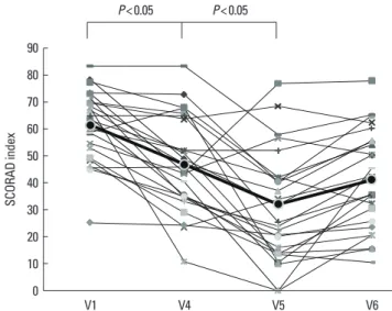
Fig. 2 presents the mean percentage improvements of clinical parameters at each visit. The SCORAD index declined until 3
months after the last IVIg injection (V5) and did not change sig- nificantly during the subsequent 3 months (V6) (V1: 61.5±13.0, V4: 46.9±17.3, V5: 32.1±19.4, and V6: 39.3±18.4; V1 vs. V5:
P<0.05) (Fig. 2, Table 2). The TBSA parameter of the SCORAD index improved until V5, but declined by V6 (V1: 50.5±12.8, V4: 40.7±11.9, V5: 29.7±14.2, and V6: 36.8±13.7; V1 vs. V5:
P<0.05) (Fig. 3A). The scores for subjective symptoms such as itching and loss of sleep did not change significantly during the
Table 1. Subjective characteristics in study
Group IVIg Placebo Total P value
No. of patients 30 10 40
Age (yr) 6.3±3.4 6.5±5.7 6.4±4.1
Sex (No.)
Male 18 4 22
Female 12 6 18
SCORAD index 61.5±13.3 42.1±9.9 55.9±15.1 >0.05
Total IgE level (IU/mL) 571.2±769.6 803.9±1134.0 692.8±922.6 >0.05
Eosinophil count (/mm3) 529.4±472.2 290.0±179.2 461.0±421.8 >0.05
IVIg, intravenous immunoglobulin.
Table 2. Comparison of SCORAD index, serum IgE, and eosinophil count between children with IVIg treatment and the controls
Visit SCORAD index Serum IgE (IU/mL) Eosinophil count (/mm3)
IVIg Control IVIg Control IVIg Control
V1 61.5±13.0* 42.1±9.9 571.2±753.4 615.2±1023.2 529.4±462.6 290±179.3
V4 46.9±17.3* 40.4±6.3 384.9±519.5 598.8±858.4 397.6±342.8 247.1±153.1
V5 32.1±19.4* 35.7±8.9 493.9±620.3 622.0±932.5 470.8±411.7 330±150.5
V6 39.3±18.4 35.9±9.6 548.9±677.2 633.9±946.7 547.6±393.3 270.0±71.3
*There shows significantly differences of SCORAD between IVIg and control at V1 vs. V4 (P=0.005) and V1 vs. V5 (P=0.17).
IVIg, intravenous immunoglobulin; V1, before therapy; V4, during therapy; V5, 3-month follow-up visit; V6, 6-month follow-up visit.
Fig. 2. SCORAD index before (V1) and during therapy (V4), and at 3 (V5) and 6 months (V6) after the last intravenous immunoglobulin (IVIg) injection. The SCO- RAD index was significantly lower at the 3-month follow-up visit (V5) compared with the index at V1, but it had increased by the 6-month follow-up visit (V6).
SCORAD index
90 80 70 60 50 40 30 20 10
0 V1 V4 V5 V6
P<0.05 P<0.05
study (V1: 14.1±2.0, V4: 12.0±2.9, V5: 10.2±3.6, and V6: 12.0±
3.2) (Fig. 3B). The SCORAD index of the control group did not improve significantly until V6.
Immunological parameters in peripheral blood
The total serum IgE concentration was initially elevated in all subjects (V0). The levels declined during IVIg treatment, were higher at the 3-month post-treatment visit (V5), and returned to initial levels by 6 months after treatment (V1: 571.2±753.4, V4: 384.9±519.5, V5: 493.9±620.3, and V6: 548.9±677.2 IU/mL) (Fig. 4A, Table 2). The total eosinophil count in peripheral blood demonstrated a similar pattern: it decreased during IVIg treat- ment, but returned to the initial level by 6 months post-treat- ment (V1: 529.4±462.6, V4: 397.6±342.8, V5: 470.8±411.7, V6:
547.6±393.3 per mm2) (Fig. 4B, Table 2). The ECP level was also decreased at 3 months post-treatment, but returned to the ini- tial level by 6 months post-treatment (V1: 91.7±49.5, V4: 62.1±
30.5, V5: 58.1.2±57.9, V6: 94.8±41.6 μg/L) (Fig. 5).
Changes in IL-5, INF-γ, and ICAM-1 levels with IVIg treatment The IL-5 concentration decreased between V1 and V4, but the difference was not statistically significant. Between V1 and V5, the IL-5 level decreased significantly (P<0.05). By 6 months post-treatment, the IL-5 level had returned to the initial con- centration (V1: 89.2±34.8, V4: 73.3±36.7, V5: 64.3±37.9, and V6: 80.1±30.7 pg/mL; V1 vs. V5: P<0.05) (Fig. 6A). The IFN-γ Fig. 3. Total body surface area of lesions (A) and subjective symptoms score (B) before (V1) and during therapy (V4), and at 3 (V5) and 6 months (V6) after the last IVIg injection. The total body surface area of lesions was significantly lower at the 3-month follow-up visit (V5) compared with the area at V1, but it had increased by the 6-month follow-up visit (V6). The subjective symptoms score did not change significantly.
Subjective symptom score
20 15
10
5
0 V1 V4 V5 V6
Total body surface area (%
) 8070
60 50 40 30 20 10
0 V1 V4 V5 V6
P<0.05
A B
A B
Serum IgE concentration (IU/mL) 3,000 2,500 2,000 1,500 1,000 500
0 V1 V4 V5 V6
Total eosinophil count (/mm3) 2,500 2,000 1,500 1,000 500
0 V1 V4 V5 V6
Fig. 4. The serum IgE concentration (A) and total eosinophil count (B) before (V1) and during therapy (V4), and at 3 (V5) and 6 months (V6) after the last IVIg injection.
These parameters did not change significantly.
Serum concentration of ECP (μg/L)
250
200
150
100
50
0 V1 V4 V5 V6
P<0.01 P<0.05 P<0.01
Fig. 5. Eosinophil cationic protein (ECP) concentration before (V1) and during therapy (V4), and at 3 (V5) and 6 months (V6) after the last IVIg injection. The ECP concentration was significantly lower (P < 0.05) at the 3-month follow-up visit (V5) compared with the concentration at V1, but it had increased signifi- cantly by the 6-month follow-up visit (V6).
concentration was higher at V4 and V5 compared with that at V1 and decreased by V6, although the differences were not sig- nificant (V1: 143.7±51.2 pg/mL, V4: 195.0±98.6 pg/mL, V5:
194.0±85.6 pg/mL, and V6: 171.2±68.8 pg/mL) (Fig. 6B). The IL-5/IFN-γ ratio, assessed for Th1 and Th2, was significantly lower at V4 and V5 compared with that at V1 and increased by V6 (V1: 0.67±0.28, V4: 0.45±0.30, V5: 0.39±0.27, and V6: 0.51±
0.23; V1 vs. V4, and V1 vs. V5: P<0.05) (Fig. 7). The concentra- tion of ICAM-1 was lower at V4 than at V1, but not significantly lower. The ICAM-1 concentration decreased significantly be- tween V1 and V5 (P<0.05), but was higher at V6, approaching the level at V1 (V1: 112.2±28.2, V4: 99.4±16.1, V5: 61.3±36.9, and V6: 94.9±31.8 pg/mL) (Fig. 8).
Safety of IVIg treatment
Of the 30 children initially treated with IVIg, five discontinued therapy because of side effects: four due to severe headache and nausea after the first (V2) and second (V3) IVIg injections, and the fifth due to low-grade fever, headache, and vomiting after the first IVIg injection. These side effects were transient and self-limiting, and arose during the first few hours after injection.
DISCUSSION
The results of this long-term study demonstrate that the clini- cal severity of AD showed improvement for 3 months (V5) after the last IVIg injection, but the improvement had declined by 6 months post-treatment (V6). Total serum IgE and eosinophil Fig. 6. The IL-5 (A) and IFN-γ (B) concentrations before (V1) and during therapy (V4), and at 3 (V5) and 6 months (V6) after the last intravenous immunoglobulin (IVIg) injection. The IL-5 level was significantly lower (P<0.05) at the 3-month follow-up visit (V5) compared with the level at V1, but it had increased by the 6-month fol- low-up visit (V6). The IFN-γ level did not change significantly.
Serum concentration of IL-5 (pg/mL) 180
160 140 120 100 80 60 40 20
0 V1 V4 V5 V6
Serum concentration of IFN-γ (pg/mL) 400
350 300 250 200 150 100 50
0 V1 V4 V5 V6
P<0.05
A B
The ratio of IL-5/IFN-γ concentration
1.4
1.2
1
0.8
0.6
0.4
0.2
0 V1 V4 V5 V6
P<0.05
Fig. 7. The IL-5/IFN-γ ratio before (V1) and during therapy (V4), and at 3 (V5) and 6 months (V6) after the last intravenous immunoglobulin (IVIg) injection.
The IL-5/IFN-γ ratio was significantly lower at the 3-month follow-up visit (V5) compared with the ratio at V1, but it had increased by the 6-month follow-up visit (V6).
Serum concentration of ICAM (pg/mL)
200 180 160 140 120 100 80 60 40 20
0 V1 V4 V5 V6
P<0.01 P<0.01
Fig. 8. Intracellular cell adhesion molecule (ICAM)-1 concentration before (V1) and during therapy (V4), and at 3 (V5) and 6 months (V6) after the last IVIg injec- tion. The ICAM-1 concentration was significantly lower at the 3-month follow- up visit (V5) compared with the concentration at V1, but it had increased by the 6-month follow-up visit (V6).
count decreased during therapy and for 3 months post-treat- ment (V5), but had returned to initial levels after another 3 months (V6). Similarly, immunological parameters, including the levels of ECP, IL-5, and ICAM-1, in peripheral blood de- creased until 3 months post-treatment (V5), but had increased by the 6-month follow-up visit (V6). The level of IFN-γ in- creased until 3 months after treatment, but decreased over the subsequent 3 months.
Cumulative toxicity and the lack of efficacy may limit systemic glucocorticoid use, and patients severely affected by AD often require immunosuppressive treatment, including IFN-γ, cyclo- sporine, and IVIg. Patients with Kawasaki disease or idiopathic thrombocytopenic purpura with concomitant AD who received IVIg showed improvement of the dermatitis.14 In that study, four patients with AD had marked improvement of dermatitis symp- toms after one dose of IVIg therapy (400 mg/kg infused for 5 days), with significant improvement on days 4–7 after treatment.
Two patients with idiopathic thrombocytopenic purpura en- tered remission for AD and did not require further treatment.
However, an open label study of IVIg in patients with severe AD or hyper-IgE syndrome showed no clinical improvement,16 and a randomized prospective study of IVIg in adults with severe AD failed to show a significant clinical effect.17 Therefore, the use of IVIg in the treatment of AD remains controversial. To date, only a few individuals (10 children and 30 adults) have been treated with IVIg, and those studies were relatively short, with durations and response times of less than 3 months.
The current study included 30 eligible children who were treat- ed with high-dose IVIg for 12 weeks and 10 children who were treated with placebo. The subjects were administered monthly injections of IVIg (2.0 g/kg body weight/month) over a 12-week period. Assessment was conducted after each treatment and at 3 and 6 months after treatment; thus, the final assessment was at 9 months after the start of the study. The improvements in AD, assessed based on clinical severity and immunological pa- rameters such as ECP, lasted for 3 months after the last IVIg treatment. However, at the long-term follow-up at 6 months post-treatment, both the clinical and immunological improve- ments showed a trend back to the initial status.
Preparations of IVIg consist of intact IgE molecules with a dis- tribution of IG subclasses corresponding to that in normal se- rum. Subclass distribution varies among preparations, and some products have lower physiological levels of IgG3 and/or IgG4.
IVIg contains small amounts of other proteins and products, such as albumin, IgA, IgE, IgM, sugars, and salts. The monomer and dimer contents also vary among preparations and include up to 3% non-active polymers.19 These proteins and other mol- ecules may affect the tolerability and half-life of IVIg infusions.
Generally, the half-life of injected IVIg is approximately 2-3 weeks, but this varies depending on the immune status of the patient. The present study demonstrated that IVIg therapy (2.0 g/kg/month for 3 months) showed efficacy at 3 months, but not
6 months, after the final injection.
Previous studies have reported elevated serum levels of solu- ble ICAM-1 and endothelial leukocyte adhesion molecule (ELAM)-1 in patients with severe atopic eczema. Moreover, E- selectin and VCAM-1 are critical adhesion molecules for the trafficking of memory T cells and eosinophils into skin lesions of AD.20-22 Serum levels of the intracellular adhesion molecules ICAM-1 and ECP in previous studies correlated with improve- ments in AD; therefore, we measured ICAM-1 and ECP as mark- ers for efficacy of IVIg in the present study. IVIg reduced the lev- els of ICAM-1 and ECP until 3 months after the final IVIg treat- ment. The decreased levels of ICAM-1 and ECP suggest that IVIg therapy may decrease the influx of inflammatory cells such as eosinophils in skin lesions.
It has been proposed that Th2 cells play a key pathogenic role in AD through regulatory T-cell modulation. There is further ev- idence that monocytes from AD patients secrete increased amounts of the Th2 cytokines IL-4, IL-5, IL-10, and IL-13. A number of studies have indicated that the immunological bal- ance shifts toward a Th2 response in AD,23,24 while several other reports have suggested that Th1, rather than Th2, cells are im- portant for AD pathogenesis. One study found high levels of IFN-γ mRNA and protein expression in 80% of the eczematous skin of AD patients.25 Further research has suggested that acute inflammation is mediated by Th2 cell cytokines, whereas chronic lesions had an increased number of cells expressing IL- 12 mRNA compared with unaffected skin.26 However, these cells were limited to the skin lesion and were not found in the periph- eral blood. In the present study, the IL-5 level was significantly decreased at 3 months post-treatment treatment, but the IFN-γ level did not change significantly during or after treatment. These findings suggest that IVIg therapy may not influence T-cells, but that Th1 cells become more dominant. The apparent switch from Th2 to Th1 dominance may explain the clinical improve- ment at 3 months after IVIg therapy and its absence 3 months later, although this requires further investigation.
There were a few minor side effects associated with IVIg infu- sion in the present study, but most were self-limiting and oc- curred only during the first few hours after infusion. The side effects may be reduced by slowing the rate of infusion or ad- ministering NSAIDs or acetaminophen prior to the next IVIg infusion. Side effects such as headache and nausea might have resulted from the high dose of IVIg or might have been a conse- quence of a low level of IgG aggregation, immune complex for- mation, or complement activation in the IVIg preparationt.27,28
It is important to consider the balance between pharmacolog- ical efficacy and drug cost with respect to time commitment and financial implications of this therapy. The present study suggests that patients may need to receive IVIg therapy every 6 months, and this cost should be compared with the estimated improvement in the quality of life. Therefore, IVIg therapy should not be introduced until after the physician and patient have
considered both the clinical efficacy and the cost of the thera- py.
ACKNOWLEDGMENTS
This study was able to be completed by a grant of Greencross Pharm Co.
REFERENCES
1. Hanifin JM, Chan SC. Monocyte phosphodiesterase abnormalities and dysregulation of lymphocyte function in atopic dermatitis. J Invest Dermatol 1995;105:84S-8S.
2. Sonenthal KR, Grammer LC, Patterson R. Do some patients with atopic dermatitis require long-term oral steroid therapy? J Allergy Clin Immunol 1993;91:971-3.
3. Sowden JM, Berth-Jones J, Ross JS, Motley RJ, Marks R, Finlay AY, Salek MS, Graham-Brown RA, Allen BR, Camp RD. Double-blind, controlled, crossover study of cyclosporin in adults with severe re- fractory atopic dermatitis. Lancet 1991;338:137-40.
4. Salek MS, Finlay AY, Luscombe DK, Allen BR, Berth-Jones J, Camp RD, Graham-Brown RA, Khan GK, Marks R, Motley RJ. Cyclospo- rin greatly improves the quality of life of adults with severe atopic dermatitis. A randomized, double-blind, placebo-controlled trial.
Br J Dermatol 1993;129:422-30.
5. Grossman RM, Chevret S, Abi-Rached J, Blanchet F, Dubertret L.
Long-term safety of cyclosporine in the treatment of psoriasis. Arch Dermatol 1996;132:623-9.
6. Kirby B, Owen CM, Blewitt RW, Yates VM. Cutaneous T-cell lym- phoma developing in a patient on cyclosporin therapy. J Am Acad Dermatol 2002;47:S165-7.
7. Sewell WA, Jolles S. Immunomodulatory action of intravenous im- munoglobulin. Immunology 2002;107:387-93.
8. Spahn JD, Leung DY, Chan MT, Szefler SJ, Gelfand EW. Mechanisms of glucocorticoid reduction in asthmatic subjects treated with in- travenous immunoglobulin. J Allergy Clin Immunol 1999;103:421-6.
9. Samuelsson A, Towers TL, Ravetch JV. Anti-inflammatory activity of IVIG mediated through the inhibitory Fc receptor. Science 2001;
291:484-6.
10. Andersson J, Skansén-Saphir U, Sparrelid E, Andersson U. Intrave- nous immune globulin affects cytokine production in T lympho- cytes and monocytes/macrophages. Clin Exp Immunol 1996;104 Suppl 1:10-20.
11. Bayry J, Lacroix-Desmazes S, Delignat S, Mouthon L, Weill B, Ka- zatchkine MD, Kaveri SV. Intravenous immunoglobulin abrogates dendritic cell differentiation induced by interferon-alpha present in serum from patients with systemic lupus erythematosus. Arthri- tis Rheum 2003;48:3497-502.
12. Bayry J, Lacroix-Desmazes S, Carbonneil C, Misra N, Donkova V, Pashov A, Chevailler A, Mouthon L, Weill B, Bruneval P, Kazatch- kine MD, Kaveri SV. Inhibition of maturation and function of den- dritic cells by intravenous immunoglobulin. Blood 2003;101:758-65.
13. Jolles S, Sewell C, Webster D, Ryan A, Heelan B, Waite A, Rustin M.
Adjunctive high-dose intravenous immunoglobulin treatment for resistant atopic dermatitis: efficacy and effects on intracellular cy- tokine levels and CD4 counts. Acta Derm Venereol 2003;83:433-7.
14. Jolles S, Hughes J, Rustin M. The treatment of atopic dermatitis with adjunctive high-dose intravenous immunoglobulin: a report of three patients and review of the literature. Br J Dermatol 2000;142:
551-4.
15. Kimata H. High dose gammaglobulin treatment for atopic derma- titis. Arch Dis Child 1994;70:335-6.
16. Wakim M, Alazard M, Yajima A, Speights D, Saxon A, Stiehm ER.
High dose intravenous immunoglobulin in atopic dermatitis and hyper-IgE syndrome. Ann Allergy Asthma Immunol 1998;81:153-8.
17. Paul C, Lahfa M, Bachelez H, Chevret S, Dubertret L. A randomized controlled evaluator-blinded trial of intravenous immunoglobulin in adults with severe atopic dermatitis. Br J Dermatol 2002;147:518- 22.
18. Hanifin JM, Rajka G. Diagnostic features of atopic dermatitis. Acta Derm Venereol Suppl 1980;114:146-8.
19. Prins C, Gelfand EW, French LE. Intravenous immunoglobulin:
properties, mode of action and practical use in dermatology. Acta Derm Venereol 2007;87:206-18.
20. Kowalzick L, Kleinheinz A, Neuber K, Weichenthal M, Köhler I, Ring J. Elevated serum levels of soluble adhesion molecules ICAM- 1 and ELAM-1 in patients with severe atopic eczema and influence of UVA1 treatment. Dermatology 1995;190:14-8.
21. Wakita H, Sakamoto T, Tokura Y, Takigawa M. E-selectin and vas- cular cell adhesion molecule-1 as critical adhesion molecules for infiltration of T lymphocytes and eosinophils in atopic dermatitis. J Cutan Pathol 1994;21:33-9.
22. Halmerbauer G, Frischer T, Koller DY. Monitoring of disease activi- ty by measurement of inflammatory markers in atopic dermatitis in childhood. Allergy 1997;52:765-9.
23. Grewe M, Bruijnzeel-Koomen CA, Schöpf E, Thepen T, Langeveld- Wildschut AG, Ruzicka T, Krutmann J. A role for Th1 and Th2 cells in the immunopathogenesis of atopic dermatitis. Immunol Today 1998;19:359-61.
24. Chan S, Henderson WR Jr, Li SH, Hanifin JM. Prostaglandin E2 con- trol of T cell cytokine production is functionally related to the re- duced lymphocyte proliferation in atopic dermatitis. J Allergy Clin Immunol 1996;97:85-94.
25. Thepen T, Langeveld-Wildschut EG, Bihari IC, van Wichen DF, van Reijsen FC, Mudde GC, Bruijnzeel-Koomen CA. Biphasic response against aeroallergen in atopic dermatitis showing a switch from an initial TH2 response to a TH1 response in situ: an immunocyto- chemical study. J Allergy Clin Immunol 1996;97:828-37.
26. Hamid Q, Naseer T, Minshall EM, Song YL, Boguniewicz M, Leung DY. In vivo expression of IL-12 and IL-13 in atopic dermatitis. J Al- lergy Clin Immunol 1996;98:225-31.
27. Gelfand EW. Differences between IGIV products: impact on clini- cal outcome. Int Immunopharmacol 2006;6:592-9.
28. Nydegger UE, Sturzenegger M. Adverse effects of intravenous im- munoglobulin therapy. Drug Saf 1999;21:171-85.