Copyrights © 2017 The Korean Society of Radiology
434
Case Report
pISSN 1738-2637 / eISSN 2288-2928 J Korean Soc Radiol 2017;76(6):434-437 https://doi.org/10.3348/jksr.2017.76.6.434
INTRODUCTION
Penetrating wounds to the extremities are common and for- eign bodies retained in the soft tissue are occasionally encoun- tered. They are most frequently seen in the hand, because it is easily exposed to injury (1, 2). The foot is the second most com- mon site (1, 3). Rarely, foreign bodies may be retained within the bone and they cause confusion among the radiologists and physicians with respect to the diagnosis. As far as the English literatures are concerned, thorns, glass, needles or wooden frag- ments are the main foreign bodies that are retained in the foot or hand (3). Most of the foreign bodies are found in the meta- tarsal bones (4, 5), and a wooden foreign body retained in the calcaneus has rarely been reported (6).
We report a rare case of an unrecognized wooden foreign body retained in the calcaneus with a review of the literatures of foreign bodies retained in the foot bones.
This case report was approved by the Institutional Review
Board of our institution and the requirement for obtaining the patient’s informed consent was waived.
CASE REPORT
A 9-year-old boy presented with pain on the plantar aspect of the right heel for 7 days. He described that his pain aggravated after running and he denied any history of trauma or injury.
Physical examination showed a healthy-looking appearance without any previous medical history. Local tenderness was ob- served over the plantar aspect of his right heel area, but there was no evidence of swelling or erythema in this region. Laboratory findings revealed a normal white blood cell count, a normal C- reactive protein level, and a slightly increased erythrocyte sedi- mentation rate (33 mm/hr; normal range: 3–13 mm/hr).
Radiographs showed a tubular geographic osteolytic lesion in the anterior body of the right calcaneus with surrounding scle- rotic change (Fig. 1A). T2-weighted MRI showed an approxi-
An Unrecognized Foreign Body Retained in the Calcaneus:
A Case Report
인지되지 못한 종골 내의 이물질: 증례 보고
Ro Woon Lee, MD
1, Soo Jung Choi, MD
1*, Jae Kwang Hwang, MD
2, Jae Hong Ahn, MD
1, Chae Hoon Kang, MD
1, Dong Rock Shin, MD
1Departments of 1Radiology, 2Orthopedic Surgery, Gangneung Asan Hospital, College of Medicine, University of Ulsan, Gangneung, Korea
We describe a case of an unrecognized foreign body retained in the calcaneus. The patient denied any history of trauma. The skin overlying the calcaneus was intact with no local signs of inflammation. The retained foreign body was not observed on the ra- diograph of the calcaneus. Magnetic Resonance Imaging showed a tubular low signal intensity lesion in the calcaneal body, surrounded by strongly enhanced soft tissue and bone marrow edema caused by a foreign body reaction. A foreign body retained in the calcaneus was suspected on the basis of these findings. Surgical exploration and curettage was performed, and a rod shaped wooden fragment was found.
Index terms Foreign Bodies
Magnetic Resonance Imaging Calcaneus
Received July 4, 2016 Revised October 7, 2016 Accepted February 12, 2017
*Corresponding author: Soo Jung Choi, MD Department of Radiology, Gangneung Asan Hospital, College of Medicine, University of Ulsan, 38 Bangdong-gil Sacheon-myeon, Gangneung 25440, Korea.
Tel. 82-33-610-3485 Fax. 82-33-610-3490 E-mail: [email protected]
This is an Open Access article distributed under the terms of the Creative Commons Attribution Non-Commercial License (http://creativecommons.org/licenses/by-nc/4.0) which permits unrestricted non-commercial use, distri- bution, and reproduction in any medium, provided the original work is properly cited.
435
Ro Woon Lee, et al
jksronline.org J Korean Soc Radiol 2017;76(6):434-437
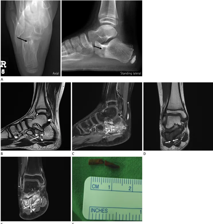
Fig. 1. A 9-year-old boy with an unrecognized foreign body retained in the calcaneus.
A. Plain radiographs with axial and standing lateral views of the right foot show a tubular geographic osteolytic lesion in the anterior body of the right calcaneus (arrows) with an ill-defined margin and surrounding sclerotic change.
B. Sagittal T2-weighted MR image shows a low signal intensity tubular lesion in the anterior body of the calcaneus (arrow) with surrounding high signal intensity tissue (arrowheads).
C. Sagittal fat-saturated enhanced T1-weighted MR image reveals non-enhancement of the tubular lesion, suggesting a foreign body (arrow), whereas the surrounding soft tissue is strongly enhanced (arrowheads), suggestive of a foreign body reaction. Note edematous bone marrow en- hancement in the body of the calcaneus (open arrows).
D, E. Coronal T1-weighted (D) and fat-saturated enhanced T1 weighted (E) MR images show a cortical defect (arrows) on the plantar surface of the calcaneal body, just below the retained foreign body. Note the focal defect with enhancement in the overlying plantar fascia (arrowheads).
F. A gross photograph of the extracted wooden foreign body.
B C D
E F
A
Axial Standing lateral
436
A Case of an Unrecognized Foreign Body Retained in the Calcaneus
jksronline.org
J Korean Soc Radiol 2017;76(6):434-437 mately 1.5 cm long low signal intensity tubular lesion in the
calcaneal body (Fig. 1B), surrounded by strongly enhanced soft tissue (Fig. 1C). MRI also showed a cortical defect on the plan- tar surface of the calcaneal body (Fig. 1D), and prominent bone marrow edema around the lesion and adjacent soft tissue signal changes (Fig. 1E). However, plantar muscles and the subcuta- neous fat layer below the retained foreign body were preserved (Fig. 1E). Radiographic diagnosis was a tubular foreign body in- side the calcaneus with surrounding foreign body granuloma for- mation and accompanying chronic osteomyelitis. However, an osteolytic tumorous condition was not completely excluded.
Surgical exploration and curettage was performed. A rod shaped wooden fragment about 1.5 cm long was found (Fig. 1F), surrounded by an inflammatory granuloma in the anterior body of the calcaneus. The foreign body was removed and curettage of the granulation tissue was performed. Pathology report showed chronic active inflammation caused by the foreign body, sugges- tive of a plant. A gram stain and AFB stain of the foreign body were negative for bacteria and yeast, and a gram stain and AFB stain of the granulation tissue were also negative.
However, the patient and his parents could not remember any occurrence of a penetration injury to the heel, in spite of inten- sive inquiry after the removal of the foreign body.
DISCUSSION
Most foreign bodies retained in the extremities are superficial and they cause temporary discomfort (7). They are most fre- quently seen in the hand, followed by the foot (1-3). Making a diagnosis of acute foreign body injury is quite easy based on the history, but it becomes difficult to diagnose it in neglected or chronic cases. Clinical manifestations can appear years after the initial injury, and the patient or the patient’s parents might not remember anything about the injury. This can lead to misdiag- nosis of the lesion and it may result in unnecessary workup and inadequate treatment for the nonexistent pathology.
Our patient also denied any history of trauma and he failed to remember the cause of the foreign body retained in the calcane- us. In addition, the skin over the injured area in the patient was normal, and the retained foreign body was a radiolucent wooden fragment. Making a diagnosis of a retained foreign body embed- ded in the bone is difficult than that of a foreign body embedded
in the soft tissue, and it is more difficult in the case of a radiolu- cent foreign body.
Most of the previously reported cases of foreign bodies re- tained in the foot bones occurred in the metatarsal bones (4, 5), and a case of a wooden foreign body retained in the calcaneus has rarely been reported (6). The soft tissue in the forefoot and the midfoot is thinner and softer than that in the heel, and there- fore, the foreign body can easily reach the bone. In addition, the- oretically, it is difficult for the foreign body to penetrate the calca- neus unless a vertically loaded strong force is applied. In the present case, the wooden foreign body was also vertically orient- ed in the calcaneus. Although in the present case we could not accurately identify the foreign body, whether it was a toothpick or just wood, since the child and his parents completely forgot about the penetration injury, we assume that the tip of the wood- en foreign body must have been sharp enough to penetrate the calcaneal cortex and there must have been a vertically oriented strong impact like weight bearing over the foreign body. There- fore, the foreign body might have been embedded momentarily within the calcaneus just after penetrating the calcaneal cortex.
According to the published literatures, most of the identified foreign bodies retained in the foot bones are thorns (8), wood (9) or toothpicks (4). Therefore, radiographic findings are usually negative except for osteolytic change. In these cases, computed tomography (CT) and MRI would help to detect the retained foreign body and would lead to appropriate treatment. In our case, the wooden fragment was not detected by the radiograph, but MRI revealed a retained foreign body with a surrounding inflammatory granuloma in the calcaneus. The foreign body was hypo-intense on T1- and T2-weighted images, and a rim-like enhanced lesion around the foreign body was seen after con- trast enhancement. Peterson et al. (10) also reported two patients with wooden foreign bodies and their MRI’s revealed a target appearance with the central foreign body appearing as a signal void or a hypointense area in contrast to the surrounding hyper- intense inflammatory tissue. However, in some cases, high signal intensity of the granulation tissue on T2-weighted image may outshine the retained foreign body and may make identification difficult (3). When the wooden foreign body is small and there is no inflammatory response, detection of the retained wooden foreign body may be difficult on MR imaging. In these cases, CT scan may be useful and it may facilitate the diagnosis of a sus-
437
Ro Woon Lee, et al
jksronline.org J Korean Soc Radiol 2017;76(6):434-437 pected wooden foreign body. Ultrasonography is also known to be useful in detecting a hidden non-radiopaque foreign body in the soft tissue, but it may not be helpful for detection of a for- eign body embedded within the bone.
In conclusion, we reported a rare case of an unrecognized wooden foreign body retained in the calcaneus and showed the usefulness of MRI in its evaluation. Radiologists need to be fa- miliar with the characteristic MRI findings and they should be able to suggest the diagnosis of a retained foreign body in spite of the unknown history.
REFERENCES
1. Reginato AJ, Ferreiro JL, O’Connor CR, Barbasan C, Arasa J, Bednar J, et al. Clinical and pathologic studies of twenty- six patients with penetrating foreign body injury to the joints, bursae, and tendon sheaths. Arthritis Rheum 1990;
33:1753-1762
2. Borgia CA. An unusual bone reaction to an organic foreign body in the hand. Clin Orthop Relat Res 1963;30:188-193 3. Dürr HR, Stäbler A, Müller PE, Refior HJ. Thorn-induced
pseudotumor of the metatarsal. A case report. J Bone Joint Surg Am 2001;83-A:580-585
4. Abu Hassan FO. Retained toothpick causing pseudotumor of the first metatarsal: a case report and literature review.
Foot Ankle Surg 2008;14:32-35
5. Madhar M, Sammous Y, Bouslous J, Messaoudi T, Chafik R, Elhaoury H, et al. Osteitis of the fourth metatarsal caused by a date palm thorn in a child: why the dorsum of the foot is the most commonly injured site. J Foot Ankle Surg 2013;52:84-87
6. Guner S, Ceylan MF, Isik D, Guner SI, Ediz L. A case of wooden foreign body retained in the calcaneus. Pak J Med Sci 2011;27:932
7. Vidyadhara S, Rao SK. Thorn prick osteomyelitis of the foot in barefoot walkers: a report of four cases. J Orthop Surg (Hong Kong) 2006;14:222-224
8. Barry M, Maffulli N, Good C. The missed thorn. Acta Orthop Belg 1992;58:468-470
9. Dhillon MS, Prasanna HM, Goni V, Nagi ON. Wooden splin- ter-induced pseudo tumour of the metatarsal. Foot Ankle Surg 2000;6:45-48.
10. Peterson JJ, Bancroft LW, Kransdorf MJ. Wooden foreign bodies: imaging appearance. AJR Am J Roentgenol 2002;
178:557-562
인지되지 못한 종골 내의 이물질: 증례 보고
이로운
1· 최수정
1* · 황재광
2· 안재홍
1· 강채훈
1· 신동락
1저자들은 인지되지 못한 종골 내의 이물질에 대한 증례를 보고하고자 한다. 환자는 외상의 기왕력이 없었고, 종골을 덮고 있 는 피부에 결손이나 국소 감염의 증거는 없었다. 이물질은 종골 단순 방사선 사진에서는 보이지 않았다. 자기공명영상에서 저신호 강도를 보이는 튜브 모양의 병변은 주위에 이물반응으로 인한 강한 조영 증강을 보이는 연조직과 골수 부종을 동반 하고 있었다. 이러한 영상 소견을 미루어 볼 때 종골 내의 이물질이 의심되었다. 외과적 소파술이 시행되었고, 막대모양의 나 무 조각이 발견되었다.
울산대학교 의과대학 강릉아산병원 1영상의학과, 2정형외과