Lower Levels of Human MOB3B Are Associated with Prostate Cancer Susceptibility and Aggressive Clinicopathological Characteristics
Mps one binder (MOB) proteins are integral components of signaling pathways that control important cellular processes, such as mitotic exit, centrosome duplication, apoptosis, and cell proliferation. However, the biochemical and cellular functions of the human MOB (hMOB) protein family remain largely unknown. The present study investigated the association between hMOB3B expression and clinicopathological characteristics of prostate cancer (PCa).Study subjects included 137 PCa patients and 137 age-matched benign prostatic hyperplasia (BPH) patients. hMOB3B expression was estimated using real-time PCR and compared with clinicopathological parameters of PCa. hMOB3B mRNA expression was significantly lower in PCa tissues than in BPH control tissues (P < 0.001). According to receiver operating characteristics curve analysis, the sensitivity of hMOB3B expression for PCa diagnosis was 84.7%, with a specificity of 86% (AUC = 0.910; 95% CI = 0.869- 0.941; P < 0.001). hMOB3B expression was significantly lower in patients with elevated prostate specific antigen (PSA) levels (≥ 10 ng/mL), a Gleason score ≥ 8, and metastatic disease (any T, N+/M+) than in those with low PSA levels, a low Gleason score, and non- metastatic disease (each P < 0.05). In conclusion, low levels of hMOB3B are closely associated with aggressive clinicopathologic features in patients with PCa. Our results suggest that hMOB3B may act as a tumor suppressor in human PCa.
Keywords: Prostatic Neoplasms; Gene Expression; Genes, Tumor Suppressor; hMOB3B Eun-Ah Kim,1 Ye-Hwan Kim,1
Ho Won Kang,1 Hyung-Yoon Yoon,1 Won Tae Kim,1 Yong-June Kim,1 Seok-Joong Yun,1 Sung-Kwon Moon,2 Yung Hyun Choi,3 Isaac Yi Kim,4 Sang-Cheol Lee,1 and Wun-Jae Kim1
1Department of Urology, College of Medicine, Chungbuk National University, Cheongju;
2Department of Food and Biotechnology, Chung-Ang University, Seoul; 3Department of Biomaterial Control, Dong-Eui University, Busan, Korea; 4Section of Urological Oncology, The Cancer Institute of New Jersey, Robert Wood Johnson Medical School, New Brunswick, New Jersey, USA Received: 24 February 2015
Accepted: 1 April 2015 Address for Correspondence:
Wun-Jae Kim, MD
Department of Urology, Chungbuk National University College of Medicine and Institute for Tumor Research, 776 1sunhwan-ro, Seowon-gu, Cheonju 362-711, Korea Tel: +82.43-269-6371, Fax: +82.43-269-6144 E-mail: [email protected]
Funding: This work was supported by the National Research Foundation of Korea (NRF) grant funded by the Korea government (MSIP) (No. NRF-2014R1A2A1A09006983, 2014R1A2A2A04007036).
http://dx.doi.org/10.3346/jkms.2015.30.7.937 • J Korean Med Sci 2015; 30: 937-942
INTRODUCTION
Prostate cancer (PCa) is the second most frequently diagnosed cancer and the sixth leading cause of cancer death in men world- wide with an approximately 14% (903,500) of total new cancer cases and 6% (258,400) of the total cancer deaths in males in 2008 (1). In Korea, PCa is the fifth most common cancer in men, and its incidence is the most rapidly increasing of all cancers (2). PCa shows an extremely heterogeneous clinical course, rang- ing from indolent to aggressive, metastatic lethal disease (3).
Consequently, there is a great need to accurately estimate the tumor characteristics of PCa so that appropriate treatment op- tions can be considered. Currently, histopathological analysis (including tumor stage and grade) and serum prostate specific antigen (PSA) levels are key determinants of therapeutic deci- sion-making. However, none of the histological criteria or bio- markers reported to date show sufficient sensitivity and speci- ficity for detecting, monitoring, or determining the prognosis of
PCa (4, 5). Thus, there is a critical need for methods capable of predicting oncologic outcomes and responses to therapy in PCa patients. The abnormal expression of certain genes in cancer cells is closely related to distinct aspects of tumor progression, including tumor growth, invasion, and metastasis. Proper cell division requires the precise coordination and execution of sev- eral events in the cell cycle, including centrosome duplication, DNA replication, mitotic spindle assembly, chromosome seg- regation, and cytokinesis (6). A failure in the execution or prop- er timing of any of these events could lead to chromosome seg- regation defects, resulting in aneuploidy or polyploidy (7-9).
Mps one binder (MOB) was originally identified in yeast as a regulator of mitotic exit and cytokinesis, and was later identi- fied as a tumor suppressor (10). hMOB1 can bind to and acti- vate mammalian large tumor suppressor (LATS) and nuclear Dbf2-related (NDR) kinases (LATS1, LATS2, NDR1, and 2) by targeting and activating these kinases at the plasma membrane (11, 12). Increased expression of LATS1 or LATS2 inhibits tumor
cell growth by inducing cell cycle arrest or apoptosis (10, 11, 13- 15). Recent studies show that hMOB1 is downregulated in color- ectal and non-small cell lung cancers (9, 12). hMOB2 binds to the same domain on NDR1/2, but does not bind to LATS1/2;
binding of hMOB2 to NDR1/2 kinases inhibits the phosphory- lation of NDR and thereby blocks kinase activation (16). hMOB2 is classified as an inhibitor of NDR signaling, whereas hMOB1 is classified as a co-activator of the NDR/LATS signaling casca- des (17, 18). The human MOB protein family consists of six mem- bers: hMOB1A, 1B, 2, 3A, 3B, and 3C (18). Unfortunately, alth- ough hMOB1 and 2 have been extensively studied, the biologi- cal roles and binding partners of hMOB3A/B/C are unknown (18). Recent studies revealed that all six hMOBs are abundantly expressed in human prostate tissue (10), and that methylation- induced gene silencing of hMOB3B occurs in PCa (19). Based on previous research, we hypothesized that hMOB3B acts as a tumor suppressor, and that its loss of function contributes to the carcinogenesis and aggressiveness of human PCa. Here, we compared the expression levels of hMOB3B in normal and pros- tate cancer tissues to examine the contribution of this gene to prostate carcinogenesis. Also, and more importantly, we inves- tigated the effect of hMOB3B on the clinicopathological charac- teristics of PCa in Korean men.
MATERIALS AND METHODS Study population
This case-control study was included 137 cases of newly diag- nosed PCa and 137 age-matched benign prostatic hyperplasia (BPH) controls. The study cases were recruited from patients with histologically confirmed primary adenocarcinoma of the prostate at our hospital. Controls were selected from a database of BPH patients who underwent transurethral resection of the prostate (TURP), and were one-to-one matched (as far as pos- sible) according to age and date of blood sampling. Controls with high PSA (serum PSA levels > 2.5 ng/mL) underwent transrec- tal prostate biopsy before TURP to rule out the presence of can- cer, and those with PSA levels > 10 ng/mL were excluded from the study. Subjects with a suspicious history of previous man- agement for PCa or incomplete medical records were also ex- cluded. Gleason grade and 2002 TNM stage were used as prog- nostic factors. Gleason grade was measured in 12-core transrec- tal biopsy, TURP, or radical prostatectomy specimens. Tumor stage was estimated from the radical prostatectomy specimens, or from computed tomography (CT), magnetic resonance im- aging (MRI), or bone scan results.
RNA extraction and construction of cDNA
TRIzol (1 mL; Invitrogen, Carlsbad, USA) was added to BPH con- trol and PCa tissues and homogenized in a 5 mL glass tube. The homogenate was transferred to a 1.5 mL tube and mixed with
200 μL chloroform. After incubating for 5 min at 4°C, the homo- genate was centrifuged for 13 min at 13,000 × g at 4°C. The up- per aqueous phase was transferred to a clean tube and 500 μL isopropanol was added, followed by incubation for 60 min at 4°C. The tube was then centrifuged for 8 min at 13,000 × g and 4°C. Next, the upper aqueous phase was removed, mixed with 500 μL of 75% ethanol, and centrifuged for 5 min at 13,000 × g and 4°C. After the upper aqueous layer was discarded, the pel- let was dried at room temperature, dissolved with diethylpyro- carbonate (DEPC)-treated water, and stored at -80°C. The qual- ity and integrity of the RNA were confirmed by agarose gel elec- trophoresis and ethidium bromide staining, followed by visual examination under ultraviolet light. cDNA was then prepared from 1 μg f RNA by random priming with a First-Strand cDNA Synthesis Kit (Amersham Biosciences Europe GmbH, Freiburg, Germany) according to the manufacturer’s protocol.
Real-time PCR
To quantify the expression of hMOB3B, real-time PCR was per- formed using a Rotor Gene 6000 instrument (Corbett Research, Mortlake, Australia). Real-time PCR assays using SYBR Premix EX Taq (TAKARA BIO INC., Otsu, Japan) were carried out in mi- cro-reaction tubes (Corbett Research, Mortlake, Australia) using hMOB3B (6,528 bp) sense (5´-GTG GCA GGA TGA TCT CAA- 3´) and antisense (5´-CGG CAC AGG ATC TTC TTG-3´) primers.
The PCR reaction was performed in a final volume of 10 μL, com- prising 5 μL of 2 × SYBR Premix EX Taq buffer, 0.5 μL of each 5´
and 3´ primer (10 pM/μL), and 1 μL of sample cDNA. The prod- ucts were purified with a QIAquick Extraction kit (QIAGEN, Hil- den, Germany), quantified in a spectrometer (Perkin Elmer MBA2000, Fremont, USA), and sequenced using an automated laser fluorescence sequencer (ABI PRISM 3100 Genetic Analyz- er, Foster City, USA). A known concentration of the PCR product was then 10-fold serially diluted from 100 pg/μL to 0.1 pg/μL and used to establish a standard curve. The real-time PCR con- ditions were as follows: 1 cycle at 96°C for 20 sec, followed by 40 cycles of 3 sec at 96°C for denaturation, 15 sec at 60°C for anneal- ing, and 15 sec at 72°C for extension. The melting program was performed at 72-95°C, with a heating rate of 1°C per 45 sec. Spec- tral data were PCatured and analyzed using Rotor Gene Real- Time Analysis Software 6.0 Build 14 (Corbett Research, Mort- lake, Australia). All samples were run in triplicate. Glyceralde- hyde-3-phosphate dehydrogenase (GAPDH) was used as an endogenous RNA reference gene. Gene expression was normal- ized to the expression of GAPDH.
Statistical analysis
Clinical variables such as age, PSA, prostate size, and BMI were compared using the Mann-Whitney U-test. To evaluate tumor characteristics, Gleason scores were classified as ≤ 7 and ≥ 8, and the clinical stage was categorized as ≤ T4 or metastatic dis-
ease (any nodal or distant metastasis). Statistical analysis was performed using SPSS 20.0 software (SPSS Inc., Chicago, IL, USA), and a P value < 0.05 was considered statistically significant.
Ethics statement
The study protocol of collection and analyses of all samples was reviewed and approved by the institutional review board of Chung- buk National University (IRB approved number 2006-01-001).
Informed consent was obtained from each subject.
RESULTS
Baseline characteristics
Table 1 lists the baseline clinical and pathologic characteristics of the 137 BPH controls and the 137 PCa patients. The mean age of the PCa patients and controls was 69.2 yr. The serum PSA level was higher in PCa patients than in the BPH controls (124.9
± 380.6 vs. 4.0 ± 5.2; P < 0.001). There was no significant differ- ence between cases and controls regarding prostate size (P = 0.037). Among the 137 PCa patients, 72 (52.6%) underwent rad- ical prostatectomy and the other 65 (47.4%) underwent pallia- tive TURP. Stage and Gleason grade at diagnosis were as follows:
91 cases had localized-to-advanced disease (T2-4N0M0) and 46 (33.6%) had metastatic disease (any T, N+/M+); the Gleason score was ≤ 7 and ≥ 8 for 64 (46.7%) and 73 (53.3%) patients, respectively.
Expression levels of hMOB3B mRNA in normal and cancer tissues
To identify whether hMOB3B is involved in prostate cancer sus- ceptibility, we compared mRNA expression levels in BPH and PCa tissues. The median hMOB3B mRNA expression level in PCa tissues was significantly lower (median 426.2 × 10⁴ copies per
μg; IQR, 197.6-637.0) than that in BPH tissues (median 1,108.5
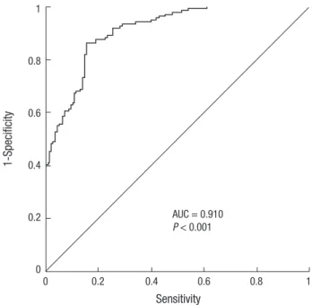
×104 copies per μg; IQR, 797.0-1,428.0) (P < 0.001, Table 1 and Fig. 1). According to receiver operating curve (ROC) statistical analysis, the sensitivity of hMOB3B expression for diagnosing prostate cancer was 84.7%, with a specificity of 86% at a refer- ence value of 737.5 × 10⁴ copies per μg (AUC, 0.910; 95% CI, 0.869- 0.941; P < 0.001) (Fig. 2).
Relationship between hMOB3B mRNA expression levels and clinicopathologic features
hMOB3B mRNA expression was significantly lower in patients with elevated PSA levels (≥ 10 ng/mL) than in those with low PSA levels (P = 0.001). In addition, hMOB3B expression was
MOBKL2B expression (×104 copies per μg)
BPH CaP
2,500
2,000
1,500
1,000
500
0
Fig. 1. hMOB3B mRNA expression levels in normal and cancer tissues. BPH, benign prostatic hyperplasia; CaP, prostate cancer.
Fig. 2. Receiver operating characteristic (ROC) curve generated to calculate the sen- sitivity and specificity of hMOB3B for detecting prostate cancer. AUC, area under the curve.
1-Specificity
Sensitivity
0 0.2 0.4 0.6 0.8 1 1
0.8
0.6
0.4
0.2
0
AUC = 0.910 P < 0.001 Table 1. Clinicopathologic characteristics of prostate cancer patients and BPH controls
Characteristics BPH PCa P value
No. 137 137
Age (yr; range) 69.2 (46-85) 69.2 (48-86) 0.994 PSA ± SD (ng/mL) 4.0 ± 5.2 124.9 ± 380.6 < 0.001 Prostate size (gram) 38.8 ± 23.0 41.3 ± 22.2 0.387 Operation (%)
TURP
Radical prostatectomy 137 (100) 65 (47.4) 72 (52.6) Gleason score, No. (%)
≤ 7
≥ 8 64 (46.7)
73 (53.3) Stage (%)
T1-4N0M0
Metastatic (any T N+/M+) 91 (66.4)
46 (33.6) hMOB3B expression, median
(IQR; × 104 copies per μg) 1,108.5
(797.0-1,428.0) 426.2
(197.6-637.0) < 0.001 P values were obtained from the Mann-Whitney U-test. BPH, benign prostatic hyper- plasia; PCa, prostate cancer; IQR, interquartile range; SD, standard deviation; TURP, transurethral resection of the prostate.
Table 2. hMOB3B mRNA expression levels according to PSA, Gleason score, and metastasis status in prostate cancer patients
Characteristics No hMOB3B expression, median
(IQR; × 104 copies/μg) P value PSA
≤ 10
> 10 38
99 545.2 (382.9-730.3) 349.6 (123.6-569.9)
0.001
Gleason score ≤ 7
≥ 8 64
73 509.8 (300.6-687.3) 320.9 (96.9-545.2)
< 0.001
Stage T1-4N0M0
any T N+/M+ 91
46 491.1 (299.2-716.4) 243.8 (96.4-448.4)
< 0.001
P values were obtained from the Mann-Whitney U-test. IQR, interquartile range; PSA, prostate specific antigen; SD, standard deviation.
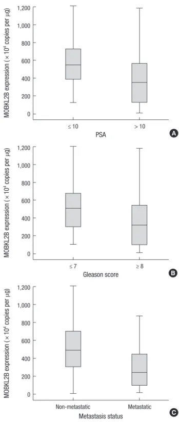
Fig. 3. Relationship between hMOB3B mRNA expression levels and clinicopathologic features in prostate cancer. (A) PSA, (B) Gleason score, and (C) metastatic status.
MOBKL2B expression (×104 copies per μg)
≤ 10 > 10
PSA 1,200
1,000
800
600
400
200
0
A
MOBKL2B expression (×104 copies per μg)
≤ 7 ≥ 8
Gleason score 1,200
1,000
800
600
400
200
0
B
MOBKL2B expression (×104 copies per μg)
Non-metastatic Metastatic
Metastasis status 1,200
1,000
800
600
400
200
0
C significantly lower in cancer tissue specimens from patients with
a higher Gleason score (≥ 8) and metastatic disease (any T, N+/
M+) than in samples from those with a lower Gleason score (≤ 7) and non-metastatic disease (T2-4N0M0) (each P < 0.001) (Ta- ble 2 and Fig. 3A-C).
DISCUSSION
In this study, we identified a relationship between hMOB3B ex- pression and an increased risk of PCa. In addition, hMOB3B ex- pression was closely associated with clinicopathologic features in patients with PCa. MOB proteins are crucial regulators of NDR family kinases and are conserved from yeast to humans. Mem- bers of the MOB protein family regulate mitosis, cell prolifera- tion, apoptosis, centrosome biology, and morphological chang- es (12, 20). Signal transduction cascades control essential bio- logical processes such as cell division, morphogenesis, cell grow- th, and controlled cell death/apoptosis (21). The vast majority of signaling cascades transmit extra- and intracellular inputs via protein kinases (11, 22). A conserved property of MOB pro- teins is its association with and activation of NDR (nuclear-Dbf2- related) kinases belonging to the AGC family, which includes PKA, PKG, and PKC (18, 23). Human cells express four related NDR kinases: NDR1 (also known as serine/threonine kinase 38 or STK38), NDR2 (or STK38L), LATS1 (large tumor suppressor-1), and LATS2 (11, 24-26). Originally, the biological roles of MOB proteins were investigated mainly using budding and fission yeast. In budding and fission yeast, MOB proteins are expressed from two independent genes. Mob1p regulates both the mitotic exit network in budding yeast and the septation initiation net- work in fission yeast by binding to and activating Dbf2p and Sid2p kinases, respectively (11, 12, 25, 27). In addition, Mob1p also plays a major role in modulating cytokinesis, the last stage of the cell cycle (28). MOB2 regulates cell polarity and daughter- specific gene expression programs during the yeast cell cycle by modulating Cbk1p and Orb6p kinases (29). The human MOB protein family consists of six distinct mem bers (hMOB1A, 1B, 2, 3A, 3B, and 3C), with hMOB1A/B being the best studied due to
their putative tumor suppressive functions, which are mediated through regulation of the NDR/LATS kinases (10, 11, 18, 24). The biological features of the other hMOBs are largely unknown.
hMOB1A binds to and activates human NDR1/2 kinases by sti- mulating autophosphorylation of the activation segment. Simi- larly, hMOB1A binds to and activates LATS1 and 2 (11, 18, 24).
By contrast, hMOB2 binds to NDR1 and NDR2, but not to LATS1 (16). hMOB3A/B/C do not associate with any NDR/LATS kina- ses, and the binding partners of hMOB3A/B/C are currently unknown. Recently, a study by Haldrup et al. (16) reported that hypermethylation of six novel genes, AOX1, C1orf114, GAS6, HAPLN3, KLF8, and MOB3B, was highly cancer specific. Al- though we did not measure the correlation between hMOB3B methylation and hMOB3B expression levels, our data support methylation-induced gene silencing of hMOB3B.
A possible limitation of our study is that we did not determine the levels of hMOB3B protein, for example, by Western blotting or immunohistochemical staining. Second, we could not evalu- ate the prognostic value of hMOB3 due to relatively small sam- ple size and inconsistent treatment modalities. Most importantly, the physiological binding partners and functions of the hMOB3 proteins remain undefined. Thus, further research is needed to identify physiological binding partners and to confirm the prog- nostic value of hMOB3 in PCa.
In conclusion, low levels of hMOB3B are closely associated with aggressive clinicopathologic features of PCa. Our results suggest a functional role for hMOB3B as a tumor suppressor in human PCa.
ACKNOWLEDGEMENT
The biospecimens for this study were provided by the Chung- buk National University Hospital, a member of the National Bio- bank of Korea, which is supported by the Ministry of Health, Wel- fare and Family Affairs. All samples derived from the National Biobank of Korea were obtained with informed consent under institutional review board-approved protocols.
DISCLOSURE
The authors have no potential financial conflicts on this subject.
AUTHOR CONTRIBUTION
Conception and design of the study: Kim EA, Kang HW, Yun SJ.
Acquisition of data: Kim EA, Kim YH, Yoon HY. Statistical anal- ysis: Kim WT, Kim YJ, Yun SJ. First draft of manuscript: Kim EA, Kang HW. Revision and critical review of the manuscript: Moon SK, Choi YH, Kim IY, Lee SC, Kim WJ. Manuscript approval: all authors.
ORCID
Eun-Ah Kim http://orcid.org/0000-0003-0637-1067 Ye-Hwan Kim http://orcid.org/0000-0002-8676-7119 Ho Won Kang http://orcid.org/0000-0002-8164-4427 Won Tae Kim http://orcid.org/0000-0002-7482-4550
Yong-June Kim http://orcid.org/0000-0001-7638-7174 Seok-Joong Yun http://orcid.org/0000-0001-7737-4746 Sung-Kwon Moon http://orcid.org/0000-0002-4514-3457 Yung Hyun Choi http://orcid.org/0000-0002-1454-3124 Isaac Yi Kim http://orcid.org/0000-0002-1967-5281 Sang-Cheol Lee http://orcid.org/0000-0002-4163-2210 Wun-Jae Kim http://orcid.org/0000-0002-8060-8926
REFERENCES
1. Jemal A, Bray F, Center MM, Ferlay J, Ward E, Forman D. Global cancer statistics. CA Cancer J Clin 2011; 61: 69-90.
2. Lee HW, Jeon HG, Jeong BC, Seo SI, Jeon SS, Choi HY, Lee HM. Com- parison of pathological and biochemical outcomes after radical prosta- tectomy in Korean patients with serum PSA ranges. J Korean Med Sci 2015; 30: 317-22.
3. Seo WI, Kang PM, Chung JI. Predictive value of the cancer of the prostate risk assessment score for recurrence-free survival after radical prostatec- tomy in Korea: a single-surgeon series. Korean J Urol 2014; 55: 321-6.
4. Kattan MW, Eastham JA, Stapleton AM, Wheeler TM, Scardino PT. A preoperative nomogram for disease recurrence following radical prosta- tectomy for prostate cancer. J Natl Cancer Inst 1998; 90: 766-71.
5. Partin AW, Kattan MW, Subong EN, Walsh PC, Wojno KJ, Oesterling JE, Scardino PT, Pearson JD. Combination of prostate-specific antigen, clin- ical stage, and Gleason score to predict pathological stage of localized prostate cancer. a multi-institutional update. JAMA 1997; 277: 1445-51.
6. Negrini S, Gorgoulis VG, Halazonetis TD. Genomic instability: an evolv- ing hallmark of cancer. Nat Rev Mol Cell Biol 2010; 11: 220-8.
7. Charames GS, Bapat B. Genomic instability and cancer. Curr Mol Med 2003; 3: 589-96.
8. Romanowski P, Madine MA. Mechanisms restricting DNA replication to once per cell cycle: MCMS, pre-replicative complexes and kinases. Trends Cell Biol 1996; 6: 184-8.
9. Sasaki H, Kawano O, Endo K, Suzuki E, Yukiue H, Kobayashi Y, Yano M, Fujii Y. Human MOB1 expression in non-small-cell lung cancer. Clin Lung Cancer 2007; 8: 273-6.
10. Chow A, Hao Y, Yang X. Molecular characterization of human homologs of yeast MOB1. Int J Cancer 2010; 126: 2079-89.
11. Hergovich A. MOB control: reviewing a conserved family of kinase regu- lators. Cell Signal 2011; 23: 1433-40.
12. Lai ZC, Wei X, Shimizu T, Ramos E, Rohrbaugh M, Nikolaidis N, Ho LL, Li Y. Control of cell proliferation and apoptosis by mob as tumor suppres- sor, mats. Cell 2005; 120: 675-85.
13. Yang X, Li DM, Chen W, Xu T. Human homologue of Drosophila lats, LATS1, negatively regulate growth by inducing G(2)/M arrest or apopto- sis. Oncogene 2001; 20: 6516-23.
14. Zhang J, Smolen GA, Haber DA. Negative regulation of YAP by LATS1 underscores evolutionary conservation of the Drosophila Hippo path- way. Cancer Res 2008; 68: 2789-94.
15. Ke H, Pei J, Ni Z, Xia H, Qi H, Woods T, Kelekar A, Tao W. Putative tumor suppressor Lats2 induces apoptosis through downregulation of Bcl-2 and Bcl-x(L). Exp Cell Res 2004; 298: 329-38.
16. Hergovich A. Regulation and functions of mammalian LATS/NDR ki- nases: looking beyond canonical Hippo signalling. Cell Biosci 2013; 3: 32.
17. Hergovich A. Mammalian Hippo signalling: a kinase network regulated by protein-protein interactions. Biochem Soc Trans 2012; 40: 124-8.
18. Kohler RS, Schmitz D, Cornils H, Hemmings BA, Hergovich A. Differ- ential NDR/LATS interactions with the human MOB family reveal a neg- ative role for human MOB2 in the regulation of human NDR kinases. Mol Cell Biol 2010; 30: 4507-20.
19. Haldrup C, Mundbjerg K, Vestergaard EM, Lamy P, Wild P, Schulz WA, Arsov C, Visakorpi T, Borre M, Høyer S, et al. DNA methylation signa- tures for prediction of biochemical recurrence after radical prostatecto- my of clinically localized prostate cancer. J Clin Oncol 2013; 31: 3250-8.
20. Hergovich A, Cornils H, Hemmings BA. Mammalian NDR protein ki- nases: from regulation to a role in centrosome duplication. Biochim Bio- phys Acta 2008; 1784: 3-15.
21. Manning G, Whyte DB, Martinez R, Hunter T, Sudarsanam S. The pro- tein kinase complement of the human genome. Science 2002; 298: 1912- 34.
22. Hergovich A, Stegert MR, Schmitz D, Hemmings BA. NDR kinases reg- ulate essential cell processes from yeast to humans. Nat Rev Mol Cell Biol 2006; 7: 253-64.
23. Tamaskovic R, Bichsel SJ, Hemmings BA. NDR family of AGC kinases- -essential regulators of the cell cycle and morphogenesis. FEBS Lett 2003;
546: 73-80.
24. Praskova M, Xia F, Avruch J. MOBKL1A/MOBKL1B phosphorylation by MST1 and MST2 inhibits cell proliferation. Curr Biol 2008; 18: 311-21.
25. Bothos J, Tuttle RL, Ottey M, Luca FC, Halazonetis TD. Human LATS1 is a mitotic exit network kinase. Cancer Res 2005; 65: 6568-75.
26. Yabuta N, Okada N, Ito A, Hosomi T, Nishihara S, Sasayama Y, Fujimori A, Okuzaki D, Zhao H, Ikawa M, et al. Lats2 is an essential mitotic regu- lator required for the coordination of cell division. J Biol Chem 2007; 282:
19259-71.
27. Hou MC, Salek J, McCollum D. Mob1p interacts with the Sid2p kinase and is required for cytokinesis in fission yeast. Curr Biol 2000; 10: 619-22.
28. Luca FC, Mody M, Kurischko C, Roof DM, Giddings TH, Winey M. Sac- charomyces cerevisiae Mob1p is required for cytokinesis and mitotic exit.
Mol Cell Biol 2001; 21: 6972-83.
29. Hou MC, Wiley DJ, Verde F, McCollum D. Mob2p interacts with the pro- tein kinase Orb6p to promote coordination of cell polarity with cell cycle progression. J Cell Sci 2003; 116: 125-35.