In vitro performance and fracture resistance of novel CAD/CAM ceramic molar crowns
loaded on implants and human teeth
Verena Preis*, Sebastian Hahnel, Michael Behr, Martin Rosentritt
Department of Prosthetic Dentistry, UKR University Hospital Regensburg, Regensburg, Germany
PURPOSE. To investigate the fatigue and fracture resistance of computer-aided design and computer-aided manufacturing (CAD/CAM) ceramic molar crowns on dental implants and human teeth. MATERIALS AND METHODS. Molar crowns (n=48; n=8/group) were fabricated of a lithium-disilicate-strengthened lithium aluminosilicate glass ceramic (N). Surfaces were polished (P) or glazed (G). Crowns were tested on human teeth (T) and implant-abutment analogues (I) simulating a chairside (C, crown bonded to abutment) or labside (L, screw channel) procedure for implant groups. Polished/glazed lithium disilicate (E) crowns (n=16) served as reference. Combined thermal cycling and mechanical loading (TC: 3000×5°C/3000×55°C; ML: 1.2×10⁶ cycles, 50 N) with antagonistic human molars (groups T) and steatite spheres (groups I) was performed under a chewing simulator. TCML crowns were then analyzed for failures (optical microscopy, SEM) and fracture force was determined. Data were statistically analyzed (Kolmogorow-Smirnov, one-way-ANOVA, post-hoc Bonferroni, α=.05). RESULTS. All crowns survived TCML and showed small traces of wear. In human teeth groups, fracture forces of N crowns varied between 1214±293 N (NPT) and 1324±498 N (NGT), differing significantly (P≤.003) from the polished reference EPT (2044±302 N). Fracture forces in implant groups varied between 934±154 N (NGI_L) and 1782±153 N (NPI_C), providing higher values for the respective chairside crowns. Differences between polishing and glazing were not significant (P≥.066) between crowns of identical materials and abutment support. CONCLUSION. Fracture resistance was influenced by the ceramic material, and partly by the tooth or implant situation and the clinical procedure (chairside/labside). Type of surface finish (polishing/glazing) had no significant influence. Clinical survival of the new glass ceramic may be comparable to lithium disilicate. [J Adv Prosthodont 2018;10:300-7]
KEYWORDS: Glass ceramic; Dental crown; Dental implant; Fatigue; Fracture resistance
INTRODUCTION
Natural esthetic appearance and longevity of the restoration combined with an efficient workflow in the laboratory and dental practice are major aims in restoring teeth or implants
in prosthetic dentistry. With increasing capabilities and accep- tance of computer-aided design and computer-aided manu- facturing (CAD/CAM) especially for single restorations, new machinable esthetic materials have been introduced. While lithium disilicate and zirconia ceramics have been successfully clinically applied for many years,1-7 new classes of zirconia- reinforced lithium silicate ceramics, ceramic-network materi- als and, recently, lithium-disilicate-strengthened lithium alu- minosilicate glass ceramics have been introduced. While most previously used ceramic materials required time-con- suming working steps performed in the dental laboratory (milling, sintering/crystallization), chairside ceramics that are easy to mill in a fully crystallized state may represent an advantageous option. Some new classes of ceramics offer chairside fabrication without subsequent crystallization, while esthetics and mechanical properties may be compara- ble to lithium disilicate. With a complete chairside workflow,
Corresponding author:
Verena Preis
Department of Prosthetic Dentistry, UKR University Hospital Regensburg Franz-Josef-Strauss-Allee 11, 93042 Regensburg, Germany
Tel. +499419446073: e-mail, [email protected]
Received January 25, 2018 / Last Revision March 7, 2018 / Accepted May 8, 2018
© 2018 The Korean Academy of Prosthodontics
This is an Open Access article distributed under the terms of the Creative Commons Attribution Non-Commercial License (http://creativecommons.
org/licenses/by-nc/3.0) which permits unrestricted non-commercial use, distribution, and reproduction in any medium, provided the original work is properly cited.
the clinical procedure of the surface finish gains impor- tance. Chairside polishing may be an alternative to glazing in the laboratory.
The advantages of a digital workflow (intraoral scanning, CAD/CAM) in combination with choosing an appropriate dental material may not only be relevant for tooth restora- tions but also for implant-supported crowns. The success of chairside cemented implant restorations may be limited by gingival and peri-implant inflammation effects caused by residual cement.8 Labside bonding of implant crowns to a titanium base and leaving a screw hole for chairside fixation may resolve this problem, but the strength of the crown may be affected by the presence of the screw hole.9-13 For both the chairside and the labside procedures of implant-sup- ported crown fabrication, an altered loading situation with increased masticatory forces by rigid implant bearing exists, and therefore a higher fatigue and fracture resistance may be required for implant-supported restorations.14-16 Previous studies have shown that CAD/CAM materials may perform differently depending on their use as implant- or tooth-sup- ported restorations.9,11
Clinical evidence for survival of new chairside CAD/
CAM materials is very limited.17,18 First in vitro results about performance and material characterization of recently intro- duced CAD/CAM materials (e.g. zirconia-reinforced lithi- um silicate ceramics, hybrid materials) are promising.19-23 However, further in vitro investigations are necessary as advanced ceramic materials as lithium disilicate strength- ened lithium aluminosilicate glass ceramics are constantly launched on the market.
Prior to routine clinical application, in vitro tests may facilitate a contemporary evaluation of new materials and restorations by combining reproducible laboratory condi- tions with basic requirements (occlusal loading, thermocy- cling) of the clinical situation.
The hypothesis of this study was that the in vitro perfor- mance and fracture resistance of lithium-disilicate-strength- ened lithium aluminosilicate glass ceramic crowns
a) are comparable to lithium disilicate crowns,
b) are affected by the surface treatment (polished or glazed),
c) depend on the tooth or implant abutment situation.
MATERIALS AND METHODS
The study investigated molar crowns fabricated of a novel lithium-disilicate-strengthened lithium aluminosilicate glass ceramic (Group N, Li2O-Al2O3-SiO2, Nice, Straumann, Freiburg, Germany) in comparison to a well-known lithium disilicate glass ceramic (Group E, Li2Si2O5, IPS e.max CAD, Ivoclar Vivadent, Schaan, Liechtenstein) as a reference mate- rial. Further details on the materials used are given in Table 1. The lithium disilicate strengthened lithium aluminosilicate glass-ceramic was manufactured by a co-crystallization of lithium disilicate and lithium aluminosilicate upon con- trolled firing of a starting glass.
The molar crowns (n = 64; n = 8/group) were tested with two different surface modifications (P = polished, G = glazed) on human teeth (T) or implant-abutment analogues (I). For the implant groups, either a chairside (C) or labside (L, with screw channel) clinical procedure was simulated.
The following eight groups were defined:
NPT: Nice polished crowns tested on human teeth.
NGT: Nice glazed crowns tested on human teeth.
EPT: E.max CAD polishedcrowns tested on human teeth (reference group).
EGT: E.max CAD glazedcrowns tested on human teeth (reference group).
NPI_L: Nice polished crowns tested on implant abut- ments with labside procedure.
NGI_L: Nice glazed crowns tested on implant abut- ments with labside procedure.
NPI_C: Nice polished crowns tested on implant abut- ments with chairside procedure.
NGI_C: Nice glazed crowns tested on implant abut- ments with chairside procedure.
For the tooth groups (T), extracted caries-free human molars (mandibular right first molar, n = 32) were collected and stored in 0.5% chloramine solution for no longer than 4 weeks. The variability of human molars was respected by preselecting teeth with comparable size and shape and by randomly dividing the teeth to the subgroups. The teeth were prepared according to ceramic guidelines respecting a circular and occlusal anatomical reduction of 1.5 mm and a preparation angle of 4 degrees. A 1 mm deep cervical circu- lar shoulder with rounded inner angles was prepared. All
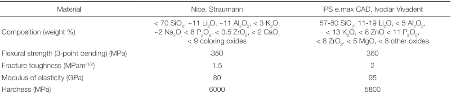
Table 1. Materials, information provided by the manufacturers
Material Nice, Straumann IPS e.max CAD, Ivoclar Vivadent
Composition (weight %)
< 70 SiO2, ~11 Li2O, ~11 Al2O3, < 3 K2O,
~2 Na2O, < 8 P2O5, < 0.5 ZrO2, < 2 CaO,
< 9 coloring oxides
57-80 SiO2, 11-19 Li2O, < 5 Al2O3,
< 13 K2O, < 8 ZnO, < 11 P2O5,
< 8 ZrO2, < 5 MgO, < 8 other oxides
Flexural strength (3-point bending) (MPa) 350 360
Fracture toughness (MPam-1/2) 1.5 2
Modulus of elasticity (GPa) 80 95
Hardness (MPa) 6000 5800
teeth were prepared by one person with identical prepara- tion equipment. Standardized preparation was performed on basis of an original model, and the preparation design was controlled with a gauge.
The roots of the teeth were coated with a 1 mm poly- ether layer (Impregum, 3M, Seefeld, Germany) to simulate the human periodontium and the resilience of the teeth.
Therefore, the roots of the teeth were dipped in wax, which was replaced by polyether in a subsequent fabrication pro- cess 24-26 before the teeth were vertically fixed in sample holders (Palapress Vario, Kulzer, Hanau, Germany).
For the implant groups (I), implant-abutment analogues (n = 32; Straumann, Freiburg, Germany, titanium grade IV, implant diameter 4.1 mm, implant length 12 mm, abutment length 6 mm, 6°) were vertically positioned in resin blocks (Palapress Vario) in order to simulate a posterior implant situation replacing the mandibular right first molar.
All abutments and prepared teeth were digitalized (Cerec Omnicam, Sirona, Bensheim, Germany) and full-contour crowns of the lithium-disilicate-strengthened lithium alumi- nosilicate glass ceramic (group N, n = 48) and the lithium disilicate glass ceramic (reference group E, n = 16) were milled (Cerec, MCXL, Sirona, Bensheim, Germany). As the lithium-disilicate-strengthened lithium aluminosilicate glass ceramic blocks were milled in a fully crystallized state, only the lithium disilicate glass crowns were crystallized after milling.
Half of the specimens were polished (OptraFine, Ivoclar Vivadent, Schaan, Liechtenstein) or glazed (group N: Vita Akzent Plus, Vita Zahnfabrik, Bad Säckingen, Germany;
group E: IPS e.max ceram glaze, Ivoclar Vivadent, Schaan, Liechtenstein) according to the manufacturers’ instructions.
Crown dimensions on human teeth (groups T) were: cir- cular and occlusal wall thickness 1.5 mm and cervical wall thickness 1 mm. All crowns were etched (5% hydrofluoric acid, 20 seconds) and silanized (silane coupling agent Monobond Plus, Ivoclar Vivadent, Schaan, Liechtenstein, 60 seconds). Before adhesive bonding (Variolink Esthetic DC, Ivoclar Vivadent, Schaan, Liechtenstein; Elipar Trilight, 3M, Seefeld, Germany, 3 × 60 seconds), the prepared teeth were treated with the bonding system Syntac classic (Syntac Primer/ Syntac Adhesive/ Heliobond, Ivoclar Vivadent, Schaan, Liechtenstein) according to the manufacturer’s instructions.
For the implant groups (I), all abutments were sand- blasted (110 µm Al2O3, 1.5 bar) and the crowns were condi- tioned analogous to the tooth groups. Identical crowns were fabricated for two situations: a) chairside procedure: the crowns were directly bonded onto the implant-abutment analogue and the excess luting material was removed; b) lab- side procedure: a screw channel was manually drilled into the central fossa of the crown with a diamond bur (red/
fine, diameter: 1.5 mm, water cooling). The crowns were bonded onto the implant-abutment analogue, the excess lut- ing material was removed, and the screw channel was restored with composite (Filtek Supreme; 3M, Seefeld, Germany; Elipar Trilight, 40 seconds).
A combined thermal cycling and mechanical loading (TCML: 3000 × 5°C/ 3000 × 55°C, 2 minutes each cycle, H2O distilled; 1.2 × 10⁶ cycles à 50 N, 1.6 Hz) was performed in a chewing simulator (eGo Kältesysteme, Regensburg, Germany). Extracted human molars were used as antago- nists in three-point-contact for crowns on human teeth.
Steatite balls (diameter: 12 mm, CeramTec, Plochingen, Germany) were used to standardize antagonists in three- point-contact on identical implant crowns. Parameters were chosen on data of zirconia and ceramic restorations simu- lating a maximum of five years of oral service.27,28
During TCML all crowns were controlled daily for fail- ures and failed crowns were excluded from further simula- tion and testing. After TCML all crowns were investigated in detail for failure analysis with optical microscopy (light microscope SV8, Olympus, Hamburg, Germany, magnifica- tion ×10) and scanning electron microscopy (SEM, Quanta FEG 400, FEI, Hillsboro, Oregon, USA, low vacuum, 10 keV, magnification ×20-100). Wear facets of the tooth groups were characterized. All crowns that survived TCML were loaded to fracture (universal testing machine Zwick 1446, Zwick, Ulm, Germany). The force was applied on the centre of the crowns using a steel ball (diameter: 12 mm, velocity: 1 mm/min) with a 1 mm tin foil (Dentaurum, Ispringen, Germany) that was inserted between crown and ball to prevent force peaks. The failure determination was set to 10% decrease of the maximum force or acoustic signal failure (crack). Power calculation (G*Power 3.1.3, HHU Düsseldorf, Germany) provided an estimated power of > 90%
using eight specimens per group. Force distribution was controlled with the Kolmogorow-Smirnov-test. Mean values and standard deviations (SD) were calculated and analysed by one-way analysis of variance (ANOVA) and the Bonferroni multiple comparison test for post hoc analysis (SPSS 23, IBM, Armonk, NY, USA, α = .05).
RESULTS
No crowns failed during TCML. All crowns showed small wear traces in their individual contact points. No chipping or fractures were found, but small cone cracking was partly observed both for polished and glazed crowns. For glazed crowns, worn inhomogeneous glaze material was found in the marginal areas of the wear facets. No strong differences were found between the lithium-disilicate-strengthened lithi- um aluminosilicate glass ceramic and the reference material.
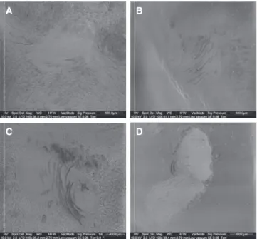
Individual surface inhomogeneities with a diameter smaller than 1 mm were found for all materials. Enamel antagonists showed scratches and inhomogeneities, with some cracks and fractures. Exemplary SEM pictures of worn ceramic surfaces are given in Fig. 1.
Fracture forces in the tooth groups (T) were 1214 ± 293 N (NPT) and 2044 ± 302 N (EPT) for polished crowns, and 1324 ± 498 N (NGT) and 1550 ± 317 N (EGT) for glazed crowns. Statistical comparison revealed significant (P = .000, ANOVA) differences between the materials. Individual differences (Bonferroni) were found between EPT and
NPT (P = .001) and NGT (P = .003). For both materials, no significantly (P > .066) different fracture results were found between glazed or polished crowns. Polished refer- ence crowns showed significantly (P = .001) higher fracture results.
Fracture values for the crowns on implants varied between 934 ± 154 N (NGI_L) and 1782 ± 153 N (NPI_
C). Chairside crowns for both types (glazed and polished) provided higher fracture results than the respective labside crowns. For both clinical procedures (L, C) lower fracture values were found for glazed crowns, but the results were not statistically different (P > .152) (Table 2).
Failures were characterized by fracture of the crown in all cases, partly combined with a complete debonding of the crown. An exemplary light microscope picture is given in Fig. 2.
DISCUSSION
Lithium-disilicate-strengthened lithium aluminosilicate glass ceramic crowns showed similar performance during in vitro TCML, but lower fracture resistance than the lithium disili- cate reference crowns, rejecting the first part of the hypoth- esis with regard to fracture resistance. Polishing or glazing did not significantly influence the results. Therefore, the second part of the hypothesis was rejected. The third part of the hypothesis was confirmed with restrictions since only chairside implant-supported crowns partly showed higher fracture resistance in comparison to labside implant- and tooth-supported crowns.
A CAD/CAM lithium disilicate ceramic has been cho- sen as reference material in this study, as it has been proven to be suitable for single tooth restorations both in vivo1,2,4,29,30
and in vitro19,31-33 for many years. Fracture forces of these reference crowns are comparable to previous results, rang- ing between about 1000 N to 2500 N,19,31,34 depending on the testing conditions. The present results showed that the lithium-disilicate-strengthened lithium aluminosilicate glass ceramic crowns had comparable resistance against fatigue as the reference crowns, but lower fracture resistance in the respective tooth groups. Nevertheless, as the fracture values exceeded maximum chewing forces, which are reported to be up to 900 N,35 all groups of molar crowns are able to withstand occlusal forces applied in the posterior region.
Differences in fracture resistance between the two materials may be explained by the material composition and the mechanical properties like fracture toughness and modulus of elasticity (Table 1). The chairside machining of fully crystallized lithium disilicate strengthened lithium alumino- silicate glass ceramic crowns may also influence fatigue and fracture resistance. A previous study about the machinability of CAD/CAM materials has found a higher susceptibility to edge chipping induced by milling for ceramic-based Table 2. Fracture forces (mean ± standard deviation) in N
Code Fracture force (N) Lowest value (N) Fracture pattern
NPT 1214 ± 293bd 911 crown fracture
NGT 1324 ± 498a 850 crown fracture
EPT 2044 ± 302ab 1613 crown fracture
EGT 1550 ± 317 1019 crown fracture
NPI_L 1340 ± 163 651 crown fracture
NGI_L 934 ± 154c 728 crown fracture
NPI_C 1782 ± 153cd 1594 crown fracture
NGI_C 1274 ± 414 825 crown fracture
• Identical letters indicate significant differences.
Fig. 1. Exemplary SEM pictures (magnification: ×100) of occlusal wear areas with cracks. (A) NP, (B) NG, (C) EP, (D) EG.
A B
C D
Fig. 2. Exemplary light microscope picture (magnification:
10×) of a fractured crown.
materials compared to polymer materials.36 Another study investigating implant-supported zirconia-reinforced lithium silicate crowns reported significantly lower fracture resis- tance if milled in a fully crystallized condition compared to milled in a presintered condition.37 These results may not be applied to other ceramic materials, but it may be assumed that any surface damage induced by hard milling can reduce fracture resistance. However, SEM evaluation did not indi- cate any superficial defects. The particular crystal morphol- ogy (co-crystallization of lithium disilicate and aluminosili- cate) embedded in a glassy matrix may be responsible for favorable post-firing milling properties of chairside ceramics like Nice. Nevertheless, further investigations on the machin- ability of fully crystallized chairside ceramics and potential detrimental effects are recommended.
The surface state of ceramics gains high importance for chairside fabrication without subsequent firing and surface finish (glazing) in the dental laboratory. Smooth and glossy surfaces might be important not only for esthetic reasons but also longevity of the restoration. Any surface irregulari- ties, roughness, or superficial defects induced by milling may act as stress concentration sites, increase wear, and cause potential origins of cracks that might propagate by water- assisted subcritical crack growth23,38 during clinical service time. Due to repeated load cycles, fatigue and wear lead to the formation and proceeding of subsurface cracks39 that may finally result in chipping failures or fracture. Because of their microstructure, complex interactions between glass and crystalline phases, and minor mechanical properties, glass ceramics are obviously more prone to microploughing and microcracking than high-strength ceramics as zirconia.
In the present study, wear facets with small cone cracking were observed both for polished and glazed crowns irrespec- tive of the glass ceramic material. Cone cracks are described as defects that appear when the antagonist slides across the ceramic surface.40,41 These cracks might not necessarily result in fatigue failures of the crown, which is in accordance with the present observations as all crowns survived TCML with- out failures. However, to keep wear and mechanical degra- dation of the material as low as possible, surface finish after milling is mandatory. The designated workflow for chairside ceramics like Nice includes polishing after milling. As an alternative, ceramics may still be glazed in the dental labora- tory. Glaze firing is supposed to be advantageous in terms of healing cracks that might have been induced by hard milling.42 Glaze may also seal deep grooves or imperfections that might not be completely reached by polishing. From roughness aspects, polishing has proven to be effective in reducing high surface roughness of glass ceramics to similar values as measured for glaze layers.41,43 Therefore, the use of chairside polishing kits is a reasonable and time-saving alter- native in clinical practice and might justify the description of Nice as genuine chairside ceramic. Present results did not indicate lower fracture resistance of polished Nice crowns compared to glazed ones. Quite the contrary, for polished implant-supported crowns, fracture forces were even higher than for their respective glazed crowns, even though not
significant. Irrespective of polished or glazed surfaces, ongoing wear in contact areas results in removal of glass/
glaze and exposure of crystalline phases (lithium disilicate, aluminosilicate) that may superpose the original surface state and enforce wear of material and antagonist.
Therefore, it is recommended to control and if necessary to repolish roughened glass ceramic surfaces at annual recall sessions at dental practice.
Different fracture values for the two clinical procedures (chairside/ labside) for implant restorations were found, showing higher values for the chairside crowns. However, this difference was only significant for the polished chair- side crown (NPI_C). A weakening effect of the screw chan- nel in labside crowns therefore existed, but it did not criti- cally affect fracture resistance. This is in accordance with previous studies that did not report any significant influence of the screw channel on the failure loads of most ceramic materials (zirconia, lithium disilicate, zirconia-reinforced lithium silicate).9,11,44 The presence of a screw channel may therefore be a critical factor for the success of implant-sup- ported crowns only if materials with lower strength, for example composites, are applied.9,11 The focus for brittle materials has to be put on the drilling process; if done care- fully, no pre-damaging of the crystalline material structure might occur and no negative effects on strength might be expected for glass ceramics. Of course it has to be consid- ered that the present study design eliminated any potential failures related to the screw in the implant-abutment con- nection, as an one-piece implant-abutment analogue with- out screwing joint was used. While it has been confirmed earlier that screwed connections are critical parts in implant- supported restorations,45,46 the present study focused on investigating a new chairside CAD/CAM material in its application as crown with regard to clinical procedures.
With fracture values up to about 1700 N, Nice implant-sup- ported crowns showed comparable fracture resistance as previously reported11,37 for zirconia-reinforced lithium sili- cate implant-supported crowns that were also milled in a fully crystallized state. Nevertheless, fracture resistance of both chairside CAD/CAM materials is still inferior to lithi- um disilicate in implant-supported restorations.11,37
Although crowns of the tooth groups were not abso- lutely identical in shape and geometry due to inevitable indi- vidual differences of the human teeth, similar fracture val- ues were found between tooth and implant groups of the lithium-disilicate-strengthened lithium aluminosilicate glass ceramic crowns. Only one implant group (NPI_C) provided significantly higher fracture values. Influences of the differ- ent geometry resulting from smaller occlusal support or higher crown thickness in the case of the implanted-sup- ported crowns were not apparent, but may have affected the results. Assuming an altered loading situation with increased chewing forces by rigid implant bearing, a higher stability of the materials is generally required for implant restora- tions.14,15,47,48 Against these expectations, the modulus of elasticity of the abutment (tooth, implant) did not negative- ly influence the results. In contrary, group NPI_C even
showed significantly higher fracture resistance than NPT.
Further tests are recommended for investigating these influ- ences.
Failure analysis after fracture testing indicated contact- induced cracks resulting in complete crown fractures in all groups. It has to be considered that fracture testing does not reflect any clinically observable failure modes and the found fracture forces exceeded physiological chewing forces and force peaks in vivo. Crown fractures were partly com- bined with debonding. Considering that none of the crowns failed during TCML, surface activation/treatment of the crown materials is considered good. As all crowns were bonded adhesively in this study, the influence of the luting material has to be discussed. The mechanical strength of the crowns may have enabled conventional cementation.
Shock absorbing capacity might have been improved although a previous study did not show any significant dif- ference in fracture strength among different luting agents used with different types of ceramics.37
Further aspects of the study design have to be evaluated critically. The use of steatite spheres and human molars as antagonists during TCML might be discussed controversial- ly. Since dental ceramics ideally achieve wear behaviour sim- ilar to that of enamel, their wear properties should be char- acterized in relation to those of human tooth antagonists.
Therefore, characterization of wear facets was only applied for the tooth groups. By contrast, steatite spheres guaran- teed a standardized antagonistic situation but might have caused different wear and damage. Nevertheless, steatite spheres have proven their suitability for testing implant-sup- ported restorations in previous studies.11,46,49 While fatigue and wear phenomena of the antagonist are wishful during TCML, fracture results may not be influenced by damage or deformation of the antagonist. Therefore, steel spheres of identical diameter were chosen for fracture testing.
The artificial periodontal mobility only allows an approx- imation of the clinical situation, and the design did not sim- ulate feedback and control of the applied loading forces as under clinical conditions.50 Considering that the contact sit- uation during TCML is influenced by wear effects and flexi- ble bearing in the tooth groups, a shift of contact points may occur. However, no different failure patterns, wear trac- es or fatigue effects were found between the groups in the failure analysis.
ConClusion
Survival of lithium-disilicate-strengthened lithium alumino- silicate glass ceramic crowns (Nice) during TCML was com- parable to lithium disilicate crowns. Fracture values of all restorations were in a range where clinical application seems not restricted. Fracture resistance was partly influenced by the tooth or implant situation and the applied clinical proce- dure, showing higher values for the chairside situation with- out screw channel.
The type of surface finish (polishing versus glazing) did not significantly influence the results; therefore polishing
seems to be a time-saving and promising alternative to enable a complete chairside workflow of the new CAD/CAM ceramic.
oRCid
Verena Preis https://orcid.org/0000-0003-2625-7835 Martin Rosentritt https://orcid.org/0000-0002-6017-5529 REFEREnCEs
1. Rauch A, Reich S, Schierz O. Chair-side generated posterior monolithic lithium disilicate crowns: clinical survival after 6 years. Clin Oral Investig 2017;21:2083-9.
2. Gehrt M, Wolfart S, Rafai N, Reich S, Edelhoff D. Clinical re- sults of lithium-disilicate crowns after up to 9 years of ser- vice. Clin Oral Investig 2013;17:275-84.
3. Toman M, Toksavul S. Clinical evaluation of 121 lithium disil- icate all-ceramic crowns up to 9 years. Quintessence Int 2015;
46:189-97.
4. Pieger S, Salman A, Bidra AS. Clinical outcomes of lithium disilicate single crowns and partial fixed dental prostheses: a systematic review. J Prosthet Dent 2014;112:22-30.
5. Cacaci C, Cantner F, Mücke T, Randelzhofer P, Hajtó J, Beuer F. Clinical performance of screw-retained and cemented im- plant-supported zirconia single crowns: 36-month results.
Clin Oral Investig 2017;21:1953-9.
6. Dogan S, Raigrodski AJ, Zhang H, Mancl LA. Prospective co- hort clinical study assessing the 5-year survival and success of anterior maxillary zirconia-based crowns with customized zir- conia copings. J Prosthet Dent 2017;117:226-32.
7. Nejatidanesh F, Moradpoor H, Savabi O. Clinical outcomes of zirconia-based implant- and tooth-supported single crowns. Clin Oral Investig 2016;20:169-78.
8. Wasiluk G, Chomik E, Gehrke P, Pietruska M, Skurska A, Pietruski J. Incidence of undetected cement on CAD/CAM monolithic zirconia crowns and customized CAD/CAM im- plant abutments. A prospective case series. Clin Oral Implants Res 2017;28:774-8.
9. Preis V, Hahnel S, Behr M, Bein L, Rosentritt M. In-vitro fa- tigue and fracture testing of CAD/CAM-materials in im- plant-supported molar crowns. Dent Mater 2017;33:427-33.
10. Rosentritt M, Raab P, Hahnel S, Stöckle M, Preis V. In-vitro performance of CAD/CAM-fabricated implant-supported temporary crowns. Clin Oral Investig 2017;21:2581-7.
11. Rosentritt M, Hahnel S, Engelhardt F, Behr M, Preis V. In vi- tro performance and fracture resistance of CAD/CAM- fabricated implant supported molar crowns. Clin Oral Investig 2017;21:1213-9.
12. Zarone F, Sorrentino R, Traini T, Di lorio D, Caputi S.
Fracture resistance of implant-supported screw- versus ce- ment-retained porcelain fused to metal single crowns: SEM fractographic analysis. Dent Mater 2007;23:296-301.
13. Al-Omari WM, Shadid R, Abu-Naba’a L, El Masoud B.
Porcelain fracture resistance of screw-retained, cement-re- tained, and screw-cement-retained implant-supported metal ceramic posterior crowns. J Prosthodont 2010;19:263-73.
14. Pjetursson BE, Thoma D, Jung R, Zwahlen M, Zembic A. A systematic review of the survival and complication rates of implant-supported fixed dental prostheses (FDPs) after a mean observation period of at least 5 years. Clin Oral Implants Res 2012;23:22-38.
15. Romeo E, Lops D, Margutti E, Ghisolfi M, Chiapasco M, Vogel G. Long-term survival and success of oral implants in the treatment of full and partial arches: a 7-year prospective study with the ITI dental implant system. Int J Oral Maxillofac Implants 2004;19:247-59.
16. Jung RE, Pjetursson BE, Glauser R, Zembic A, Zwahlen M, Lang NP. A systematic review of the 5-year survival and com- plication rates of implant-supported single crowns. Clin Oral Implants Res 2008;19:119-30.
17. Zimmermann M, Koller C, Mehl A, Hickel R. Indirect zirconia- reinforced lithium silicate ceramic CAD/CAM restorations:
Preliminary clinical results after 12 months. Quintessence Int 2017;48:19-25.
18. Dirxen C, Blunck U, Preissner S. Clinical performance of a new biomimetic double network material. Open Dent J 2013;
7:118-22.
19. Preis V, Behr M, Hahnel S, Rosentritt M. Influence of cemen- tation on in vitro performance, marginal adaptation and frac- ture resistance of CAD/CAM-fabricated ZLS molar crowns.
Dent Mater 2015;31:1363-9.
20. Schwindling FS, Rues S, Schmitter M. Fracture resistance of glazed, full-contour ZLS incisor crowns. J Prosthodont Res 2017;61:344-9.
21. Belli R, Wendler M, de Ligny D, Cicconi MR, Petschelt A, Peterlik H, Lohbauer U. Chairside CAD/CAM materials. Part 1: Measurement of elastic constants and microstructural char- acterization. Dent Mater 2017;33:84-98.
22. Wendler M, Belli R, Petschelt A, Mevec D, Harrer W, Lube T, Danzer R, Lohbauer U. Chairside CAD/CAM materials. Part 2: Flexural strength testing. Dent Mater 2017;33:99-109.
23. Ramos Nde C, Campos TM, Paz IS, Machado JP, Bottino MA, Cesar PF, Melo RM. Microstructure characterization and SCG of newly engineered dental ceramics. Dent Mater 2016;
32:870-8.
24. Scharnagl P, Behr M, Rosentritt M, Leibrock A, Handel G.
Simulation of physiological tooth mobility in in-vitro stress examination of dental restorations in the masticator. J Dent Res 1998;77:1260. Abstract 431.
25. Rosentritt M, Behr M, Gebhard R, Handel G. Influence of stress simulation parameters on the fracture strength of all- ceramic fixed-partial dentures. Dent Mater 2006;22:176-82.
26. Rosentritt M, Behr M, Scharnagl P, Handel G, Kolbeck C.
Influence of resilient support of abutment teeth on fracture resistance of all-ceramic fixed partial dentures: an in vitro study. Int J Prosthodont 2011;24:465-8.
27. Rosentritt M, Behr M, van der Zel JM, Feilzer AJ. Approach for valuating the influence of laboratory simulation. Dent Mater 2009;25:348-52.
28. Rosentritt M, Siavikis G, Behr M, Kolbeck C, Handel G.
Approach for valuating the significance of laboratory simula- tion. J Dent 2008;36:1048-53.
29. Fasbinder DJ, Dennison JB, Heys D, Neiva G. A clinical eval-
uation of chairside lithium disilicate CAD/CAM crowns: a two-year report. J Am Dent Assoc 2010;141:10S-4S.
30. Reich S, Schierz O. Chair-side generated posterior lithium dis- ilicate crowns after 4 years. Clin Oral Investig 2013;17:1765- 72.
31. Yang R, Arola D, Han Z, Zhang X. A comparison of the fracture resistance of three machinable ceramics after thermal and mechanical fatigue. J Prosthet Dent 2014;112:878-85.
32. Zesewitz TF, Knauber AW, Nothdurft FP. Fracture resistance of a selection of full-contour all-ceramic crowns: an in vitro study. Int J Prosthodont 2014;27:264-6.
33. Carvalho AO, Bruzi G, Giannini M, Magne P. Fatigue resis- tance of CAD/CAM complete crowns with a simplified ce- mentation process. J Prosthet Dent 2014;111:310-7.
34. Seydler B, Rues S, Müller D, Schmitter M. In vitro fracture load of monolithic lithium disilicate ceramic molar crowns with different wall thicknesses. Clin Oral Investig 2014;18:
1165-71.
35. Varga S, Spalj S, Lapter Varga M, Anic Milosevic S, Mestrovic S, Slaj M. Maximum voluntary molar bite force in subjects with normal occlusion. Eur J Orthod 2011;33:427-33.
36. Chavali R, Nejat AH, Lawson NC. Machinability of CAD- CAM materials. J Prosthet Dent 2017;118:194-9.
37. Weyhrauch M, Igiel C, Scheller H, Weibrich G, Lehmann KM.
Fracture strength of monolithic all-ceramic crowns on titani- um implant abutments. Int J Oral Maxillofac Implants 2016;
31:304-9.
38. Salazar Marocho SM, Studart AR, Bottino MA, Bona AD.
Mechanical strength and subcritical crack growth under wet cyclic loading of glass-infiltrated dental ceramics. Dent Mater 2010;26:483-90.
39. Mair LH, Stolarski TA, Vowles RW, Lloyd CH. Wear: mecha- nisms, manifestations and measurement. Report of a work- shop. J Dent 1996;24:141-8.
40. Kim JW, Kim JH, Thompson VP, Zhang Y. Sliding contact fa- tigue damage in layered ceramic structures. J Dent Res 2007;
86:1046-50.
41. Preis V, Behr M, Handel G, Schneider-Feyrer S, Hahnel S, Rosentritt M. Wear performance of dental ceramics after grinding and polishing treatments. J Mech Behav Biomed Mater 2012;10:13-22.
42. Aurélio IL, Dorneles LS, May LG. Extended glaze firing on ceramics for hard machining: Crack healing, residual stresses, optical and microstructural aspects. Dent Mater 2017;33:226- 40.
43. Preis V, Grumser K, Schneider-Feyrer S, Behr M, Rosentritt M. Cycle-dependent in vitro wear performance of dental ce- ramics after clinical surface treatments. J Mech Behav Biomed Mater 2016;53:49-58.
44. Hussien AN, Rayyan MM, Sayed NM, Segaan LG, Goodacre CJ, Kattadiyil MT. Effect of screw-access channels on the fracture resistance of 3 types of ceramic implant-supported crowns. J Prosthet Dent 2016;116:214-20.
45. Rosentritt M, Hagemann A, Hahnel S, Behr M, Preis V. In vi- tro performance of zirconia and titanium implant/abutment systems for anterior application. J Dent 2014;42:1019-26.
46. Preis V, Kammermeier A, Handel G, Rosentritt M. In vitro
performance of two-piece zirconia implant systems for ante- rior application. Dent Mater 2016;32:765-74.
47. de Kok P, Kleverlaan CJ, de Jager N, Kuijs R, Feilzer AJ.
Mechanical performance of implant-supported posterior crowns. J Prosthet Dent 2015;114:59-66.
48. Pjetursson BE, Karoussis I, Bürgin W, Brägger U, Lang NP.
Patients’ satisfaction following implant therapy. A 10-year prospective cohort study. Clin Oral Implants Res 2005;16:
185-93.
49. Kammermeier A, Rosentritt M, Behr M, Schneider-Feyrer S, Preis V. In vitro performance of one- and two-piece zirconia implant systems for anterior application. J Dent 2016;53:94- 101.
50. Ferrario VF, Sforza C, Zanotti G, Tartaglia GM. Maximal bite forces in healthy young adults as predicted by surface electro- myography. J Dent 2004;32:451-7.