190
rotation of a permanent incisor and diastema. Rarely, it is as- sociated with a dentigerous cyst3.
In this study, we investigated the characteristics of recent mesiodens cases and compared them with mesiodens re- search by Kim and Lee4 in 2003.
II. Materials and Methods
This research was conducted on patients who underwent mesiodens extraction at Chosun University Dental Hospital between January 2009 and September 2013. All patients were examined clinically and radiologically and were assessed in terms of gender, age, main reason for the visit to the hospital, effects on permanent incisors, shape of the supernumerary tooth, and presence of dentigerous cysts. In all patients, me- siodenses were extracted surgically. A comparative analysis was carried out between these data and the results of research published in 20034.
I. Introduction
The etiology of supernumerary teeth is unclear. The most frequent type of supernumerary tooth is the mesiodens, lo- cated between the central incisors in the anterior maxilla1,2.
Mesiodenses are more common in males than in females.
A mesiodens, which is often located on the palatal side of the root of a deciduous tooth, can interfere with the eruption of the permanent incisor or can cause malocclusion such as
ORIGINAL ARTICLE
Su-Gwan Kim
Department of Oral and Maxillofacial Surgery, School of Dentistry, Chosun University, 303 Pilmun-daero, Dong-gu, Gwangju 61452, Korea
TEL: +82-62-220-3815 FAX: +82-62-228-7316 E-mail: sgckim@chosun.ac.kr
ORCID: http://orcid.org/0000-0002-0424-9984
This is an open-access article distributed under the terms of the Creative Commons Attribution Non-Commercial License (http://creativecommons.org/licenses/by-nc/4.0/), which permits unrestricted non-commercial use, distribution, and reproduction in any medium, provided the original work is properly cited.
CC
A comparative analysis of patients with mesiodenses:
a clinical and radiological study
Sung-Suk Lee1, Su-Gwan Kim1, Ji-Su Oh1, Jae-Seek You1, Kyung-In Jeong1, Young-Kyun Kim2, Sang-Ho Lee3, Nan-Young Lee3
1Department of Oral and Maxillofacial Surgery, School of Dentistry, Chosun University, Gwangju,
2Department of Oral and Maxillofacial Surgery, Section of Dentistry, Seoul National University Bundang Hospital, Seongnam,
3Department of Pediatric Dentistry, School of Dentistry, Chosun University, Gwangju, Korea
Abstract(J Korean Assoc Oral Maxillofac Surg 2015;41:190-193)
Objectives: A mesiodens appears most commonly as a supernumerary tooth impacted in the anterior maxilla. The purpose of this study is analyze mesiodens clinically.
Materials and Methods: Gender, crown form, direction of impaction, relation to permanent incisors, and chief complaints of patients with extracted mesiodens were analyzed.
Results: Patients were analyzed for motivation to visit the hospital; 85.4% of the patients were referred from other hospitals. Mesiodens was more common in males than in females (3.7:1), and 70.1% of patients had only one mesiodens, while 29.6% had two mesiodenses. Of the mesiodenses, 61.4% were of the aconical form, and the most common direction was upward (62.4%), followed by the normal position (26.0%) and the horizontal position (11.6%). The mesiodenses caused orthodontic problems with the permanent incisors in 46.3% of cases. Mesiodens associated with dentigerous cyst was rarely observed in our patient group.
Conclusion: Mesiodens is more common in males than in females and often affects the permanent incisors. Thus, careful clinical and radiological evaluations of mesiodenses are important.
Key words: Supernumerary tooth, Tooth abnormalities
[paper submitted 2015. 3. 11 / revised 1st 2015. 5. 7, 2nd 2015. 5. 26 / accepted 2015. 5. 27]
Copyright Ⓒ 2015 The Korean Association of Oral and Maxillofacial Surgeons. All rights reserved.
http://dx.doi.org/10.5125/jkaoms.2015.41.4.190 pISSN 2234-7550·eISSN 2234-5930
A comparative analysis of patients with mesiodenses: a clinical and radiological study
191 their visit to the hospital for another treatment.(Table 1)
We radiologically examined 492 supernumerary teeth for impaction type and found that 307 teeth (62.4%) were impacted in the opposite direction, 128 teeth (26.0%) were impacted in the normal direction, and 57 teeth (11.6%) were impacted horizontally.(Table 2)
On the basis of clinical examinations, 200 patients (52.9%) had no specific symptoms due to the presence of mesiodens, 94 patients (24.9%) had diastema, 59 patients (15.6%) had delayed eruption of a permanent incisor, and 22 patients (5.8%) had an angled incisor due to the mesiodens. Inflam- mation caused by the mesiodens was observed in three (0.8%)
III. Results
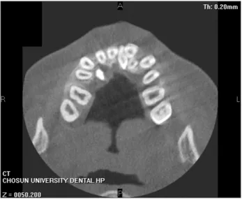
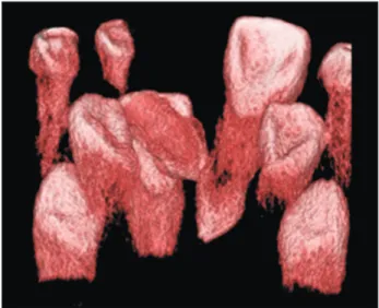
In total, 378 patients visited the hospital with mesiodens between January 2009 and September 2013.(Fig. 1-5) There were 298 males and 80 females, showing a male-to-female ratio of 3.7:1. Their ages varied from 3 to 75 years, with a mean age of 8.4 years.
Of the patients, 265 patients (70.1%) had one supernumer- ary tooth, 112 patients (29.6%) had two supernumerary teeth, and one patient (0.3%) had three supernumerary teeth. In patients with more than one supernumerary tooth, all super- numerary teeth were extracted at the same visit.
Patients were analyzed for motivation to visit to the hospi- tal. Of the 378 enrolled patients, 323 patients (85.4%) were referred from other hospitals specifically for removal of the supernumerary tooth. In 15 patients (4.0%) cases, an orth- odontic problem was caused by the mesiodens; in 40 patients (10.6%) cases, the mesiodens was found by chance during
Fig. 1. A mesiodens found in an oral panoramic X-ray.
Sung-Suk Lee et al: A comparative analysis of patients with mesiodenses: a clinical and radiological study. J Korean Assoc Oral Maxillofac Surg 2015
Fig. 2. An impacted conical-shaped mesiodens.
Sung-Suk Lee et al: A comparative analysis of patients with mesiodenses: a clinical and radiological study. J Korean Assoc Oral Maxillofac Surg 2015
Fig. 3. Two horizontally impacted mesiodenses.
Sung-Suk Lee et al: A comparative analysis of patients with mesiodenses: a clinical and radiological study. J Korean Assoc Oral Maxillofac Surg 2015
Fig. 4. A mesiodens impacted on the palatal side of the central incisor.
Sung-Suk Lee et al: A comparative analysis of patients with mesiodenses: a clinical and radiological study. J Korean Assoc Oral Maxillofac Surg 2015
J Korean Assoc Oral Maxillofac Surg 2015;41:190-193
192
siodenses. Orhan et al.5 reported that 76% to 86% of patients had only one mesiodens, 12% to 23% had two, and fewer than 1% had three or more.
Ray et al.6 observed that mesiodenses appeared twice as frequently in males than females, and Rajab and Hamdan7 re- ported a male-to-female ratio of 2.2:1 in Jordanian children.
Both our previous and present results suggest a higher male- to-female ratio. We found 4:1 in our previous study and 3.7:1 in this study.
Morphologically, mesiodenses can have various shapes8. The most common shapes are conical, incisor-like, and tuber- cular types. The conical type was seen in both our previous study and in the present study, at frequencies of 66.0% and 61.4%, respectively4.
In some patients, the mesiodens erupts ‘normally’ but is mostly impacted or is pointed in the opposite direction. This is probably because the supernumerary tooth has an ab- normal eruption path or erupts ectopically9,10. An impacted mesiodens can interfere with the eruption of the adjacent permanent teeth or can cause malocclusion11. Comparing im- paction direction between our previous study and this study showed that 52.0% and 62.4% were impacted in the opposite direction, 38.0% and 26.0% were in the normal direction, and 10.0% and 11.6% were impacted horizontally, respectively4. No specific effect on permanent incisors was consistently found in either the previous study or in this study (64.0% and 52.9%, respectively). Delayed eruption was found in 20.0%
patients.(Table 3)
Following extraction, the crown shape was analyzed. A conical shape was found in 302 crowns (61.4%), a round shape in 62 crowns (12.6%), a tubercular type in 53 crowns (10.8%), a shape similar to a canine in 40 crowns (8.1%), and a shape similar to an incisor in 35 crowns (7.1%).(Table 4) In two patients (0.5%), dentigerous cyst was found radiologi- cally.
IV. Discussion
In many cases, a mesiodens is a single impacted tooth.
Comparing results of our previous study with the present study, we found that 75.0% and 70.1% patients had only one mesiodens, and 25.0% and 29.6%, respectively4, had two me-
Fig. 5. A mesiodens impacted in the opposite direction.
Sung-Suk Lee et al: A comparative analysis of patients with mesiodenses: a clinical and radiological study. J Korean Assoc Oral Maxillofac Surg 2015
Table 1. Chief complaint (n=378)
Chief complaint Number (%) of cases Referred from another clinic
Orthodontic problem Other treatment
323 (85.4) 15 (4.0) 40 (10.6)
Sung-Suk Lee et al: A comparative analysis of patients with mesiodenses: a clinical and radiological study. J Korean Assoc Oral Maxillofac Surg 2015
Table 2. Position of mesiodens according to year
Position 2003 (n=50) 2013 (n=492)
Upward Downward Horizontal
26 (52.0) 19 (38.0) 5 (10.0)
307 (62.4) 128 (26.0) 57 (11.6) Values are presented as number (%).
Sung-Suk Lee et al: A comparative analysis of patients with mesiodenses: a clinical and radiological study. J Korean Assoc Oral Maxillofac Surg 2015
Table 3. Effects on permanent maxillary incisors according to year
Effect 2003 (n=50) 2013 (n=378)
Non-specific Delayed eruption
Axial rotation or inclination of erupted permanent incisors Diastema
Intraoral infection
32 (64.0) 10 (20.0) 4 (8.0) 2 (4.0) 2 (4.0)
200 (52.9) 59 (15.6) 22 (5.8) 94 (24.9)
3 (0.8) Values are presented as number (%).
Sung-Suk Lee et al: A comparative analysis of patients with mesiodenses: a clinical and radiological study. J Korean Assoc Oral Maxillofac Surg 2015
Table 4. Shapes of mesiodens according to year
Shape 2003 (n=50) 2013 (n=492)
Conical Canine-like Incisior-like Tuberculated Round
33 (66.0) 9 (18.0) 4 (8.0) 2 (4.0) 2 (4.0)
302 (61.4) 40 (8.1) 35 (7.1) 53 (10.8) 62 (12.6) Values are presented as number (%).
Sung-Suk Lee et al: A comparative analysis of patients with mesiodenses: a clinical and radiological study. J Korean Assoc Oral Maxillofac Surg 2015
A comparative analysis of patients with mesiodenses: a clinical and radiological study
193 Ji-Su Oh, http://orcid.org/0000-0002-8369-5025
Jae-Seek You, http://orcid.org/0000-0001-7638-9583 Kyung-In Jeong, http://orcid.org/0000-0002-8922-0290 Young-Kyun Kim, http://orcid.org/0000-0002-7268-3870 Sang-Ho Lee, http://orcid.org/0000-0003-2513-6871 Nan-Young Lee, http://orcid.org/0000-0002-4738-9389
References
1. von Arx T. Anterior maxillary supernumerary teeth: a clinical and radiographic study. Aust Dent J 1992;37:189-95.
2. Lustmann J, Bodner L. Dentigerous cysts associated with supernu- merary teeth. Int J Oral Maxillofac Surg 1988;17:100-2.
3. Dinkar AD, Dawasaz AA, Shenoy S. Dentigerous cyst associated with multiple mesiodens: a case report. J Indian Soc Pedod Prev Dent 2007;25:56-9.
4. Kim SG, Lee SH. Mesiodens: a clinical and radiographic study. J Dent Child (Chic) 2003;70:58-60.
5. Orhan AI, Ozer L, Orhan K. Familial occurrence of nonsyndromal multiple supernumerary teeth. A rare condition. Angle Orthod 2006;76:891-7.
6. Ray D, Bhattacharya B, Sarkar S, Das G. Erupted maxillary conical mesiodens in deciduous dentition in a Bengali girl--a case report. J Indian Soc Pedod Prev Dent 2005;23:153-5.
7. Rajab LD, Hamdan MA. Supernumerary teeth: review of the litera- ture and a survey of 152 cases. Int J Paediatr Dent 2002;12:244-54.
8. Gallas MM, García A. Retention of permanent incisors by me- siodens: a family affair. Br Dent J 2000;188:63-4.
9. Fernández Montenegro P, Valmaseda Castellón E, Berini Aytés L, Gay Escoda C. Retrospective study of 145 supernumerary teeth.
Med Oral Patol Oral Cir Bucal 2006;11:E339-44.
10. Meighani G, Pakdaman A. Diagnosis and management of super- numerary (mesiodens): a review of the literature. J Dent (Tehran) 2010;7:41-9.
11. Asaumi JI, Shibata Y, Yanagi Y, Hisatomi M, Matsuzaki H, Ko- nouchi H, et al. Radiographic examination of mesiodens and their associated complications. Dentomaxillofac Radiol 2004;33:125-7.
and 15.6% of cases, diastema in 4% and 24.9%, and rota- tion of a permanent incisor in 8.0% and 5.8% between the previous study and this study, respectively. Malocclusion of a permanent incisor was found in 32.0% and 46.3% of previ- ous and current patients, respectively, and inflammation was observed in 4% and 0.8% of cases4.
Only two patients (0.5%) in the present study were radio- logically observed to have dentigerous cyst associated with a mesiodens, and Dinkar et al.3 have reported a correlation between dentigerous cyst and a mesiodens, although such a finding is rare.
V. Conclusion
It is important to determine both the number and location of mesiodenses in a patient. It is also necessary to carefully examine the impaction direction, the effects on adjacent teeth, and the formation of dentigerous cyst. When a pathologic condition is found related to the presence of a mesiodens, ex- traction of the mesiodens should be considered.
Conflict of Interest
No potential conflict of interest relevant to this article was reported.
ORCID
Sung-Suk Lee, http://orcid.org/0000-0002-4564-6302 Su-Gwan Kim, http://orcid.org/0000-0002-0424-9984