www.jpis.org
pISSN 2093-2278 eISSN 2093-2286 Copyright © 2011 Korean Academy of PeriodontologyThis is an Open Access article distributed under the terms of the Creative Commons Attribution Non-Commercial License (http://creativecommons.org/licenses/by-nc/3.0/).
Effect of globular adiponectin on interleukin-6 and interleukin-8 expression in periodontal ligament and gingival fibroblasts
Hong Gyu Park1, Eun Jung Bak2, Ji Hye Kim1,3,4, Yang-Sin Lee1, Seong-Ho Choi3,5, Jeong-Heon Cha1,2,3,4, Yun-Jung Yoo1,3,4,*
1Department of Oral Biology, BK21 Project, Oral Science Research Center, Yonsei University College of Dentistry, Seoul, Korea
2Oral Cancer Research Institute, Yonsei University College of Dentistry, Seoul, Korea
3Research Center for Orofacial Hard Tissue Regeneration, Yonsei University College of Dentistry, Seoul, Korea
4Department of Applied Life Science, Yonsei University Graduate School, Seoul, Korea
5Department of Periodontology, Research Institute for Periodontal Regeneration, Yonsei University College of Dentistry, Seoul, Korea
Purpose: Globular adiponectin (gAd) is a type of adipocytokine, which is mainly produced by adipose tissue. It has been report
ed that gAd acts as a pro as well as an antiinflammatory factor. Interleukin (IL)6 and IL8 are proinflammatory cytokines. To investigate the role of gAd on periodontal tissues, the expression of adiponectin receptor 1 (AdipoR1) and the effect of gAd on the expression of IL6 and IL8 were investigated in periodontal ligament (PDL) and gingival fibroblasts.
Methods: PDL and gingival fibroblasts were cultured from human periodontal tissues. gAd derived from Escherichia coli and murine myeloma cells were used. The expression of AdipoR1 was estimated by reverse transcriptionpolymerase chain reac
tion and western blot. The expression of cytokines was measured by enzymelinked immunosorbent assay.
Results: PDL and gingival fibroblasts expressed both mRNA and protein of AdipoR1. gAd derived from E. coli increased the production of IL6 and IL8, but polymyxin B, an inhibitor of lipopolysaccharide (LPS), inhibited IL6 and IL8 production in
duced by gAd in both types of cells. gAd derived from murine myeloma cells did not induce IL6 and IL8 production in those cells. gAd derived from E. coli contained higher levels of LPS than gAd derived from murine myeloma cells. LPS increased pro
duction of IL6 and IL8 in PDL and gingival fibroblasts, but pretreatment of cells with gAd derived from murine myeloma cells did not inhibit LPSinduced IL6 and IL8 expression.
Conclusions: Our results suggest that PDL and gingival fibroblasts express AdipoR1 and that gAd does not act as a modulator of IL6 and IL8 expression in PDL and gingival fibroblasts.
Keywords: Adiponectin, Periodontal ligament, Fibroblasts, Receptors, Cytokines.
INTRODUCTION
Periodontitis is an inflammatory disease caused by bacteria and the inflammatory/immune response to bacteria. Epide
miological studies have suggested that periodontitis is inti
mately related with obesity [12] and diabetes [3]. Several stu
dies have reported that adipocytokines, which are produced
by adipocytes, can modulate inflammation [4] and insulin sen
sitivity [5]. Therefore, the role of adipocytokines in the patho
genesis of periodontitis has become an area of interest in re
search.
Adiponectin is a type of adipocytokine, produced mainly by adipose tissue [6,7]. Studies have shown that adiponectin is also expressed in skeletal muscle cells and cardiac and syno
Received: May 4, 2011; Accepted: May 21, 2011
*Correspondence: Yun-Jung Yoo
Department of Oral Biology, Yonsei University College of Dentistry, 134 Sinchon-dong, Seodaemun-gu, Seoul 120-752, Korea E-mail: [email protected], Tel: +82-2-2228-3060, Fax: +82-2-2227-7903
fulllength adiponectin (fAd) and a globular adiponectin (gAd) [6]. fAd consists of an Nterminal signal peptide, a variable domain, a collagenlike domain, and a Cterminal C1qlike globular domain [4]. gAd consists of a globular domain that is cleaved from fAd by proteolysis [7]. Two receptors for adi
ponectin have been reported: adiponectin receptor 1 (Adi
poR1) is a high affinity receptor for gAd, and AdipoR2 is an intermediate affinity receptor for gAd and fAd [10].
Until now, the role of adiponectin in inflammation has not been clear. Some studies have demonstrated that adiponec
tin has a proinflammatory effect, but others showed an anti
inflammatory effect or did not show any effects. The proin
flammatory effects of adiponectin have been suggested by demonstrating its cytokineinduction activity. Research has shown that fAd stimulates the production of interleukin (IL)
8 and monocyte chemoattractant protein1 in monocytes and endothelial cells [11]. In addition, fAd has been shown to stimulate production of IL6 and IL8 by rheumatoid synovi
al fibroblasts, suggesting that adiponectin is involved in the pathogenesis of rheumatoid arthritis [12,13]. Studies have also shown that gAd induces the production of IL1β, IL6, IL8, and TNFα in macrophages [14,15] and IL6 in cardiac fibro
blasts [16]. However, other studies have demonstrated that adiponectin has an antiinflammatory effect by showing its induction of tolerance to lipopolysaccharide (LPS) and inhi
bition of phagocytosis in macrophages. Pretreatment with gAd reduced LPSinduced production of IL6 and TNFα in macrophages, suggesting that adiponectin causes macro
phages to become resistant to LPS [14]. Treatment of macro
phages with adiponectin inhibited macrophage phagocyto
sis, suggesting that adiponectin suppresses macrophage func
tion and that inhibitionby adiponectin of macrophages may contribute to termination of the inflammatory reaction [17].
However, other studies have not been able to demonstrate the induction of IL6 and TNFα and the suppression of LPS
induced IL6 and TNFα production by adiponectin in mac
rophages, suggesting that some adiponectin effects, such as the inhibition and stimulation of cytokine (IL6 and TNFα) production in macrophages, may be confused by LPS con
tamination [18,19].
Interestingly, adiponectin plasma levels are high in healthy humans and decreased in individuals with obesity and type 2 diabetes [20,21]. One study found a trend toward decreased serum levels of adiponectin in periodontitis patients [22]. More
over, infection with periodontopathogen such as Porphyromo- nas gingivalis was shown to cause decreased serum levels of adiponectin in type 2 diabetic mice [23]. In another study, an
timicrobial periodontal treatment (APT) not only ameliorated periodontitis but also increased serum levels of adiponectin
amelioration of periodontitis by APT might cause an increase in serum levels of adiponectin in type 2 diabetic subjects [24].
Pretreatment of mice with gAd was shown to suppress Ag- gregatibacter actinomycetemcomitans LPSinduced TNFα ex
pression in a murine macrophage cell line [25]. gAd was also found to suppress osteoclast formation induced by LPS of A.
actinomycetemcomitans [26]. Those reports suggest that, in periodontitis, adiponectin may function as an inhibitor of both LPSmediated cytokine expression in macrophages and LPSmediated osteoclast formation. Periodontal ligament (PDL) and gingival fibroblasts play an important role in the pathogenesis of periodontitis by producing proinflammato
ry cytokines such as IL6 and IL8 [27]. To this date, however, the role of adiponectin in PDL and gingival fibroblasts has not been clearly determined. In this study, the expression of adiponectin AdipoR1 and the effect of gAd on the expression of IL6 and IL8 were investigated in PDL and gingival fibro
blasts.
MATERIALS AND METHODS
Culture of cells
Human PDL fibroblasts were prepared from extracted teeth for orthodontic treatment as described previously [28]. The protocol was approved by the Institutional Review Board of Yonsei Dental Hospital (IRB No. 220100005). Using blades, PDL tissue fragments were removed from the middle one third of the extracted tooth roots and washed three times with αmodified Eagle’s medium (αMEM, Gibco BRL, San Diego, CA, USA) containing antibiotics of 300 mg/mL strep
tomycin, 300 unit/mL penicillin, and 0.75 mg/mL amphoteri
cinB (Gibco BRL). PDL tissue fragments were placed in 100
mm dishes containing αMEM supplemented with the anti
biotics of 100 mg/mL streptomycin, 100 unit/mL penicillin, 0.25 mg/mL amphotericinB, and 20% fetal bovine serum (FBS, Gibco BRL). The culture media was replaced every 2 days until cells grew from the fragments, after which the cultures were maintained with αMEM containing antibiotics and 10% FBS. Upon reaching 70% confluence, the cells were re
moved with 0.25% trypsin and passaged to 100mm dishes.
Gingival fibroblasts were prepared from gingival tissues ex
cised for crown lengthening. Gingival tissues were cut into small fragments, and then the tissue fragments were incu
bated in 2.4 U dispase II (Roche, Mannheim, Germany) and 0.25% collagenase (Roche) for 1 hour at 37°C. The tissue frag
ments were then separated into epithelium and connective tissue. Connective tissue fragments were placed in 100mm dishes containing Dulbecco’s modified Eagle’s medium (DM
EM, Gibco BRL) supplemented with antibiotics and 10% FBS
fibroblasts. The cells used in this study had been passaged 5 times. THP1 monocytes and MDAMB231 breast cancer cells were maintained in Roswell Park Memorial Institute 1640 medium (Gibco BRL) and DMEM each containing both anti
biotics and 10% FBS.
Western blotting
PDL fibroblasts, gingival fibroblasts or MDAMB231 (5×10⁵ cells/well) were cultured in 6well culture plates. THP1 mono
cytes (5×10⁵ cells/well) were plated in 6well plates and treat
ed with phorbol 12myristate 13acetate (PMA, 100 ng/mL, SigmaAldrich Co., St. Louis, MO, USA) for 24 hours to induce the differentiation of monocytes into macrophages. After reaching confluence, the whole cells were lysed with lysis buffer (20 mM TrisHCl, pH7.5, 150 mM NaCl, 1 mM Na2ED
TA, 1% Triton, 2.5 mM sodium pyrophosphate, 1 mM βgly
cerophosphate, 1 mM Na3VO4, 1 μg/mL leupeptin, and 1 mM phenylmethylsulfonyl fluoride; Cell Signaling Technology Inc., Danvers, MA, USA). After centrifugation at 12,000×g at 4°C, the supernatant was used for the protein assay. Protein concentrations were determined with bovine serum albu
min protein assay kits (BioRad Laboratories Inc., Hercules, CA, USA). Forty micrograms of protein per sample of cell ex
tract were run on a 10% SDSpolyacrylamide gel electropho
resis and then electroblotted onto a polyvinylidene fluoride membrane (BioRad Laboratories Inc.). The membranes were blocked with 5% skim milk in trisbuffered saline (10 mM TrisHCl, 166 mM NaCl, pH 7.4) for 1 hr, rinsed, and then in
cubated overnight with primary antibodies against adipo
nectin (R&D Systems Inc., Minneapolis, MN, USA), AdipoR1 (Abcam plc, Cambri dge, UK), or actin (SantaCruz Biotechnol
ogy, Santa Cruz, CA, USA) at a dilution of 1:1,000. Secondary antibody, peroxidaseconjugated antirabbit antibody (Jack
son ImmunoResearch Laboratories Inc., West Grove, PA, USA), was used at 1:2,500. Protein bands were visualized by an ECL kit (Amersham, Buckinghamshire, UK).
Reverse transcription-polymerase chain reaction (RT-PCR) PDL fibroblasts or gingival fibroblasts (5×10⁵ cells/well) were cultured in 6well culture plates. THP1 monocytes (5×10⁵ cells/well) were plated in 6well plates and treated with PMA (100 ng/mL) for 24 hours to induce the differentiation of mo
nocytes into macrophages. After reaching confluence, total RNA in PDL fibroblasts, gingival fibroblasts, or THP1 mac
rophages was isolated with Trizol reagent (Invitrogen, Carls
bad, CA, USA). Two micrograms of total RNA was converted to cDNA using an RT premix kit (Bioneer, Seoul, Korea) in ac
cordance with the manufacturer’s instructions. The cDNA was amplified with iStarTaq (Intron Biotech, Seongnam, Ko
ense primer, 5´CCTTTCCCCAAGCTGAAGCTGC3´ and an
tisense primer, 5´CCTTGACAAAGCCCTCAGCGAT3´; GAP
DH sense primer, 5´CGGAGTCAACGGATTTGGTCGTAT3´
and antisense primer, 5´AGCCTTCTCCATGGTGGTGAAG
AC3´. For AdipoR1, the reaction mixtures were subjected to 35 cycles of denaturation and annealing at 60°C. For GAPDH, the reaction mixtures were subjected to 28 cycles of denatur
ation and annealing at 56°C. The PCR products were then re
solved by electrophoresis in a 1% agarose gel containing eth
idium bromide.
3-(4,5-dimethylthiazol-2-yl)-2,5-diphenyltetrazolium (MTT) assay
Cell viability was measured using an MTT assay. PDL fibro
blasts or gingival fibroblasts (1.7×10⁵ cells/well) were seeded in 24well plates and maintained in media containing antibi
otics and 10% FBS. After being maintained for 7 hours, the cells were starved in serumfree media for 20 hours and then treated with the indicated concentration of polymyxin B for 24 hours. After treatment, 20 μL of MTT solution (5 mg/mL in PBS, SigmaAldrich Co.) was added to each well. After in
cubation for 4 hours, the medium was aspirated, and 200 μL of dimethylsulfoxide was added to each well to dissolve MTTformazan crystals. The plates were shaken for 5 min
utes, and the absorbance was measured on an MRX II micro
plate rea der (Dynatech Laboratories Inc., Chantilly, VA, USA) at 570 nm.
Enzyme-linked immunosorbent assay (ELISA)
PDL fibroblasts or gingival fibroblasts (1.7×10⁵ cells/well) were plated in 24well culture plates and maintained in me
dia containing antibiotics and 10% FBS for 7 hours. THP1 monocytes were plated in 24well plates and treated with PMA for 24 hours to induce the differentiation of monocytes into macrophages. After being maintained for the indicated time, the cells were starved in serumfree media for 20 hours and then treated with myeloma cellderived recombinant gAd (MgAd, R&D systems), Escherichia coliderived gAd (EgAd, Phoenix Pharmaceuticals, Burlingame, USA), or LPS (Sigma
Aldrich Co.) for 24 hours. For polymyxin B treatment, cells were pretreated with polymyxin B for 1 hour and then treated with MgAd, EgAd, or LPS for 24 hours. For estimating the induction of tolerance to LPS by gAd, the cells were pretreat
ed with gAd for 24 hours and then treated with LPS for an additional 24 hours. After the treatment, the levels of IL6 or IL8 were measured in the culture supernatants of PDL fi
broblasts, gingival fibroblasts, or THP1 macrophages using an ELISA kit (BioLegend, San Diego, CA, USA) in accordance with the manufacturer’s instructions.
The results were analyzed using oneway analysis of vari
ance and Tu key’s test. A Pvalue of <0.05 was considered to indicate statistical significance.
RESULTS
Expression of adiponectin and adiponectin receptor
Expression of adiponectin was estimated by western blot.
Subcutaneous adipose tissue from the inguinal region ex
pressed adiponectin, but PDL fibroblasts, gingival fibroblasts, and THP1 macrophages did not show adiponectin expres
sion (Fig. 1A). Expression of AdipoR1 was estimated by RTPCR and western blot. Like THP1 macrophages, which have been known to express AdipoR1, PDL and gingival fibroblasts ex
pressed mRNA of AdipoR1 (Fig. 1B) [29]. Similar to THP1 ma
crophages and the breast cancer cell line MDAMB231,which are positive controls for the expression of AdipoR1, PDL and gingival fibroblasts also expressed the protein of AdipoR1 (Fig. 1C) [30].
and gingival fibroblasts
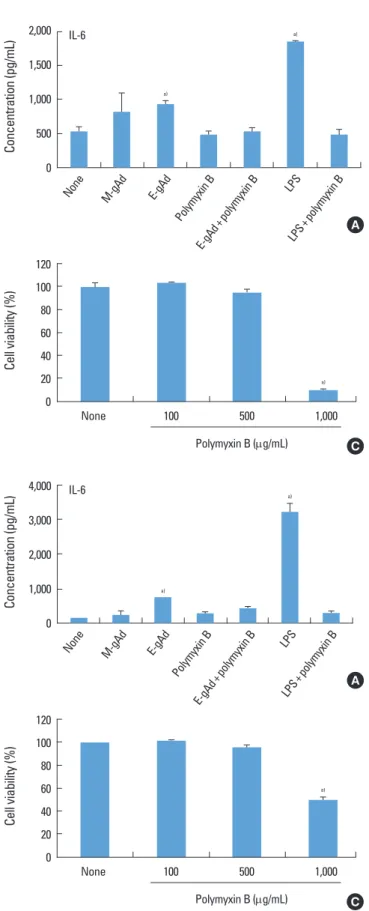
The effect of gAd on the expression of IL6 and IL8 was estimated in PDL and gingival fibroblasts by ELISA. Treat
ment with E. coliderived gAd increased the expression of IL6 and IL8 in PDL fibroblasts, but treatment with myelo
ma cell linederived gAd did not increase the expression of IL6 and IL8 (Fig. 2A, B). Polymyxin B is an inhibitor of LPS.
Polymyxin B blocked IL6 and IL8 expression induced by E.
coliderived gAd at a concentration of 100 μg/mL which poly
myxin B inhibited LPS activity (Fig. 2A, B). Treatment of gin
gival fibroblasts with E. coli or myeloma cellderived gAd showed results similar to those of PDL fibroblasts (Fig. 3A, B).
Polymyxin B did not have a cytotoxic effect on PDL and gin
gival fibroblasts at concentrations less than or equal to 500 μg/mL (Figs. 2C, 3C). LPS increased IL6 and IL8 expression in THP1 macrophages, but myeloma cellderived gAd did not stimulate expression of those cytokines (Fig. 4).
Effect of gAd on LPS-induced IL-6 and IL-8 expression To estimate the induction of tolerance to LPS by gAd, PDL and gingival fibroblasts were pretreated with myeloma cell
derived gAd before exposure to LPS. LPS increased the ex
pression of IL6 and IL8 in PDL fibroblasts, but pretreatment of cells with gAd did not decrease or increase the expression of IL6 and IL8 induced by LPS (Fig. 5A). Gingival fibroblasts showed results similar to those of PDL fibroblasts (Fig. 5B).
DISCUSSION
The alteration in serum levels of adiponectin according to periodontal status and the effect of adiponectin on periodon
topathogeninduced cytokine expression by macrophages and osteoclast formation have been estimated [2325], but few studies have addressed the effect of adiponectin on the cells of periodontal tissue such as PDL and gingival fibro
blasts. In this study, it was shown that PDL and gingival fi
broblasts express AdipoR1 and that gAd does not affect the production of IL6 and IL8 in those cells.
Although the main source of adiponectin is adipocytes, sy
novial and cardiac fibroblasts also express adiponectin [4,8,31].
In this study, we also found expression of adiponectin in adi
pocytes, but PDL and gingival fibroblasts did not express adi
ponectin like synovial and cardiac fibroblasts. AdipoR1 is most highly expressed in skeletal muscle [4], but its expression has also been detected in synovial and cardiac fibroblasts [8,12,13].
As a result, we looked for the expression of AdipoR1 in PDL and gingival fibroblasts. Similar to synovial and cardiac fi
broblasts, PDL and gingival fibroblasts expressed not only the mRNA but also the protein of AdipoR1.
Adipose
tissue PDL
Ad
Actin
GF THP-1
PDL AdipoR1
GAPDH
GF THP-1
MDA PDL GF THP-1
AdipoR1
Actin
A
B
C Figure 1. Expression of adiponectin and adiponectin receptor 1 in periodontal ligament and gingival fibroblasts. Expression of adipo
nectin (Ad) in adipose tissue, periodontal ligament (PDL) fibroblasts, gingival fibroblasts (GF), and THP1 macrophages was estimated by western blot (A). Expression of adiponectin receptor 1 (AdipoR1) in PDL fibroblasts, GF, MDAMB231 breast cancer cell line (MDA), and THP1 macrophages was estimated by reverse transcriptionpoly
merase chain reaction (B) and western blot (C).
IL6 induces the differentiation of B cells to antibodyse
creting plasma cells and stimulates bone resorption [32,33].
IL8 stimulates the attraction and activation of neutrophils
and bone resorption [32]. The IL6 and IL8 levels of periodon
titis patients are higher than those of healthy patients [32].
That evidence suggests that IL6 and IL8 play an important
Concentration (pg/mL)
1,500 1,000
500 0
None M-gAd
E-gAd Polymyxin B
E-gAd+polymyxin B
LPS+polymyxin B LPS
a)
Concentration (pg/mL)
400 300 200 100 0
None M-gAd
E-gAd Polymyxin B
E-gAd+polymyxin B
LPS+polymyxin B LPS
a)
Figure 2. Effect of globular adiponectin on the expression of inter
leukin (IL)6 and IL8 in periodontal ligament fibroblasts. Periodon
tal ligament fibroblasts were treated with murine myeloma cellde
rived globular adiponectin (MgAd, 15 μg/mL), Escherichia colide
rived gAd (EgAd, 15 μg/mL), or lipopolysaccharide (LPS) (100 ng/
mL) in the presence or absence of polymyxin B (100 μg/mL) for 24 hours. Levels of IL6 (A) and IL8 (B) in culture supernatants were assayed by enzymelinked immunosorbent assay. Cell viability treat
ed with polymyxin B was estimated by an 3(4,5dimethylthiazol2
yl)2,5diphenyltetrazolium assay (C). a)Significantly different from untreated cells (P<0.05).
A
Cell viability (%)
120 100 80 60 40 20 0
None 100 500 1,000
Polymyxin B (μg/mL)
a)
C
B
Cell viability (%)
120 100 80 60 40 20
0 None 100 500 1,000
Polymyxin B (μg/mL)
a)
C
Figure 3. Effect of globular adiponectin on the expression of inter
leukin (IL)6 and IL8 in gingival fibroblasts. Gingival fibroblasts were treated with murine myeloma cellderived globular adiponec
tin (MgAd, 15 μg/mL), Escherichia coliderived gAd (EgAd, 15 μg/
mL), or lipopolysaccharide (LPS) (100 ng/mL) in the presence or ab
sence of polymyxin B (100 μg/mL) for 24 hours. Levels of IL6 (A) and IL8 (B) in culture supernatants were assayed by enzymelinked immunosorbent assay. The effect of gAd on the expression of cyto
kines was compared with LPS. Cell viability treated with polymyxin B was estimated by an 3(4,5dimethylthiazol2yl)2,5diphenyltet
razolium assay (C). a)Significantly different from untreated cells (P<
0.05).
Concentration (pg/mL) 4,000 3,000 2,000
1,000 0
None M-gAd
E-gAd Polymyxin B
E-gAd+polymyxin B
LPS+polymyxin B LPS IL-6
a)
a)
A
Concentration (pg/mL) 4,000 3,000 2,000
1,000 0
None M-gAd
E-gAd Polymyxin B
E-gAd+polymyxin B
LPS+polymyxin B LPS IL-8
a)
a)
B
role in the pathogenesis of periodontitis. In this study, PDL and gingival fibroblasts expressed AdipoR1, which is a recep
tor for gAd [10]. The serum level of adiponectin was found to be 12.4±5.1 μg/mL in humans with healthy gingival [34]. As a result, we investigated the effect of gAd on IL6 and IL8 ex
pression of those cells at a concentration of 15 μg/mL. Sourc
es of commercially available recombinant gAd are E. coli and murine myeloma cells. Therefore, we investigated the effect of E. coli and murine myeloma cellderived gAd on the ex
pression of those cytokines. E. coliderived gAd increased IL6 and IL8 production in PDL and gingival fibroblasts. How
ever, polymyxin B, an inhibitor of LPS, blocked not only the production of IL6 and IL8 induced by LPS, but also the pro
duction of those cytokines induced by E. coliderived gAd.
Analysis of E. coliderived gAd by a limulus amebocyte lysate assay revealed contamination with LPS (data not shown). That suggests that the effect of E. coliderived gAd is due to con
taminated LPS. Murine myeloma cellderived gAd contained lower levels of LPS than E. coliderived gAd (data not shown).
To confirm the effect of gAd, the effect of murine myeloma cellderived gAd was estimated. Murine myeloma cellde
rived gAd did not show an increase of IL6 and IL8 produc
Figure 4. Effect of globular adiponectin on the production of interleukin (IL)6 and IL8 in THP1 macrophages. THP1 macrophages were cultured for 24 hours in the presence of murine myeloma cellderived globular adiponectin (MgAd) or lipopolysaccharide (LPS). The levels of IL6 (A) and IL8 (B) in culture supernatants were assayed by enzymelinked immunosorbent. a)Significantly different from untreated cells (P<0.05).
Concentration (pg/mL) 4,000 3,000 2,000 1,000 0
None M-gAd LPS
A B
Concentration (pg/mL) 500 400 300 200 100 0
None M-gAd LPS
Figure 5. Effect of globular adiponectin on the production of interleukin (IL)6 and IL8 induced by lipopolysaccharide (LPS) in periodontal ligament and gingival fibroblasts. Periodontal ligament fibroblasts (A) and gingival fibroblasts (B) were cultured for 24 hours in the presence of murine myeloma cellderived globular adiponectin (MgAd, 15 μg/mL) and then stimulated for 24 hours with LPS (100 ng/mL). The levels of IL6 and IL8 in the culture supernatant were assayed by enzymelinked immunosorbent. a)Significantly different from untreated cells (P<0.05).
Concentration (pg/mL) 18,00015,000 12,000 9,000 6,000 3,000
0 None M-gAd LPS M-gAd+LPS
IL-6 a)
a)
Concentration (pg/mL) 15,000 12,000 9,000 6,000 3,000
0 None M-gAd LPS M-gAd+LPS
IL-8
a) a)
Concentration (pg/mL) 15,000 12,000 9,000 6,000 3,000
0 None M-gAd LPS M-gAd+LPS
IL-8 a)
a)
Concentration (pg/mL) 20,000 15,000 10,000 5,000
0 None M-gAd LPS M-gAd+LPS
IL-6
a) a)
A B
gAd does not affect IL6 and IL8 production in PDL and gin
gival fibroblasts.
gAd induced IL6 and IL8 secretion by THP1 macropha
ges [14,15]. However, in this study, gAd derived from murine myeloma cells did not stimulate those cytokines in the same cell lines. Similar to our results, the outcome of one study showed that fAdinduced IL6 production was blocked by polymyxin B in macrophages, suggesting that fAd does not induce IL6 induction and that evaluation of certain effects of adiponectin may be entangled by LPS contamination [19].
Our results also support the proposal that LPS contamination be considered when estimating the function of adiponectin in inflammation.
It has been reported that pretreatment of macrophages with adiponectin induces tolerance to LPS, suggesting that adipo
nectin has antiinflammatory properties [14]. However, in another study, the tolerance effect of adiponectin on LPSin
duced cytokine expression was not found [18]. Our results showed that pretreatment of PDL and gingival fibroblasts with murine myeloma cellderived gAd did not decrease LPS
induced IL6 and IL8 expression, suggesting that gAd does not induce tolerance to LPS in PDL and gingival fibroblasts.
Previous reports have shown that adiponectin stimulates the production of IL6 and IL8 by rheumatoid synovial fi
broblasts, indicating that the proinflammatory effects of ad
iponectin might play a role in the pathogenesis of rheuma
toid arthritis [9,12,13]. In this study, however, we could not find the proinflammatory effect of adiponectin in PDL and gingival fibroblasts in periodontal tissue. Recently, Yamagu
chi et al. [35] reported expression of AdipoR1 in gingival fibro
blasts, but they did not estimate either the expression of Adi
poR1 in PDL cells or the effect of adiponectin on the expres
sion of cytokines in those cells. This study is meaningful in its demonstration of the expression of AdipoR1 in both PDL and gingival fibroblasts, its estimation of the effect of gAd on IL6 and IL8 expression, and its suggestion of the signifi
cance of LPS contamination in estimating the function of gAd.
In conclusion, our results showed that PDL and gingival fi
broblasts expressed AdipoR1, and gAd did not affect the ex
pression of IL6 and IL8 in those cells. Although gAd did not have a modulating effect on IL6 and IL8 expression in those cells, expression of AdipoR1 indicates that gAd may have oth
er effects on PDL and gingival fibroblasts, which will be stud
ied in the future.
CONFLICT OF INTEREST
No potential conflict of interest relevant to this article was
ACKNOWLEDGEMENTS
This research was supported by Basic Science Research Pro
gram through the National Research Foundation of Korea (NRF) funded by the Ministry of Education, Science and Tech
nology (KRF2008313E00584).
REFERENCES
1. Ekuni D, Yamamoto T, Koyama R, Tsuneishi M, Naito K, Tobe K. Relationship between body mass index and peri
odontitis in young Japanese adults. J Periodontal Res 2008;
43:41721.
2. Han DH, Lim SY, Sun BC, Paek DM, Kim HD. Visceral fat areadefined obesity and periodontitis among Koreans. J Clin Periodontol 2010;37:1729.
3. Mealey BL, Rose LF. Diabetes mellitus and inflammatory periodontal diseases. Curr Opin Endocrinol Diabetes Obes 2008;15:13541.
4. Sun Y, Xun K, Wang C, Zhao H, Bi H, Chen X, et al. Adipo
nectin, an unlocking adipocytokine. Cardiovasc Ther 2009;
27:5975.
5. Pittas AG, Joseph NA, Greenberg AS. Adipocytokines and insulin resistance. J Clin Endocrinol Metab 2004;89:447
52.
6. Tilg H, Moschen AR. Adipocytokines: mediators linking adipose tissue, inflammation and immunity. Nat Rev Im
munol 2006;6:77283.
7. Lago F, Dieguez C, GómezReino J, Gualillo O. Adipokines as emerging mediators of immune response and inflam
mation. Nat Clin Pract Rheumatol 2007;3:71624.
8. Huang D, Yang C, Wang Y, Liao Y, Huang K. PARP1 sup
presses adiponectin expression through poly(ADPribo
syl)ation of PPAR gamma in cardiac fibroblasts. Cardio
vasc Res 2009;81:98107.
9. Tan W, Wang F, Zhang M, Guo D, Zhang Q, He S. High adiponectin and adiponectin receptor 1 expression in sy
novial fluids and synovial tissues of patients with rheu
matoid arthritis. Semin Arthritis Rheum 2009;38:4207.
10. Yamauchi T, Kamon J, Ito Y, Tsuchida A, Yokomizo T, Kita S, et al. Cloning of adiponectin receptors that mediate anti
diabetic metabolic effects. Nature 2003;423:7629.
11. Rovin BH, Song H. Chemokine induction by the adipo
cytederived cytokine adiponectin. Clin Immunol 2006;
120:99105.
12. Tang CH, Chiu YC, Tan TW, Yang RS, Fu WM. Adiponectin enhances IL6 production in human synovial fibroblast via an AdipoR1 receptor, AMPK, p38, and NFkappa B path
13. Kitahara K, Kusunoki N, Kakiuchi T, Suguro T, Kawai S. Adi
ponectin stimulates IL8 production by rheumatoid syno
vial fibroblasts. Biochem Biophys Res Commun 2009;378:
21823.
14. Tsatsanis C, Zacharioudaki V, Androulidaki A, Dermitzaki E, Charalampopoulos I, Minas V, et al. Adiponectin induc
es TNFalpha and IL6 in macrophages and promotes tol
erance to itself and other proinflammatory stimuli. Bio
chem Biophys Res Commun 2005;335:125463.
15. Tsatsanis C, Zacharioudaki V, Androulidaki A, Dermitzaki E, Charalampopoulos I, Minas V, et al. Peripheral factors in the metabolic syndrome: the pivotal role of adiponectin.
Ann N Y Acad Sci 2006;1083:18595.
16. Liao W, Yu C, Wen J, Jia W, Li G, Ke Y, et al. Adiponectin in
duces interleukin6 production and activates STAT3 in adult mouse cardiac fibroblasts. Biol Cell 2009;101:26372.
17. Yokota T, Oritani K, Takahashi I, Ishikawa J, Matsuyama A, Ouchi N, et al. Adiponectin, a new member of the family of soluble defense collagens, negatively regulates the grow
th of myelomonocytic progenitors and the functions of macrophages. Blood 2000;96:172332.
18. Neumeier M, Weigert J, Schäffler A, Wehrwein G, Müller
Ladner U, Schölmerich J, et al. Different effects of adipo
nectin isoforms in human monocytic cells. J Leukoc Biol 2006;79:8038.
19. Turner JJ, Smolinska MJ, Sacre SM, Foxwell BM. Induc
tion of TLR tolerance in human macrophages by adipo
nectin: does LPS play a role? Scand J Immunol 2009;69:
32936.
20. Arita Y, Kihara S, Ouchi N, Takahashi M, Maeda K, Miya
gawa J, et al. Paradoxical decrease of an adiposespecific protein, adiponectin, in obesity. Biochem Biophys Res Commun 1999;257:7983.
21. Hotta K, Funahashi T, Arita Y, Takahashi M, Matsuda M, Okamoto Y, et al. Plasma concentrations of a novel, adi
posespecific protein, adiponectin, in type 2 diabetic pa
tients. Arterioscler Thromb Vasc Biol 2000;20:15959.
22. Furugen R, Hayashida H, Yamaguchi N, Yoshihara A, Oga
wa H, Miyazaki H, et al. The relationship between peri
odontal condition and serum levels of resistin and adipo
nectin in elderly Japanese. J Periodontal Res 2008;43:556
62.
23. Nishihara R, Sugano N, Takano M, Shimada T, Tanaka H, Oka S, et al. The effect of Porphyromonas gingivalis infec
tion on cytokine levels in type 2 diabetic mice. J Periodon
tal Res 2009;44:30510.
N, Aizawa Y, et al. Effect of antimicrobial periodontal treat
ment and maintenance on serum adiponectin in type 2 diabetes mellitus. J Clin Periodontol 2009;36:1428.
25. Kamio N, Akifusa S, Yamaguchi N, Nonaka K, Yamashita Y.
Antiinflammatory activity of a globular adiponectin func
tion on RAW 264 cells stimulated by lipopolysaccharide from Aggregatibacter actinomycetemcomitans. FEMS Immunol Med Microbiol 2009;56:2417.
26. Yamaguchi N, Kukita T, Li YJ, Martinez Argueta JG, Saito T, Hanazawa S, et al. Adiponectin inhibits osteoclast forma
tion stimulated by lipopolysaccharide from Actinobacillus actinomycetemcomitans. FEMS Immunol Med Microbi
ol 2007;49:2834.
27. Scheres N, Laine ML, de Vries TJ, Everts V, van Winkelhoff AJ. Gingival and periodontal ligament fibroblasts differ in their inflammatory response to viable Porphyromonas gingivalis. J Periodontal Res 2010;45:26270.
28. Lee YS, Bak EJ, Kim M, Park W, Seo JT, Yoo YJ. Induction of IL8 in periodontal ligament cells by H(2)O (2). J Microbiol 2008;46:57984.
29. Chinetti G, Zawadski C, Fruchart JC, Staels B. Expression of adiponectin receptors in human macrophages and re
gulation by agonists of the nuclear receptors PPARalpha, PPARgamma, and LXR. Biochem Biophys Res Commun 2004;314:1518.
30. Dos Santos E, Benaitreau D, Dieudonne MN, Leneveu MC, Serazin V, Giudicelli Y, et al. Adiponectin mediates an anti
proliferative response in human MDAMB 231 breast can
cer cells. Oncol Rep 2008;20:9717.
31. Ehling A, Schäffler A, Herfarth H, Tarner IH, Anders S, Dis
tler O, et al. The potential of adiponectin in driving arthri
tis. J Immunol 2006;176:446878.
32. Okada H, Murakami S. Cytokine expression in periodon
tal health and disease. Crit Rev Oral Biol Med 1998;9:248
66.
33. Preshaw PM, Taylor JJ. How has research into cytokine in
teractions and their role in driving immune responses impacted our understanding of periodontitis? J Clin Peri
odontol 2011;38 Suppl 11:6084.
34. Saito T, Yamaguchi N, Shimazaki Y, Hayashida H, Yone
moto K, Doi Y, et al. Serum levels of resistin and adiponec
tin in women with periodontitis: the Hisayama study. J Dent Res 2008;87:31922.
35. Yamaguchi N, Hamachi T, Kamio N, Akifusa S, Masuda K, Nakamura Y, et al. Expression levels of adiponectin recep
tors and periodontitis. J Periodontal Res 2010;45:296300.