Natarajan Chellappa
Department of Oral and Maxillofacial Surgery, Government Dental College and Hospital, P D'Mello Road, St. George Hospital, Near Chhatrapati Shivaji Terminus Area, Fort, Mumbai 400001, India
TEL: +91-022-2262-0668 E-mail: natarajan.balaji.c@gmail.com ORCID: https://orcid.org/0000-0003-4052-3319
This is an open-access article distributed under the terms of the Creative Commons Attribution Non-Commercial License (http://creativecommons.org/
licenses/by-nc/4.0/), which permits unrestricted non-commercial use, distribution, and reproduction in any medium, provided the original work is properly cited.
CC
A working paradigm for managing mandibular fractures under regional anesthesia
Natarajan Chellappa1, Vikas Meshram1, Prajwalit Kende1, Jayant Landge1, Neha Aggarwal1, Manish Tiwari2
1Department of Oral and Maxillofacial Surgery, Government Dental College and Hospital, Mumbai,
2Private Practitioner, Bengaluru, India
Abstract(J Korean Assoc Oral Maxillofac Surg 2018;44:275-281)
Objectives: Isolated mandibular fractures contribute to approximately 45% of maxillofacial traumas. Improper management of mandibular fractures can cause myriad potential complications and can lead to serious functional and aesthetic sequelae. The objective of the study is to design a stepwise approach for managing isolated mandibular fractures using open reduction and internal fixation (ORIF) with regional anesthesia on outpatient basis.
Materials and Methods: Patients with isolated mandibular fractures presenting to the department of maxillofacial surgery were selected for ORIF under regional anesthesia based on occlusion, age, socioeconomic status, general condition, habits, and allied medical ailments. Standard preoperative, intraoperative, and postoperative protocols were followed. All patients were followed up for a minimum of 4 weeks up to a maximum of 1 year.
Results: Of 23 patients who received regional anesthesia, all but one had good postoperative functional occlusion. One patient was hypersensitive and had difficulty tolerating the procedure. Two patients developed an extraoral draining sinus, one of whom was managed with local curettage, while the other required hardware removal. One patient, who was a chronic alcoholic, returned 1 week after treatment with deranged fracture segments after he fell while intoxicated.
Conclusion: With proper case selection following a stepwise protocol, the majority of mandibular fractures requiring ORIF can be managed with re- gional anesthesia and yield minimal to no complications.
Key words: Mandibular fractures, Regional anesthesia, Complications, Perioperative management, Champy’s lines
[paper submitted 2017. 12. 9 / revised 2018. 1. 17 / accepted 2018. 1. 22]
Copyright © 2018 The Korean Association of Oral and Maxillofacial Surgeons. All rights reserved.
I. Introduction
Maxillofacial surgical procedures require special attention to patient airways, especially because the airway space must be shared by the surgeons and the anesthetist throughout the procedure. General anesthesia (GA) with endotracheal intu- bation is the most common technique in maxillofacial surger- ies. However, this technique exposes patients to the stress of being awake during airway manipulation, as well as to
potential postoperative complications from GA. All the risks associated with a difficult intubation and GA are avoidable if surgery is performed under regional anesthesia.
Previously, regional anesthesia has been successfully used for various minor oral surgical procedures; currently it has gained popularity for use among patients with inherent risks for GA. Reports have described successful temporomandibu- lar joint (TMJ) surgeries being carried out under regional anesthesia1. The aim of this article is to assess the effective- ness of managing isolated mandibular fracture patients under regional anesthesia on an outpatient basis.
The mandible is the most commonly fractured bone in the facial skeleton2 and mandible fractures are twice as common as mid-face fractures3. Proper functioning of the mandible is essential for speech and mastication. Mandibular fractures can sometimes also result in facial contour defects. Because of action by the supra-hyoid and masticatory muscles, frac- ture fragments are often displaced. Mandibular fracture patients also present with acute trismus due to pain, muscle spasm, or mechanical obstruction of the overriding fracture
segments.
Because of the complex anatomy and multifarious compli- cations4 associated with mandibular fractures ranging from hematoma, irreversible nerve injury, non-union, mal-union, and TMJ disorders, to life threatening complications like in- fection and osteomyelitis, it is imperative that surgeons inter- vene early.
Currently, open reduction and internal fixation (ORIF) us- ing mini-plates via a transoral approach is the most frequently performed treatment modality5 for mandibular fractures be- cause it allows early restoration of jaw function and obviates the need for extended periods of maxillomandibular fixation (MMF), which is troublesome for patients.
II. Materials and Methods
Herein, we review 23 patients who had isolated mandibular fractures reporting to the Department of Oral and Maxil- lofacial Surgery, Government Dental College and Hospital (Mumbai, India) from January 2, 2016 to November 24, 2016. The objectives were to evaluate: 1) the versatility of re- gional anesthesia for managing isolated fractures; 2) various intraoral and extraoral surgical approaches that can be carried out under regional anesthesia; 3) ease of surgical site expo- sure and internal fixation with minimal patient discomfort;
and 4) postoperative stability and functional restoration and various complications encountered.
Inclusion criteria:
• Patients presenting with isolated mandibular fractures (≤2 weeks old), either single or multiple fractures with indica- tions for ORIF
Exclusion criteria:
• Patients with condylar fractures requiring ORIF
• Fractures that were more than 2 weeks old
• Malunion fractures
• Patients with uncontrolled systemic diseases
• Patients not willing to consent to the procedure 1. Preoperative procedures
Routine blood examinations, including complete blood count, bleeding time, clotting time, and blood sugar levels were performed for all patients, and complete histories were taken to rule out any active communicable infectious dis- eases.
We used an orthopantomograph (OPG; Planmeca, Helsinki, Finland) for all radiographs, irrespective of mandibular frac-
ture type. For some individual cases, additional radiographs were taken using cone-beam computed tomography (Planme- ca Romexis; Planmeca) of the following views: mandibular occlusal view, mandibular lateral oblique view, and mandibu- lar posteroanterior view.
Tetanus toxoid 0.5 mL (Tetanus Toxoid [adsorbed]; Se- rum Institute of India, Pune, India) was administered intra- muscularly for patients who had not received a vaccination within the past year. Oral prophylaxis was administered to all patients. Upper and lower Erich arch bars were secured pre- operatively to aid peri-surgical occlusion after reducing the fracture fragments until the fixation. Afterward, patients were not routinely placed in MMF, except for those with condylar fractures who were placed in intermaxillary fixation (IMF) for 1 week.
A 20-gauge intravenous (IV) catheter was placed in all patients just before procedure initiation and dextrose normal saline infusion was started at a rate of 150 mL/h and was con- tinued throughout the surgical procedure. Oxygen saturation and heart rate were monitored with a pulse oximeter through- out the procedure.
All patients were preoperatively given IV Augmentin 1.2 g (GlaxoSmithKline, Mumbai, India), IV metronidazole 100 mL, IV diclofenac 75 mg, and IV dexamethasone 8 mg.
2. Operative procedure
All patients were prepared, scrubbed, and draped according to standard aseptic surgical protocols.
A bilateral conventional inferior alveolar nerve block was given with a 1:1 mixture of 2% lignocaine with 1:200,000 adrenalines and 0.5% bupivacaine. In patients with severe trismus and restricted mouth opening, a closed-mouth Akino- si-Vazirani technique was used to achieve bilateral mandibu- lar nerve block.
In addition to nerve blocks, the surgical site was infiltrated with anesthetic solution and extraoral infiltrations were also made at the lower border of the mandible to reduce pain when handling muscular structures for adequate exposure.
3. Reduction and fixation
Among the 23 patients, 21 patients had their fracture site exposed through a standard mandibular vestibular approach.
Two patients had extraoral lacerations that allowed exposure of the fracture. No difficulty was encountered in reducing the fracture segments due muscle spasm.
As per AO principle, the dentate segment was fixed first. Twelve patients had a single fracture (5 angle frac- tures, 7 parasymphysis fractures) and 11 patients had more than one fracture (8 angle+parasymphysis fractures, 1 parasymphysis+sub-condylar fracture; 1 parasymphysis+sub- condylar+coronoid fracture, and 1 comminuted angle+body fracture).
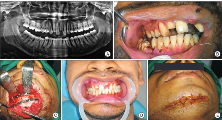
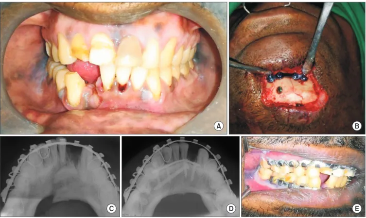
Among the 17 parasymphysis fracture sites, two patients received 2-mm profile, 4-hole titanium mini-plates (Ortho Max mandible plating system; Ortho Max Manufacturing Company, Vadodara, India) with gaps in 15 sites. One pa- tient had a comminuted right parasymphysis fracture that was fixed using a 2-mm profile, 7-hole continuous titanium mini-plate (Ortho Max mandible plating system) at the lower border and a 2-mm profile, 5-hole continuous titanium mini- plate placed 4 to 5 mm above the lower plate.(Fig. 1) The triangular segment was fixed with a 12-mm long, 2-mm pro- file titanium screw. In another patient, a single lag screw that was 20-mm long with a 2.5-mm profile was used at the lower border and a 2-mm profile, 4-hole titanium mini-plate with a gap was placed above the lag screw.(Fig. 2)
Of the 14 angle fracture sites, 13 were fixed with a single upper border plate with a 2-mm profile and 4-hole titanium
mini-plate (Ortho Max mandible plating system) with a gap.
One angle fracture site was comminuted and was fixed using a 2-mm profile, 2-hole titanium mini-plate with a gap at the upper border and a 2.5-mm profile and 4-hole titanium plate on the lateral surface.(Fig. 3) While fixing the angle fracture, the most proximal screws were fixed after the release of the MMF for ease of access, as suggested by Ellis and Walker6. Of 14 angle fracture sites, 4 required removal of the third molar while 3 had a missing third molar, and the remaining 7 third molars were retained.
The surgical sites were closed in layers according to site characteristics and patients were discharged after being ob- served for 1 hour. Patients were not placed in MMF, except for those with condylar fractures who were placed in IMF for 1 week. Patients were administered Augmentin 625 mg (twice a day), metronidazole 400 mg tab. (three times a day), and diclofenac 50 mg (three times a day) for 5 days with nutrition supplements for 1 month and were advised to maintain strict oral hygiene. All patients had follow-up visits at regular in- tervals: on the 1st postoperative day, 1st week, 4th week, and 6th week. After 6 weeks, the arch bars were removed and pa- tients were seen 3 months, 6 months, and 1 year after surgery.
A B
C D E
Fig. 1. Extraoral approach for management of right parasymphysis fracture combined with right condylar and right coronoid fracture under regional anesthesia. A. Preoperative orthopantomograph showing fracture. B. Deranged occlusion. C. Extraoral exposure through lacera- tion. D. Wound closure. E. Postoperative occlusion after 6 weeks.
Natarajan Chellappa et al: A working paradigm for managing mandibular fractures under regional anesthesia. J Korean Assoc Oral Maxillofac Surg 2018
A B
C D E
Fig. 2. Displaced right parasymphysis fracture managed through extraoral approach under regional anesthesia with lag screw and 4-hole titanium miniplate. A. Deranged occlusion. B. Fracture site exposed through extraoral laceration and fixation done. C. Preoperative man- dibular occlusal view. D. Postoperative mandibular occlusal view. E. Postoperative occlusion.
Natarajan Chellappa et al: A working paradigm for managing mandibular fractures under regional anesthesia. J Korean Assoc Oral Maxillofac Surg 2018
A B
C D E
Fig. 3. Comminuted right body and angle fracture managed under regional anesthesia. A. Right lateral oblique mandible. B. Orthopanto- mograph (OPG). C. Intraoral exposure and fixation of fracture segments. D. Postoperative OPG. E. Postoperative occlusion after 6 weeks.
Natarajan Chellappa et al: A working paradigm for managing mandibular fractures under regional anesthesia. J Korean Assoc Oral Maxillofac Surg 2018
III. Results
A total of 23 patients with 32 fracture sites were included in this review; 20 patients were male and 3 were female. Pa- tient ages ranged from 19 to 49 years.
Among the 23 patients, 1 had a left angle and right para- symphysis fracture that was intolerant to the procedure.
This patient was given 10 mg of IV Valium (Cipla, Mumbai, India), which did not help to calm the patient. It was then de- cided to limit treatment to a single mini-plate at the superior border of the angle and a single mini-plate at the parasym- physis.
In general, the reductions and fixations obtained were stable. Recovery was uneventful in all 23 patients with stable reproducible occlusion even after 6 months; this included patients with condylar fractures who were managed with conservative closed reduction. Two patients developed an ex- traoral draining sinus. One patient was managed with incision and drainage, while the other required hardware removal af- ter 3 months. One alcoholic patient came back with deranged occlusion 1 week after surgery, because the patient fell while intoxicated and was managed with MMF for 6 weeks.
IV. Discussion
All patients in this review underwent ORIF for their man- dibular fractures, while condylar fractures were managed conservatively with closed reduction. A simple straightfor- ward method should always be chosen over a tedious compli- cated one, provided it offers a similar outcome. With respect to fixation methods, rigid fixation was employed in all cases except two parasymphysis and body fractures fixed with 2 mini-plates and an Erich arch bar. All the angle fractures were managed with semi-rigid fixation employing a single upper border mini-plate. Internal fixation methods for mul- tiple mandibular fractures are controversial. Champy et al.5, in their landmark 1978 article, outlined the ideal approach for osteosynthesis. Many authors, including Ellis and Walker6, supported the use of a single mini-plate at the upper border for angle fractures6,7. Champy et al.5 stated that placement of a single mini-plate at the upper border in an angle fracture was sufficient for attaining functional stability, irrespective of whether the patient had a single or multiple mandibular fracture. In 2013, Ellis8 advocated that at least one rigid fixa- tion should be required in multiple fracture cases to reduce complications. Ellis8 stated that placement of two mini-plates along with an arch bar provided rigid fixation to the fracture
segments because it allowed no movement of the fracture site.
All fractures in this study were either single or multiple, except for those with condylar fractures, and all were man- aged with patients under regional anesthesia.
Although various techniques for inferior alveolar nerve block have been described in the literature9, a conventional inferior nerve block technique was used for patients in this study, because the operator was fluent with the technique. For patients with severe trismus, a closed-mouth Akinosi-Vazira- ni technique for mandibular nerve block was employed. Lo- cal infiltration over the surgical site and the lower border of the mandible was performed to minimize oozing because the anesthetic contained adrenaline to aid in painless handling of muscular structures. No difficulty was encountered with regard to fracture reduction from muscle spasm.
All patients were successfully managed under regional anesthesia without any intraoperative uncontrolled events, with one exception, which arose from a judgment error in patient selection, who was very apprehensive about the surgi- cal procedure and who had a decreased pain threshold, which prompted us to place a single mini-plate at the upper border of the left angle and a single mini-plate in the parasymphysis region.
Because the majority of mandibular fracture patients pres- ent with trismus, which makes preoperative anesthetic as- sessment difficult10, the anesthetist must proceed under the assumption that the mouth opening will increase after induc- tion, but this assumption could lead to serious consequences if the anticipated mouth opening is not achieved. To avoid this, the anesthetist may resort to performing awake fiber- optic intubation, which is very unpleasant for patients and also introduces serious complications11. Associated mid-face fractures can sometimes preclude naso-tracheal intubation.
Following mandibular fracture treatment, mandibular nerve blocks have been frequently used for postoperative analge- sia12. To eliminate the inherent risks associated with GA, regional anesthesia has been successfully used in traumatic maxillofacial cases13.
The advantage of regional over GA is that the patient is conscious and has control over the musculature that main- tains the airway, controls gastric secretions, and aids in condylar positioning. Although condylar sag has not been re- ported in the literature as a main cause of deranged occlusion following mandibular fracture treatment, it is nevertheless an established causative factor for postoperative malocclusion.
Previous studies suggested that patients under GA should
be awakened intraoperatively to check the condyle position within the fossa14. Occlusion is a dynamic relationship that depends on dentoalveolar architecture, TMJ articulation, and masticatory muscles. Thus, it is important to emphasize the role of muscle tone, muscular activity, and proprioception to maintain condylar position, which is not affected by regional anesthesia, and to prevent postoperative malocclusion.
Additionally, regional anesthesia has a clear advantage over GA in that the patient is conscious during the procedure, which can warn the surgeon of impending complications.
Under regional anesthesia, patients require less postopera- tive nursing care and have a shorter recovery time. All our patients were discharged 1 hour after the procedure. Regional anesthesia negates other GA complications, like atelectasis, pulmonary edema, nausea, and vomiting and it provided bet- ter pain postoperative relief. Also, regional anesthetic proce- dures are cheaper, which is particularly important in lower- middle income countries, like India, where the majority of people with maxillofacial trauma are of lower economic status.
Complications are very common when managing mandibu- lar fractures and can occur during any phase of treatment4,15. Surprisingly, only two of our 23 patients, accounting for 32 fracture sites, developed infection. The lower infection incidence was due to our emphasis on strict aseptic surgical protocol and because we placed patients on IV antibiotics during their surgery. Of the two patients who experienced infections, one had very poor oral hygiene, which contributed to infection of surgical site, and the other had a contaminated extraoral laceration, which was responsible for the infection.
No incidence of non-union or mal-union was observed in our patients. One patient returned a week later with deranged occlusion due to a fall while intoxicated. Alcohol and maxil- lofacial trauma have a well-established relationship in the literature16.
All our patients were prescribed protein and vitamin sup- plements. By default, oral intake by mandibular fracture pa- tients was reduced, despite the fact that trauma patients have elevated metabolic demands17, which must be supplemented.
We infer that regional anesthesia is an excellent intraopera- tive alternative to GA for ORIF for mandibular fractures in selected cases following stringent perioperative protocols.
V. Conclusion
To the best of our knowledge, this is the first work to em- phasize the reliability of regional anesthesia for treating mul-
tiple mandibular fractures in an outpatient procedure using both intraoral and extraoral approaches. We hope this work expands treatment opportunities for a wide spectrum of max- illofacial diseases that can be managed with regional anesthe- sia.
ORCID
Natarajan Chellappa, https://orcid.org/0000-0003-4052- 3319
Vikas Meshram, https://orcid.org/0000-0003-4052-707X Prajwalit Kende, https://orcid.org/0000-0001-5688-6371 Jayant Landge, https://orcid.org/0000-0002-1913-1401 Neha Aggarwal, https://orcid.org/0000-0001-7757-7834 Manish Tiwari, https://orcid.org/0000-0003-1642-373X
Authors’ Contributions
N.C. participated in performing the surgical procedure, data collection and wrote the manuscript. V.M. participated surgi- cal procedure and proof reading. P.K., J.L., N.A., and M.T.
participated in proof reading. All authors read and approved the final manuscript.
Ethics Approval and Consent to Participate
We would like to notify it that the ethical approval was waived by the ethics committee of the institution because this study is a retrospective study. All the surgical procedures were performed prior (January to November 2016), and man- uscript process was started at the culmination of surgical pro- cedures. All the surgical procedures were in accordance with the published literature that is in current practice throughout the world. Also, we obtained the informed consent from the patients for the procedure. Patients’ identity is not disclosed under any circumstances.
Consent for Publishing Photographs
Written informed consent was obtained from the patients for publication of this article and accompanying images.
Conflict of Interest
No potential conflict of interest relevant to this article was reported.
References
1. Gajiwala KJ. Surgery of temporomandibular joint under local an- aesthesia. Indian J Plast Surg 2008;41:175-82.
2. Manodh P, Prabhu Shankar D, Pradeep D, Santhosh R, Murugan A.
Incidence and patterns of maxillofacial trauma-a retrospective anal- ysis of 3611 patients-an update. Oral Maxillofac Surg 2016;20:377- 3. Ellis E 3rd, Moos KF, El-Attar A. Ten years of mandibular frac-83.
tures: an analysis of 2,137 cases. Oral Surg Oral Med Oral Pathol 1985;59:120-9.
4. Zweig BE. Complications of mandibular fractures. Atlas Oral Max- illofac Surg Clin North Am 2009;17:93-101.
5. Champy M, Loddé JP, Schmitt R, Jaeger JH, Muster D. Mandibular osteosynthesis by miniature screwed plates via a buccal approach.
J Maxillofac Surg 1978;6:14-21.
6. Ellis E 3rd, Walker LR. Treatment of mandibular angle fractures using one noncompression miniplate. J Oral Maxillofac Surg 1996;54:864-71; discussion 871-2.
7. Kumaran PS, Thambiah L. Versatility of a single upper border miniplate to treat mandibular angle fractures: a clinical study. Ann Maxillofac Surg 2011;1:160-5.
8. Ellis E 3rd. Open reduction and internal fixation of combined angle and body/symphysis fractures of the mandible: how much fixation
is enough? J Oral Maxillofac Surg 2013;71:726-33.
9. Khalil H. A basic review on the inferior alveolar nerve block tech- niques. Anesth Essays Res 2014;8:3-8.
10. Heard AM, Green RJ, Lacquiere DA, Sillifant P. The use of man- dibular nerve block to predict safe anaesthetic induction in patients with acute trismus. Anaesthesia 2009;64:1196-8.
11. Woodall NM, Harwood RJ, Barker GL. Complications of awake fibreoptic intubation without sedation in 200 healthy anaesthetists attending a training course. Br J Anaesth 2008;100:850-5.
12. Singh B, Bhardwaj V. Continuous mandibular nerve block for pain relief. A report of two cases. Can J Anaesth 2002;49:951-3.
13. El-Anwar MW, Hegab A. Internal fixation of single mandibular fracture under mandibular nerve block. Oral Maxillofac Surg 2016;20:57-61.
14. Politi M, Toro C, Costa F, Polini F, Robiony M. Intraoperative awakening of the patient during orthognathic surgery: a method to prevent the condylar sag. J Oral Maxillofac Surg 2007;65:109-14.
15. Ellis E 3rd. Treatment methods for fractures of the mandibular angle. Int J Oral Maxillofac Surg 1999;28:243-52.
16. Elledge RO, Elledge R, Aquilina P, Hodson J, Dover S. The role of alcohol in maxillofacial trauma: a comparative retrospective audit between the two centers. Alcohol 2011;45:239-43.
17. Falender LG, Leban SG, Williams FA. Postoperative nutritional support in oral and maxillofacial surgery. J Oral Maxillofac Surg 1987;45:324-30.