Photodynamic hyperthermal chemotherapy with indocyanine green: a novel cancer therapy for 16 cases of malignant soft tissue sarcoma
7
0
0
전체 글
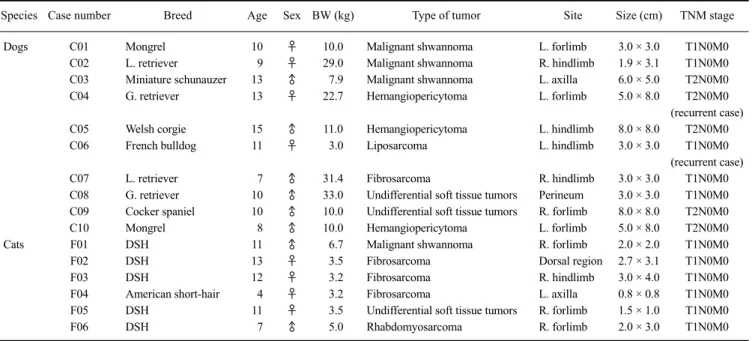
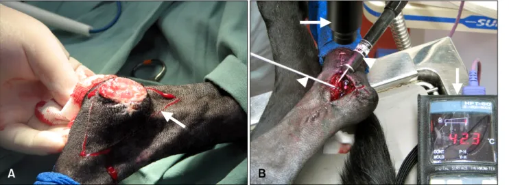
(2) 118 Masaki Onoyama et al.. Table 1. Data for the clinical cases Species Case number Dogs. Cats. Breed. Age. Sex BW (kg). Type of tumor. C01 C02 C03 C04. Mongrel L. retriever Miniature schunauzer G. retriever. 10 9 13 13. ♀ ♀ ♂ ♀. 10.0 29.0 7.9 22.7. Malignant shwannoma Malignant shwannoma Malignant shwannoma Hemangiopericytoma. C05 C06. Welsh corgie French bulldog. 15 11. ♂ ♀. 11.0 3.0. C07 C08 C09 C10 F01 F02 F03 F04 F05 F06. L. retriever G. retriever Cocker spaniel Mongrel DSH DSH DSH American short-hair DSH DSH. 7 10 10 8 11 13 12 4 11 7. ♂ ♂ ♂ ♂ ♂ ♀ ♀ ♀ ♀ ♂. 31.4 33.0 10.0 10.0 6.7 3.5 3.2 3.2 3.5 5.0. Site. Size (cm). TNM stage. L. forlimb R. hindlimb L. axilla L. forlimb. 3.0 × 3.0 1.9 × 3.1 6.0 × 5.0 5.0 × 8.0. Hemangiopericytoma Liposarcoma. L. hindlimb L. hindlimb. 8.0 × 8.0 3.0 × 3.0. Fibrosarcoma Undifferential soft tissue tumors Undifferential soft tissue tumors Hemangiopericytoma Malignant shwannoma Fibrosarcoma Fibrosarcoma Fibrosarcoma Undifferential soft tissue tumors Rhabdomyosarcoma. R. hindlimb Perineum R. forlimb L. forlimb R. forlimb Dorsal region R. hindlimb L. axilla R. forlimb R. forlimb. 3.0 × 3.0 3.0 × 3.0 8.0 × 8.0 5.0 × 8.0 2.0 × 2.0 2.7 × 3.1 3.0 × 4.0 0.8 × 0.8 1.5 × 1.0 2.0 × 3.0. T1N0M0 T1N0M0 T2N0M0 T2N0M0 (recurrent case) T2N0M0 T1N0M0 (recurrent case) T1N0M0 T1N0M0 T2N0M0 T2N0M0 T1N0M0 T1N0M0 T1N0M0 T1N0M0 T1N0M0 T1N0M0. BW: body weight, TNM: T, size of the primary tumor; N, condition of the regional lymph nodes; M, absence/presence of distant metastasis.. Hospital of Tottori University (six dogs and four cats; Tottori, Japan), Takayama Pet Clinic (two dogs; Osaka, Japan), Aino Animal Hospital (one dog and two cats; Shizuoka, Japan) and Tokyo Animal Medical Center (one dog; Tokyo, Japan), respectively. The animals were diagnosed with STS based on preoperative biopsies. The ages of the animals ranged from 4 to 15 years. The dogs included two Labrador retrievers, two golden retrievers, one cocker spaniel, one Welsh corgi, one miniature schnauzer, one French bulldog, and two mongrels. The cats included five domestic shorthair (DSH) cats and one American shorthair. We measured the original tumor size by caliper before surgery. All cases were classified according to TNM (T, size of the primary tumor; N, condition of the regional lymph nodes; M, absence/presence of distant metastasis) stage [21]. Tumors in five dogs and six cats were classified as T1 (≤ 5 cm in diameter at the greatest dimension), and T2 (> 5 cm in diameter at greatest dimension) in five dogs. Among the dogs, the tumor types included three cases of malignant schwannoma, three cases of hemangiopericytoma, one case of liposarcoma, one case of fibrosarcoma, and two cases of undifferentiated soft tissue tumors. In the cats, the tumor types included three cases of fibrosarcoma, one case of malignant schwannoma, one case of rhabdomyosarcoma, and one case of an undifferentiated soft tissue tumor. The tumor sites included the trunk in four cases (one in the dorsal region, two in the axilla, and one in the perineum) and the limbs in 12 cases. No lung metastasis was observed by radiography in any case.. We explained the risk of recurrence and treatment options, including surgery, radiation, and chemotherapy, to the animal owners. When tumors had developed in the limbs, we proposed amputation as the first choice of treatment to the owners. However, the owners did not desire amputation and radiation. We then proposed to all owners other treatments including the combination of PHCT and surgery. We explained that PHCT is an experimental therapy, and all owners of the pets enrolled in this clinical trial provided informed consent.. Surgical treatment For tumors that had developed in the limbs, we maintained sufficient skin for skin closure with skin sutures after the tumors were removed because the owners did not want us to perform reconstructive surgery such as skin grafting. As a result, we could not obtain sufficient surgical margins. We removed the underlying fascia of the tumors. In some cases, we could not perform perfect skin closure due to the presence of skin ulcers (Fig. 1A). In some cases (cases C06, C09, and F04), we removed the tumors with an ultrasonic aspiration device (Qucer; M & M, Japan). For case F02, only PHCT was performed because the owner did not wish the animal to undergo surgery. PHCT ICG (25 mg/vial, Giagnogreen; Daiich Sankyo, Japan) was dissolved in 9 mL of saline with an adjusted pH of 5.0. As an anti-tumor drug, 1 mL of bleomycin (1 mg/mL, Buleo;.
(3) A new adjuvant therapy for malignant soft tissue sarcoma 119. Fig. 1. (A) Skin incision (arrow) in case C07. (B) PHCT was performed after surgery. The arrow indicates the broadband light source while the arrowheads indicate the thermometer and thermometer sensor.. Nippon Kayaku, Japan) or carboplatin (10 mg/mL; Nippon Kayaku) was added to the ICG solution. For some cases, a small volume (0.1 or 0.2 mL) of paclitaxel (Bristol-Myers Squibb, USA.) was added to the ICG solution. For one case (case C04), no anti-tumor drugs were administered in accordance with the owner’s request. A broadband light source (Super Lizer 5000; Tokyo Iken, Japan) emitting a wavelength spectrum from 600 to 1,600 nm with a 5,000 mW maximum output power was used because ICG responses to light at wavelength of 600∼800 nm. For each case, the tumors were resected and ICG solution was injected into the resected area 3-dimensionally, including the skin surgical margin (3 cm). One mL of the 2 ICG solution was administered per cm of the wound bed. Irradiation was administered at a distance of 10 cm from 2 2 the resected area (irradiation area: 113 cm , 40 mW/cm ) 2 2 for 20 min per 113 cm (48 J/cm ; Fig. 1B) immediately after the ICG solution was injected. The temperature at the o surface of the resected area was kept under 45 C by moving light source near and away from the skin surface and monitored with a thermometer. The first round of PHCT was performed immediately after skin suturing following surgery. The treatment interval between the second and fourth round of PHCT was generally 1 week, and then the treatment was performed at intervals of 2 to 4 weeks. At a minimum, treatment was continued for 3 months after surgery. At that point, we continued the treatment if the owner desired. For the second and subsequent rounds of PHCT, the treatments were performed with all animals under sedation. In some cases, local anesthesia was induced by Lidocaine of 15∼50 mg/head (Xylocaine; AstraZeneca, Japan).. Follow-up after PHCT In all cases, follow-up examinations for STS recurrence and metastasis were performed at intervals of several months for 1 year after the first round of PHCT. Thereafter, follow-up examinations were performed once a year for 5 years.. Results Table 2 presents a summary of the study results. PHCT was performed three to 21 times. The treatment frequency depended on the wishes of the owner. No severe side effects, including severe skin burns, necrosis, or rupture of skin sutures, were observed in any of the animals although skin redness and minor skin burns occurred. The overall canine survival time (ST) except for 1 case of amputation (case C06) ranged from 225 to 1,901 days (median survival time: 767 days). In seven out of 10 dogs (70.0%), no recurrence was observed during the follow-up periods ranging from 238 to 1,901 days. In five of these seven dogs, no recurrence was observed after more than 2 years. The remaining three dogs (two with undifferentiated soft tissue sarcomas and one with liposarcoma) experienced recurrence or metastasis over intervals ranging from 72 to 162 days after surgery. One case with liposarcoma (case C06) experienced local recurrence occurring within 1 month after the first surgery. In this animal, local recurrence was also observed 72 days after the first round of PHCT following the second surgery. The disease-free time (DFT) was prolonged by the use of PHCT. However, amputation was performed in accordance with the owner’s desire, and the dog is currently alive. One dog (case C08) of the remaining three dogs died due to tumor progression. In case 08, metastasis to the sublumbar lymph nodes was.
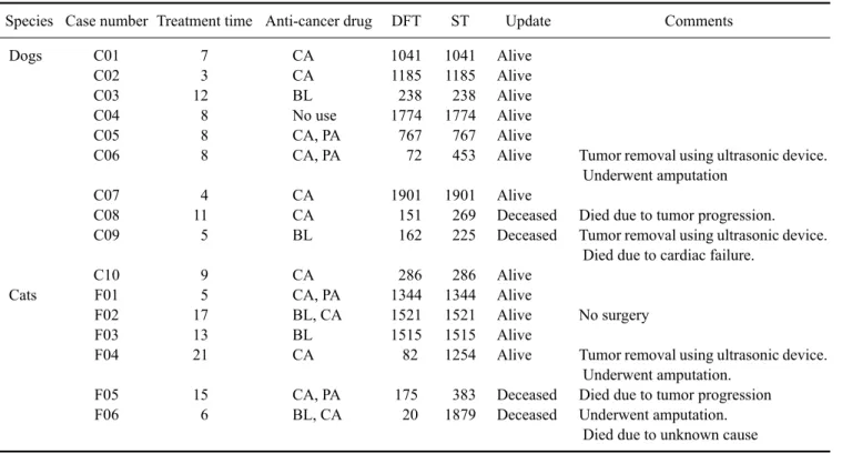
(4) 120 Masaki Onoyama et al.. Table 2. Summary of treatment outcomes Species Case number Treatment time Anti-cancer drug Dogs. Cats. DFT. ST. Update. C01 C02 C03 C04 C05 C06. 7 3 12 8 8 8. CA CA BL No use CA, PA CA, PA. 1041 1185 238 1774 767 72. 1041 1185 238 1774 767 453. Alive Alive Alive Alive Alive Alive. C07 C08 C09. 4 11 5. CA CA BL. 1901 151 162. 1901 269 225. Alive Deceased Deceased. C10 F01 F02 F03 F04. 9 5 17 13 21. CA CA, PA BL, CA BL CA. 286 1344 1521 1515 82. 286 1344 1521 1515 1254. Alive Alive Alive Alive Alive. F05 F06. 15 6. CA, PA BL, CA. 175 20. 383 1879. Deceased Deceased. Comments. Tumor removal using ultrasonic device. Underwent amputation Died due to tumor progression. Tumor removal using ultrasonic device. Died due to cardiac failure.. No surgery Tumor removal using ultrasonic device. Underwent amputation. Died due to tumor progression Underwent amputation. Died due to unknown cause. DFT: disease-free time, ST: survival time, CA: carbopulatin, BL: bleomycin, PA: paclitaxel.. observed. In that animal, no local recurrence was found. In case C09, local recurrence was observed 162 days after the first round of PHCT. PHCT was repeated for this animal five times at intervals of 1 to 2 weeks. Thereafter, the dog was only followed up. Sixty-seven days after the fifth round of PHCT, local recurrence was observed. The dog eventually died of cardiac failure 225 days after the first round of PHCT. The overall feline ST, excluding two cases of amputation (cases F04 and F06), ranged from 383 to 1,521 days (median survival time: 1,344 days). In three out of the six cats (50.0%), no recurrence was observed over the follow-up periods ranging from 1,344 to 1,521 days. The remaining three cats (one case of fibrosarcoma, one case of rhabdomyosarcoma, and one case of undifferentiated STS) experienced recurrence over intervals ranging from 20 to 175 days after surgery. In two (cases F04 and F06) out of the remaining three cases, amputation was performed on one cat (case F04) that is currently alive and one cat (case F06) that died due to unknown cause. The remaining cat died due to tumor progression. In case F02 for which no surgery was performed, we periodically assessed tumor status using fine needle aspiration (FNA). No living tumor cells were observed by FNA during the eighth round of PHCT. We diagnosed the tumor as degenerative following PHCT, and the degenerative tumor tissue was removed using an ultrasonic aspiration. device (Sono Cure; Tokyo Iken, Japan). Thereafter, PHCT was performed for 3 months. This cat had no recurrence for about 5 years.. Discussion Malignant STSs rarely metastasize; however, they are locally invasive [3]. If the surgical margin is insufficient, the rate of recurrence is 10 times of that when the surgical margin is sufficient [20]. Therefore, complete resection with a sufficient surgical margin is necessary. In general, adjuvant therapies including radiation therapy and chemotherapy are administered to cases of insufficient surgical margins [13,21,22,27]. In particular, radiation therapy plays an important role in the management of STS. In the present study, the surgical margins were insufficient in all cases. However, 70.0% (7/10) of the dogs and 50.0% (3/6) of the cats did not experience recurrence over follow-up periods ranging from 238 to 1,901 days. These results suggest that PHCT decreases the risk of recurrence. As an alternative to radiation therapy, we developed a new therapy, PHT, by combining Photodynamic therapy (PDT) and hyperthermia with ICG using a broadband light source. Furthermore, we administered PHT in combination with local chemotherapy and designated this technique “PHCT”. No previous studies have demonstrated the efficacy of the combination of ICG and use of a broadband light source in.
(5) A new adjuvant therapy for malignant soft tissue sarcoma 121. cancer therapy. It has been suggested that fibrosarcomas, malignant schwannomas, and hemangiopericytomas are sensitive to PHCT considering the relationship between tumor type and PHCT efficacy. The local control rate of fibrosarcoma, malignant schwannoma, and hemangiopericytoma was 100% in the present study. However, PHCT is not effective for other tumor types including liposarcoma, rhabdomyosarcoma, and undifferentiated STSs occurring in the limbs. In these cases, the tumors recurred ranging from 20 to 175 days after the first PHCT. These results suggest that the effects of PHCT might differ depending on tumor type. Further investigation into this possibility is necessary. In the present study, no relationship between treatment frequency and recurrence was observed. In one case (case C09), local recurrence was observed 63 days after the final round of PHCT. This phenomenon suggests that it is important to periodically continue treatment with PHCT. Additionally, no relationship was observed between the use of anti-cancer drugs and outcomes. In two out of three canine and two out of three feline recurrence cases, the tumor cells had infiltrated deeply. We speculate that the light used for PHCT did not reach the tumor cells located in deep tissues. In a preliminary experiment, we found that the temperature at a depth of 2 o cm from the skin surface did not reach 40 C (data not shown), indicating that PHCT is not effective for treating tissues beyond this depth. Additionally, the ICG solution was not equally distributed. These drawbacks will be investigated in the future. Hyperthermia is not a common treatment in veterinary medicine because the required device is expensive and few fundamental data related to the efficacy of hyperthermia are available. On the other hand, there are reports of PDT being used to treat spontaneous tumors in canines and felines, and the efficiency of this technique has been recognized [11,28-30,33-35]. However, the photosensitizer for PDT is very expensive and a special diode laser is needed. Therefore, PDT is also rarely administered by veterinarians. ICG was developed as a drug to promote liver and bile duct function, and it has been used medically since 1956. This drug is safe for both human and animals [8]. In particular, ICG is widely used in ophthalmology for hyperthermia therapy with an 808-nm diode laser to treat chorioretinopathy [12]. ICG is also used for sentinel biopsies [17,19]. Some reports have suggested that ICG induces the formation of oxygen radicals [1,2,4,10,23]. Most of these findings are from in vitro studies of different human cell lines. Bozkulak et al. [4] reported that ICG with 2 2 near-infrared light (809 nm, 60 mW/cm , 24 J/cm ) is very effective for eliminating human breast cancer cells. In this study, the temperature of the medium was controlled to o ensure that it remained at 37 C. The authors concluded that. ICG acts as a new photosensitizer. Hirano et al. [18] was the first to demonstrate that ICG induces the formation of oxygen radicals in response to irradiation with light at a wavelength of 600∼800 nm. The concentration of oxygen radicals produced by ICG dissolved in ethanol is similar to that generated by other photosensitizers. Oxygen radicals are formed even if ICG is dissolved in water although the concentration is lower. The total volume of oxygen radicals produced is thought to result from the accumulation of oxygen radicals produced with each wavelength. Therefore, a broadband light emitting light over a range of 600∼800 nm would induce the formation of more oxygen radicals than a diode laser. Additionally, a broadband light that has an emission range of 600∼800 nm would stimulate ICG to simultaneously induce heat and oxygen radical production in tumor tissues. We previously investigated the effects of PHT on B16F10 murine melanoma cells in vitro [31,32]. Our results demonstrated that PHT induces early morphological changes in tumor cells that promote more cell death than hyperthermia alone. Furthermore, PHT induces early apoptosis and cell arrest. These in vitro data support the present in vivo results. Tumor cells are more sensitive to heat under acidic conditions [14]. Therefore, saline with a pH adjusted to 5.0 with acetic acid was used as a solvent for ICG. In a preliminary experiment, we found that saline with a pH of 4.0 induced tissue inflammation (data not shown). The effects of anti-tumor drugs are enhanced by heat [15,16,24,25]. Furthermore, tumor tissues have a tendency to be acidic [36]. Therefore, we selected bleomycin as an anti-tumor drug because it is effective in acidic environments [15]. Platinum-based drugs such as cisplatin and carboplatin are also useful anti-tumor reagents. In the present study, we were concerned that PHCT would affect the stability of the anti-tumor drug. However, our preliminary data showed that the anti-tumor effects of PHCT were enhanced by the addition of an anti-tumor drug (data not shown). This means that PHCT does not only affect the stability of the anti-tumor drug but also enhances its effects. In conclusion, PHCT is a simple procedure, is not associated with any severe side effects, and requires no special facilities. However, further investigation is necessary to establish PHCT as a therapeutic technique due to some associated problems involving treatment times and intervals, and selection of anti-cancer drugs. Nevertheless, this modality is expected to become a useful alternative to radiation therapy for treating superficial tumors such as STS in companion animals.. References 1. Abels C, Karrer S, Bäumler W, Goetz AE, Landthaler M,.
(6) 122 Masaki Onoyama et al.. 2.. 3. 4.. 5.. 6. 7.. 8.. 9. 10. 11. 12. 13. 14. 15.. 16. 17.. 18.. Szeimies RM. Indocyanine green and laser light for the treatment of AIDS-associated cutaneous Kaposi’s sarcoma. Br J Cancer 1998, 77, 1021-1024. Bäumler W, Abels C, Karrer S, Weiβ T, Messmann H, Landthaler M, Szeimies RM. Photo-oxidative killing of human colonic cancer cells using indocyanine green and infrared light. Br J Cancer 1999, 80, 360-363. Bostock DE, Dye MT. Prognosis after surgical excision of canine fibrous connective tissue sarcomas. Vet Pathol 1980, 17, 581-588. Bozkulak O, Yamaci RF, Tabakoglu O, Gulsoy M. Photo-toxic effects of 809-nm diode laser and indocyanine green on MDA-MB231 breast cancer cells. Photodiagnosis Photodyn Ther 2009, 6, 117-121. Chen WR, Adams RL, Heaton S, Dickey DT, Bartels KE, Nordquist RE. Chromophore-enhanced laser-tumor tissue photothermal interaction using an 808-nm diode laser. Cancer Lett 1995, 88, 15-19. Chen WR, Adams RL, Bartels KE, Nordquist RE. Chromophore-enhanced in vivo tumor cell destruction using an 808-nm diode laser. Cancer Lett 1995, 94, 125-131. Chen WR, Adams RL, Higgins AK, Bartels KE, Nordquist RE. Photothermal effects on murine mammary tumors using indocyanine green and an 808-nm diode laser: an in vivo efficacy study. Cancer Lett 1996, 98, 169-173. Cherrick GR, Stein SW, Leevy CM, Davidson CS. Indocyanine green: observations on its physical properties, plasma decay, and hepatic extraction. J Clin Invest 1960, 39, 592-600. Dernell WS, Withrow SJ, Kuntz CA, Powers BE. Principles of treatment for soft tissue sarcoma. Clin Tech Small Anim Pract 1998, 13, 59-64. Diven DG, Pohl J, Motamedi M. Dye-enhanced diode laser photothermal ablation of skin. J Am Acad Dermatol 1996, 35, 211-215. Dougherty TJ, Thoma RE, Boyle DG, Weishaupt KR. Interstitial photoradiation therapy for primary solid tumors in pet cats and dogs. Cancer Res 1981, 41, 401-404. Dzurinko VL, Gurwood AS, Price JR. Intravenous and indocyanine green angiography. Optometry 2004, 75, 743-755. Ettinger SN. Principles of treatment for soft-tissue sarcomas in the dog. Clin Tech Small Anim Pract 2003, 18, 118-122. Gerweck LE. Modification of cell lethality at elevated temperatures: the pH effect. Radiat Res 1977, 70, 224-235. Hahn GM, Braun J, Har-Kedar I. Thermochemotherapy: synergism between hyperthermia (42-43o) and adriamycin (or bleomycin) in mammalian cell inactivation. Proc Natl Acad Sci U S A 1975, 72, 937-940. Hahn GM. Potential for therapy of drugs and hyperthermia. Cancer Res 1979, 39, 2264-2268. Hirche C, Murawa D, Mohr Z, Kneif S, Hünerbein M. ICG fluorescence-guided sentinel node biopsy for axillary nodal staging in breast cancer. Breast Cancer Res Treat. 2010, 121, 373-378. Hirano T, Kohno E, Gohto Y, Obana A. Singlet oxygen generation due to ICG irradiation. Photomed Photobiol 2006, 28, 15-16.. 19. Hojo T, Nagao T, Kikuyama M, Akashi S, Kinoshita T. Evaluation of sentinel node biopsy by combined fluorescent and dye method and lymph flow for breast cancer. Breast 2010, 19, 210-213. 20. Kuntz CA, Dernell WS, Powers BE, Devitt C, Straw RC, Withrow SJ. Prognostic factors for surgical treatment of soft-tissue sarcomas in dogs: 75 cases (1986-1996). J Am Vet Med Assoc 1997, 211, 1147-1151. 21. Liptak JM, Forrest LJ. Soft tissue sarcoma. In: Withrow SJ, Vail DM, Page RL (eds.). Small Animal Clinical Oncology. 5th ed. pp. 356-380, Elsevier Saunders, St. Louis, 2012. 22. McChesney SL, Gillette EL, Dewhirst MW, Withrow SJ. Influence of WR 2721 on radiation response of canine soft tissue sarcomas. Int J Radiat Oncol Biol Phys 1986, 12, 1957-1963. 23. Mamoon AM, Gamal-Eldeen AM, Ruppel ME, Smith RJ, Tsang T, Miller LM. In vitro efficiency and mechanistic role of indocyanine green as photodynamic therapy agent for human melanoma. Photodiagnosis Photodyn Ther 2009, 6, 105-116. 24. Marmor JB. Interactions of hyperthermia and chemotherapy in animals. Cancer Res 1979, 39, 2269-2276. 25. Newell KJ, Tannock IF. Reduction of intracellular pH as a possible mechanism for killing cells in acidic regions of solid tumors: effects of carbonylcyanide-3 chlorophenylhydrazone. Cancer Res 1989, 49, 4477-4482. 26. Ogilvie GK, Reynolds HA, Richardson RC, Withrow SJ, Norris AM, Henderson RA, Klausner JS, Fowler JD, McCaw D. Phase II evaluation of doxorubicin for treatment of various canine neoplasms. J Am Vet Med Assoc 1989, 195, 1580-1583. 27. Ogilvie GK, Obradovich JE, Elmslie RE, Vail DM, Moore AS, Straw RC, Dickinson K, Cooper MF, Withrow SJ. Efficacy of mitoxantrone against various neoplasms in dogs. J Am Vet Med Assoc 1991, 198, 1618-1621. 28. Okamoto Y, Ogura K, Okamura Y, Ishii H, Sakata I, Hakamada K, Miyaki S, Nakajima S, Minami S. Canine hemangiopericytoma treated by combination of surgical resection and photodynamic therapy with novel photosensitizer, PAD-S31. Jap J Vet Anesth Surg 2005, 36, 69-73. 29. Osaki T, Takagi S, Hoshino Y, Okumura M, Kadosawa T, Fujinaga T. Efficacy of antivascular photodynamic therapy using benzoporphyrin derivative monoacid ring A (BPD-MA) in 14 dogs with oral and nasal tumors. J Vet Med Sci 2009, 71, 125-132. 30. Peaston AE, Leach MW, Higgins RJ. Photodynamic therapy for nasal and aural squamous cell carcinoma in cats. J Am Vet Med Assoc 1993, 202, 1261-1265. 31. Radzi R, Osaki T, Tsuka T, Imagawa T, Minami S, Okamoto Y. Morphological study in B16F10 murine melanoma cells after photodynamic hyperthermal therapy with indocyanine green (ICG). J Vet Med Sci 2012, 74, 465-472. 32. Radzi R, Osaki T, Tsuka T, Imagawa T, Minami S, Nakayama Y, Okamoto Y. Photodynamic hyperthermal therapy with indocyanine green (ICG) induces apoptosis and cell cycle arrest in B16F10 murine melanoma cells. J Vet.
(7) A new adjuvant therapy for malignant soft tissue sarcoma 123. Med Sci 2012, 74, 545-551. 33. Reeds KB, Ridgway TD, Higbee RG, Lucroy MD. Non-coherent light for photodynamic therapy of superficial tumours in animals. Vet Comp Oncol 2004, 2, 157-163. 34. Roberts WG, Klein MK, Loomis M, Weldy S, Berns MW. Photodynamic therapy of spontaneous cancers in felines, canines, and snakes with chloro-aluminum sulfonated phthalocyanine. J Natl Cancer Inst 1991, 83, 18-23. 35. Tanabe S, Yamamaguchi M, Iijima M, Nakajima S,. Sakata I, Miyaki S, Takemura T, Furuoka H, Kobayashi Y, Matsui T, Uzuka Y, Sarashina T. Fluorescence detection of a new photosensitizer, PAD-S31, in tumour tissues and its use as a photodynamic treatment for skin tumours in dogs and a cat: a preliminary report. Vet J 2004, 167, 286-293. 36. Wike-Hooley JL, van den Berg AP, van der Zee J, Reinhold HS. Human tumour pH and its variation. Eur J Cancer Clin Oncol 1985, 21, 785-791..
(8)
수치
관련 문서
Median overall survival of 28 multiple myeloma patients treated with Cyclophosphamide-Prednisone combination regimens as a primary therapy was 115 weeks.. Median
For patients treated for chin augmentation using either genioplasty with Medpor Ⓡ or osteotomy, the predictive value of the correlation of the hard and
In addition, if the patients had a high MPV level (cut-off value of 7.95 fL) without low-dose aspirin therapy, they were at risk for ischemic stroke, especially in
Kameoka, “Sentinel lymph node biopsy for breast cancer patients using fluorescence navigation with indocyanine green,” World Journal of Surgical Oncology, Vol..
Dombret H, Sutton L, Duarte M, Daniel MT, Leblond V, Castaigne S, Degos L: Combined therapy with all-trans retinoic acid and high-dose chemotherapy in
a prospective analysis of 50 cases. Preoperative staging of non-small-cell lung cancer with positron-emission tomography. Staging of non- small-cell lung cancer
• Hormone Therapy With or Without Combination Chemotherapy in Treating Women Who Have Undergone Surgery for Node-Negative Breast Cancer (The TAILORx Trial)..
(Background) The standard triple therapy used as the first-line treatment for Helicobacter pylori infection are a combination of proton pump inhibitor(PPI),
