Ⅰ. 서 론
진단을 위해 방사선을 이용한 영상 검사는 계속해서 증가하 고 있으며[1], 전리방사선으로 인한 확률적 영향(stochastic effect)이나, 결정적 영향(deterministic effect)에 대한 위험
요소 또한 증가하고 있다[2]. 조직에 방사선이 균일 또는 불균일 하게 조사되는 경우, 조직별 상대적인 위험도의 차이를 반영하 여 평가한 양을 유효 선량(effective dose)이라고 한다. 유효선 량의 계산에 사용되는 조직 가중계수(tissue weighting factor)는 조직의 상대적 위험도를 나타낸다. 국제 방사선 방호
<원저>
진단용 X선을 이용한 방사선검사에서 차폐체 모양과 부가필터 변화에 따른 난소의 선량 분석
최준호
강원대학교병원 영상의학과
A Dose Analysis on the Ovary According to the Type of Shielding Material and the Change of Additional Filter in Radiography Using Diagnostic X-ray
Choi Joon-Ho
Department of Radiology, Kangwon National University Hospital
Abstract The gonads are directly affected by radiation exposure during radiography of the pelvis, abdomen, and spine.
Exposure of the gonads to radiation can cause genetic mutations and can result in the occurrence of malignant tumors.
In this study, we created three types of shielding material shapes for shielding of the ovaries, which are the gonads of female during radiography of the pelvis, and comparative evaluations using shadow shielding methods. The source surface distance(SSD) was 100 cm and the field size was 42 cm × 43 cm. The three types of shielding material shapes(type 1, 2 and 3) were assessed and the entrance surface dose in the ovaries were measured. The thickness of the shielding materi- al was expanded from 0.3 mm to 2.4 mm and after five repetitions, radiation values were measured and mean values were calculated. The mean dose were 3.09 mGy for type 1, 3.54 mGy for type 2, and 3.19 mGy for type 3, indicating that the measurements were the lowest for type 1. When an additional filter of 0.2 Cu + 1 Al was used, the dose were 3.72 mGy for type 1, 5.43 mGy for type 2, and 4.05 mGy for type 3, indicating that the measurements were the lowest for type 1. The results show that, even if the shielding material is not thick, in other words, even with a thickness of 2.94 mGy for the SN 3(0.9 mm) of type 1, shielding can be achieved, with a patient dose lower than the diagnostic ref- erence level(3.42 mGy). Additionally, among the three types of shielding material, the type 1 appeared to be the most appropriate shielding material. It is thought that the use of shielding material could reduce the risk factors for stochastic effects or critical effects of ionizing radiation during pelvic or lumbar radiography.
Key Words : Radiation, Gonad, Shadow Shielding, Entrance Surface Dose, Dose Area Product, Diagnostic Reference Level 중심 단어 : 방사선, 생식선, 그림자 차폐, 입사표면선량, 면적선량, 진단참조준위
This paper was a product of research results gained through the support of the Ministry of Trade and Industry’s Industrial Technology Innovation Project(10049785, ‘Radiation or Non-ionization diagnosis and treatment equipment’ specializing R&D platform and medical equipment technology development)
Corresponding author: Joon-Ho Choi, Department of Radiology, Kangwon National University Hospital, 156, Baengnyeong-ro, Chuncheon-si, Gangwon-do, 24289, Republic of Korea / Tel: +82-33-258-2329 / E-mail: [email protected]
Received 22 October 2019; Revised 16 November 2019; Accepted 22 December 2019
위원회(international commission on radiological protection;
ICRP)는 전통적으로 생식선이 장기 중 조직 가중계수가 가장 높은 방사선 감수성 기관으로 간주해 왔다[3]. 최근 생식선 (gonad)의 조직 가중계수가 0.2에서 0.08로 하양 조정되었지 만 골반 내에 있는 생식선은 여전히 방사선에 민감한 부위 중 하나이다. 생식선의 생식 세포(germ cell)에 방사선이 조사 되면 유전적인 돌연변이의 원인이 될 수 있고, 악성 종양을 발생시키는 위험을 초래 할 수 있다[4-6]. 생식선(gonad) 차폐 (shielding)로 인하여 고환(testicle)은 약 95%의 선량을 줄일 수 있고, 난소(ovary)는 약 50%의 선량을 줄일 수 있기 때문에 상당히 효과적이라고 할 수 있다[7-9]. 생식선 차폐로 인한 난소의 선량 감소가 고환보다 낮은 이유는 골반에서 난소의 위치가 크게 퍼져 있어 차폐가 쉽지 않기 때문이다[10]. 현재 식품의약품안전청에 의해 제작 배포된 일반영상의학검사 의 환자선량 권고량 또는 환자선량 진단참조준위(diagnostic reference level; DRL) 가이드라인[11]은 입사표면선량 (entrance surface dose; ESD)으로 측정되어 표시되며, 방사선방호의 최적화를 위한 지표로 활용되고 있다. 실제 성인 여성의 골반 검사 시 난소를 차폐하기 위해 난소의 위치를 찾는 것에 어려움이 있고[10,12,13], 차폐체(shielding material)로 인하여 검사하고자 하는 위치에서 해부학적 부위 가 영상에 보이지 않을 수 있다. 이러한 이유 때문에 반복적인 검사로 인하여 환자에게 더 많은 방사선이 조사될 수 있다.
본 논문에서는 차폐체를 직접 신체 위에 올려놓는 것이 아니라 그림자 차폐(shadow shielding) 방법을 이용하여 신체 접촉 없이 난소의 위치에 맞게 차폐체를 조절하여 기존에 사용하였던 생식선 차폐체와의 유용성을 비교해 보았다.
Ⅱ. 대상 및 방법
1. 대상 및 장비
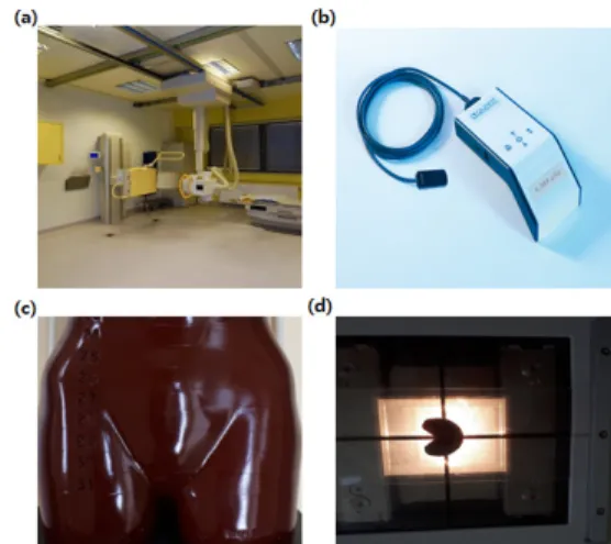
본 실험에 사용된 장비는 DR(digital radiography) 시스템 (Digital Diagnost, Philips, Germany, 장비규격은 150 KV, 812 mA)과 선량계(dosimeter, QUART didoEAS, Germany), 그리고 인체조직과 등가인 물질로 구성된 선량측정 팬톰 인 rando phantom(alderson research laboratories, USA)을 사용하였다. 팬톰은 female rando phantom으로 신장 150 cm, 무게 50 kg이다. 검출기(detector)는 43 cm
× 43 cm 넓이의 pixium 검출기를 사용하였고, 검출기 안에는 amplimat 이온화 챔버가 5개 장착되어 있다. 차폐체는 유연성 이 있는 납 재질로 연당량 0.3mm를 사용하였다.
Fig. 1. The figure shows the equipment used in the experiment: (a) digital radiography system; (b) dosimeter; (c) the pelvis of Rando phantom; and (d) shielding material
2. 측정방법
선원과 피부와의 거리(source skin distance, SSD) 100 cm, 조사범위(field size)는 42 cm × 43 cm로 하였다. Fig.
2와 같이 차폐체를 직접 난소 부위에 올려놓지 않고, 그림자 차폐(shadow shielding)방법을 사용하였다[14-16]. 차폐체 를 아크릴판에 부착한 다음, X-ray tube의 부가 필터를 끼울 수 있는 부분(filter assembly)에 차폐체를 부착시킨 아크릴판 을 끼우고 측정을 진행하였다.
Fig. 2. The image illustrates gonad shielding used shadow shielding method. The method entails attaching the shielding material to the acrylic plate and inserting the plate into the filter assembly. The source skin distance (SSD) was 100 cm
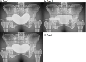
Fig. 3. The image of ovarian shielding in female rando phantom using three different shielding materials
Fig. 3에서와 같이 일반적으로 생식선 차폐에 사용하는 차 폐체의 모양 중 종류를 3가지로 분류하여 난소의 선량을 측정 하였다. 차폐체 두께를 0.3 mm에서 2.4 mm까지 늘려가며 5회 반복하여 선량값을 측정하고, 그 값에 대한 평균을 구하 였다. 자동노출제어(auto exposure control, AEC) 모드 (mode)를 이용하여 입사피부선량(entrance skin dose, ESD)을 mGy 단위로 그리고 면적선량(dose area product, DAP)을 mGy・cm2 단위로 측정하였다.
Fig. 4. Illustration of the zones used to classify the position of the ovary. The position of the ovaries was identified in at least two different planes and recorded according to the three zones. The yellow part (zone 1) corresponds to the true pelvis.
The red part (zone 2) indicates that is located at the center of the sacroiliac joint in the true pelvis. The blue part (zone 3) indicates that it is located on the side of the true pelvis
Fig. 4는 난소의 위치를 나타낸 것으로서 노랑색(zone 1)은 진골반(true pelvis)에 해당되는 부분으로, 치골결합(symphysis pubis)과 골반(pelvis)의 측면 그리고 천장관절(sacroiliac joint) 하한선에 의해 정의되는 원형으로 된 부분을 말한다.
빨간색 부분(zone 2)은 진골반에서 천장관절의 중앙에 위치하
는 경우를 나타내고 있다. 파랑색 부분(zone 3)은 진골반에서 옆쪽으로 위치하는 경우를 나타내고 있다[17].
Ⅲ. 결 과
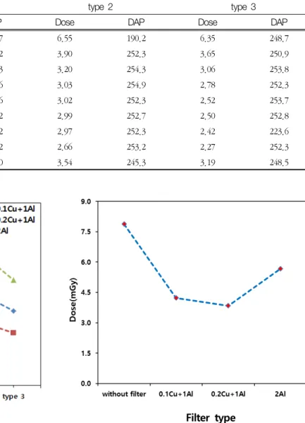
Table 1은 차폐체 모양에 따라 차폐체 두께를 증가시키며 측정한 난소(ovary)의 선량을 나타낸 것이다. 차폐체 두께를 SN 8(2.4mm)로 하였을 때, type 1에서 선량은 2.26 mGy, type 3에서 선량은 2.27 mGy, 그리고 type 2 에서는 2.66 mGy로 type 1에서 제일 낮게 측정되었음을 알 수 있었다.
type 1에서 전체 평균 선량은 3.09 mGy, type 2에서는 3.54 mGy, 그리고 type 3에서는 3.19 mGy로 측정되어 type 1에서 난소에 조사된 선량이 제일 낮게 측정되었음을 알 수 있었다.
또한 차폐체 1개일 때 보다 2개를 사용하였을 때 급격한 선량 감소를 보이는 것을 알 수 있었다. type 2에서 SN 1일 때 선량은 6.55 mGy, SN 2에서는 3.90 mGy로 2.65 mGy의 선량이 감소하였음을 알 수 있었다. 그리고 SN 5에서부터는 선량감소의 큰 변화는 없었음을 알 수 있었다. 또한 세 종류의 차폐체 모두 SN 3부터 골반의 환자선량 권고량(3.42 mGy)보다 낮은 선량이 측정되었음을 알 수 있었다. type 1은 SN 8(2.26 mGy)이 SN 1(5.97 mGy)대비 62%, type 2에서는 SN 8(2.66 mGy)이 SN 1(6.55 mGy)대비 59%, 그리고 type 3은 SN 8(2.27 mGy)이 SN 1(6.35 mGy)대비 64%의 선량이 감소함을 알 수 있었다. 난소가 받는 면적선량은 세 종류의 차폐체 대부분에서 247.7 mGy・cm2 ~ 254.3 mGy・cm2 범위에 포함되었지만, type 2의 SN 1에서는 190.1 mGy・cm2, type 3의 SN 7에서는 223.6 mGy・cm2으로 낮게 측정되었음을 알 수 있었다.
Fig. 5는 차폐체 모양과 부가 필터 종류에 따른 난소의 선 량 변화를 나타낸 그래프이며, 차폐체의 두께는 SN 1(0.3 mm)에 해당되는 두께를 사용하였다. without shielding에서 는 0.2 Cu + 1 Al의 부가 필터를 사용하였을 때 3.84 mGy로 제일 낮은 선량이 측정되었다. Type 1에서 0.1 Cu + 1 Al는 5.19 mGy, 0.2 Cu + 1 Al은 3.72 mGy 그리고 2 Al은 7.37 mGy로 type 2나 type 3보다 낮게 측정되었음을 알 수 있었다.
type 2에서 0.1 Cu + 1 Al은 without shielding일 때 보다 3.7 mGy, 0.2 Cu + Al은 1.59 mGy 그리고 2 Al은 5.9 mGy 더 높게 측정되었음을 알 수 있었다.
Fig. 6은 차폐체를 사용하지 않고 부가 필터만을 사용했을 때 선량을 나타낸 것으로서 필터가 없을 때는 7.88 mGy로 측정되었다. 0.1 Cu + 1 Al의 부가 필터를 사용했을 때는 필터를 사용하지 않았을 때 보다 46%(3.65 mGy)의 선량이 감소하였고, 0.2 Cu + 1 Al의 부가 필터를 사용했을 때
51%(4.04 mGy)의 선량이 감소하였음을 알 수 있었다. 그리 고 2 Al의 부가 필터를 사용하였을 때는 선량이 28%(2.21 mGy)로 감소하여, 0.2 Cu + 1 Al의 부가 필터를 사용하였을 때 제일 많은 선량이 감소함을 알 수 있었다.
Ⅳ. 고 찰
Fawceft SL 등은 Fig. 2에서와 같이 난소가 zone 1(노랑 색)에 위치할 수 있는 경우는 80.2%, zone 2(빨강색)에 위치 할 수 있는 경우는 17.1% 그리고 zone 3(파랑색)에 위치할 Table 1. The entrance surface dose (mGy) and the dose area product (mGy・cm2) of the ovaries according to the shielding material thickness increases with the use of three types of shielding material with different shapes. When the thickness of the shielding material was SN 8 (2.4 mm), the dose was 2.26 mGy in type 1, 2.27 mGy in type 3, and 2.66 mGy in type 2 shielding material, the dose which was the lowest in type 1 shielding material
Shield number
type 1 type 2 type 3
Dose DAP Dose DAP Dose DAP
SN 1(0.3 mm) 5.97 247.7 6.55 190.2 6.35 248.7
SN 2(0.6 mm) 3.64 250.2 3.90 252.3 3.65 250.9
SN 3(0.9 mm) 2.94 245.3 3.20 254.3 3.06 253.8
SN 4(1.2 mm) 2.69 252.6 3.03 254.9 2.78 252.3
SN 5(1.5 mm) 2.45 251.6 3.02 252.3 2.52 253.7
SN 6(1.8 mm) 2.39 250.2 2.99 252.7 2.50 252.8
SN 7(2.1 mm) 2.36 252.2 2.97 252.3 2.42 223.6
SN 8(2.4 mm) 2.26 250.2 2.66 253.2 2.27 252.3
Aver. 3.09 250.0 3.54 245.3 3.19 248.5
Shield type
Fig. 5. When the shield was not used and the three types of shielding material (types 1, 2, and 3) were used, the dose of the ovaries was measured by changing the additional filter (0.1 Cu + 1 Al, 0.2 Cu + 1 Al, and 2 Al). In the absence of shielding, the lowest dose of 3.84 mGy was measured using 0.2 Cu + 1 Al additional filters. The thickness of the shielding material was fixed at SN 1 (0.3 mm).
Filter type
Fig. 6. The dose of the ovaries was measured when three types of additional filters (0.1 Cu + 1 Al, 0.2 Cu + 1 Al, and 2 Al) were used and when an additional filter was not used.
The shielding material was not used. When the 2 Al additional filter was used, the dose was reduced to 28% (2.21 mGy).
This indicated that the greatest decrease in the dose was achieved with the use of the 0.2 Cu + 1 Al additional filter.
수 있는 경우는 1.5%라고 말하고 있다[17]. 본 실험에서도 zone 1의 모양과 유사한 형태의 차폐체를 만들어 사용하였다.
차폐체 모양과 부가 필터 종류에 따른 난소의 선량 변화를 측정하였을 때 2 Al에서 선량이 높게 측정된 이유는 Al의 경 우 두께에 따라 1차 방사선보다 산란선(scatter ray)이 더 많 이 생성되기 때문이다[18-20]. 환자 선량 권고량((DRL)은 진 단 영상의학 검사 시 환자가 받는 방사선량을 측정하고 평가 하여 진단에 참고할 수 있도록 권고하는 선량 준위를 말한다.
환자 선량 권고량은 임상에서 문제가 되지 않는 적정범위에서 필요한 최소한의 선량을 사용하도록 하고 있다[21]. 2008년 국제방사선방호위원회(ICRP)는 publication 103을 발간하 여 의료영상을 목적으로 수행하는 방사선 검사에 대해서는 환자방어를 위한 최적화를 위해 환자 선량 권고량을 적용하도 록 권고하고 있다[22,23]. 국제원자력기구(international atomic energy agency, IAEA) 등 6개 국제기구가 공동으로 전리방사선의 방어를 위한 국제기본 안전기준(basic safety standards, BSS) No.115를 마련하였고, 여기에서 권고하는 성인 환자의 골반선량은 10 mGy이었다[22,23]. 영국 보건국 (health protection agency, HPA)은 2010년 골반의 환자 선량을 3.9 mGy로 권고하였다[22]. 미국은 식품의약국(food and drug administration, FDA)의 의료기기를 담당하는 CDRH(center for devices and radiological health)에서 2002년 요천추 부위의 선량을 3.21 mGy로 권고하였다 [24,25]. 우리나라의 식품의약품안전청에서 2012년도 발표 한 일반영상의학검사에 대한 환자 선량 권고량 가이드라인에 따르면 전후면(anterio-posterio, AP)검사 시 골반의 환자 선량 권고량은 3.42 mGy, 사위면(oblique)검사일 때 권고량 은 6.32 mGy이다[11,21]. 본 실험에서 SN 3(0.9 mm)의 차폐 체 두께에서 세 종류의 차폐체 모두 국제방사선방호기구(10 mGy), 영국 보건국(3.9 mGy), 미국의 CDRH(3.21 mGy)에 서 권고하는 골반부위의 선량보다 낮게 측정(3.07 mGy) 되었 음을 알 수 있었다. 그림자 차폐방법이 직접 차폐체를 차폐할 부위에 올려놓는 방법보다는 유용하다고 할 수 있지만, 정확 한 난소의 위치를 찾는 것에 어려움이 따를 수 있을 것으로 생각된다.
Ⅴ. 결 론
골반(pelvis)의 방사선 검사 시 여성의 생식선인 난소 (ovary)의 차폐를 위해 차폐체 모양을 세 종류로 만들고 그 림자 차폐방법을 이용하여 비교 평가하였다. type 1이 SN 1(5.97 mGy)과 SN 8(2.26 mGy)에서 type 2와 type 3보다
선량이 낮게 측정되었음을 알 수 있었다. 차폐체 type 1(3.09 mGy)이 type 2(3.54 mGy)대비 15%, type 3(3.19 mGy)대비 3%의 선량이 더 낮게 측정되었음을 알 수 있었 다. 세 종류의 차폐체 모두 SN 3의 차폐체 두께에서 환자 선량 권고량(3.42 mGy)보다 낮게 측정되었음을 알 수 있었 고, type 1이 제일 차폐가 잘 되었음을 알 수 있었다. 0.1 Cu + 1 Al의 부가 필터를 사용했을 때 필터를 사용하지 않 았을 때 보다 46%(3.65 mGy)의 선량이 감소하였고, 0.2 Cu + 1 Al의 부가 필터를 사용했을 때 51%(4.04 mGy)의 선량이 감소하였음을 알 수 있었다. 그리고 2 Al의 부가 필 터를 사용하였을 때는 선량이 28%(2.21 mGy)로 감소하여, 0.2 Cu + 1 Al의 부가 필터를 사용하였을 때 제일 많은 선 량이 감소함을 알 수 있었다. 본 연구의 결과로 차폐체의 두 께를 두껍게 하지 않아도 즉 Type 1의 경우 SN 3(0.9 mm) 의 두께(2.94 mGy)로도 환자선량 권고량(3.42 mGy)보다 낮게 차폐할 수 있음을 알 수 있었다. 또한 그림자 차폐방법 이 기존에 사용하고 있는 직접 차폐 부위에 올려 차폐하는 방법보다 유용함을 알 수 있었다. 이런 차폐체의 사용으로 골반이나, 요추 검사 시 난소에 조사되는 전리 방사선으로 인한 확률적 영향이나, 결정적 영향에 대한 위험요소를 줄 일 수 있을 것으로 생각된다.
Acknowledgement
본 논문은 산업통상자원부 산업기술혁신사업으로 지원된 연구결과입니다(10049785, ‘방사선 또는 비전리 진단·치료 장치’ 특화 R&D 플랫폼 및 의료기기 기술개발).
REFERENCES
[1] Holmberg O, Malone J, Rehani M, McLean D, Czarwinski R. Current issues and actions in radia- tion protection of patients. Eur J Radiol. 2010;
76(1):15-9.
[2] Linet MS, Slovis TL, Miller DL, Kleinerman R, Lee C, Rajaraman P, et al. Cancer risks associated with external radiation from diagnostic imaging procedures.
CA Cancer J Clin. 2012;62(2):75-100.
[3] Doolan A, Patrick CB, Louise AR, Healy J. Gonad protection for the anterior-posterior projection of the pelvis in diagnostic radiography in Dublin
hospitals. Radiography. 2004;10:15-21.
[4] International Commission on Radiological Protection.
Radiological protection in medicine. ICRP pub- lication 105. Ann ICRP. 2007;37(6).
[5] Sikand M, Stinchcombe S, Liversley P. Study on the use of gonadal protection shields during paediatric pelvis X-rays. Ann R Coll Surg Engl. 2003;85:422-5.
[6] Wainwright AM. Shielding reproductive organs of orthopaedic patients during pelvic radiography.
Ann R Coll Surg Eng. 2000;82;318-21.
[7] International Commission on Radiological Protection.
Protection of the patient in diagnostic radiology.
ICRP Publication 34. Ann ICRP. 1982;9:2-3.
[8] Frantzen MJ, Robben S, Postma AA, Zoetelief J, Wildberger JE, Kemerink GJ. Gonad shielding in paediatric pelvic radiography: Disadvantages pre- vail over benefit. Insights Imaging. 2012;3:23-32.
[9] Jackson G, Brennan PC. Radio-protective aprons during radiological examinations of the thorax: an optimum strategy. Radiat Prot Dosimetry. 2006;121:
391-4.
[10] Bardo DM, Black M, Schenk K, Zaritzky MF.
Location of the ovaries in girls from newborn to 18 years of age: Reconsidering ovarian shielding.
Pediatr Radiol. 2009;39:253-9.
[11] Korea Food and Drug Administration. Guideline on patient dose suggestion of general radiology.
2012;30:4-7.
[12] Abram E, Wilkinson D, Hodson C. Gonadal pro- tection from X radiation for the female. Br J Radiol.
1958;31:335-6.
[13] Sforza C, Vizzotto L, Ferrario VF, et al. Position of follicles in normal human ovary during definitive histogenesis. Early Hum Dev. 2003;74:27-32.
[14] Bailey E, Anderson V. Syllabus on radiography ra- diation protection. Sacramento, California: California Department of Health services, 1995:72-6.
[15] Dowd SB, Tilson ER, Adams L. Practical radiation
protection and applied radiobiology. Philadelphia:
WB Saunders; 1999.
[16] Raissaki MT. Pediatric radiation protection.
European Radiology Supplements. 2004;14(S1):74-8.
[17] Fawcett SL, Gomez AC, Barter SJ, Ditchfield M, Set P. More harm than good? The anatomy of mis- guided shielding of the ovaries. Br J Radiol. 2012 Aug;85:1016.
[18] Odeh D, Ogbanje G, Jonah SA. X-Rays and scatter- ing from filters used in diagnostic radiology. Int J Sci Res Publ. 2013;3(7):1-3.
[19] Sutton DG, JMartin C, Peet D, Williams JR. The characterization and transmission of scattered ra- diation resulting from X-ray beams filtered with zero to 0.99 mm copper. J Radiol Prot. 2012;32:
117-20.
[20] Vlachos I, Tsantilas X, Kalyvas N, Delis H, Kandarakis I, Panayiotakis G. Measuring scatter- ing radiation in diagnostic x rays for radiation pro- tection purpose. Radiation Protection Dosimetry.
2015;165(1-4):382-5.
[21] Investigation of patient dose for diagnostic refer- ence levels(DRL) in radiographic examination:
National Survey in Korea. Ministry of Food and Drug Safety, 2011:14-28.
[22] Hart D, Hillier MC, Shrimpton PC. Doses to pa- tients from radiographic and fluoroscopic X-ray imaging procedures in the UK-2010 Review.
Health Protection Agency, 2012:57-61.
[23] International basic safety standards for protection against ionizing radiation and for the safety of ra- diation sources. IAEA Safety Series No.115. 1996.
[24] Nationwide Evaluation of X-ray Trends(NEXT):
https://www.fda.gov/radiation-emit- ting-products/radiation-safety/nation- wide-evaluation-x-ray-trendsnext.
[25] Conference of Radiation Control Program Directors (CRCPD): https://www.crcpd.org/page/Publications.
구분 성명 소속 직위
단독 최준호 강원대학교병원 방사선사