Received: November 20, 2015 / Revised: December 22, 2015 / Accepted: December 24, 2015 Corresponding Autor: Yong Jin Lee
Molecular Imaging Research Center, Korea Institute of Radiological and Medical Sciences (KIRAMS), 75 Nowon-ro, Nowon-gu, Seoul 01812, Korea Tel +82-2-970-1364, Fax: +82-2-970-1341, E-mail: [email protected]
Copyright ⓒ 2015, The Korean Society of Radiopharmaceuticals and Molecular Probes
J R MP
J Radiopharm Mol Probes 2015;1(2):123-129ISSN 2384-1583
ORIGINAL ARTICLE
Intratumoral distribution of 64 Cu-ATSM and 18 F-FDG in VX2 tumor xenografted rabbit
Ran Ji Yoo
1,3, Ji Woong Lee
1, Kyo Chul Lee
1, Gwang Il An
1, In Ok Ko
1, Wee Sup Chung
1, Ji Ae Park
1, Kyeong Min Kim
1, Yang-Kyu Choi
3, Joo Hyun Kang
1, Sang Moo Lim
2, and Yong Jin Lee
1,*1Molecular Imaging Research Center, Korea Institute of Radiological & Medical Sciences (KIRAMS), Seoul, Korea;
2Department of Nuclear Medicine, Korea Institute of Radiological & Medical Sciences, Seoul, Korea;
3Department of Laboratory Animal Medicine, College of Veterinary Medicine, Konkuk University, Seoul, Korea
ABSTRACT 64Cu-labeled diacetyl-bis(N4-methylthiosemicarbazone) is a promising agent for internal radiation therapy and imaging of hypoxic tissues. In the study, we confirmed hypoxia regions in VX2 tumor implanted rabbits with injection 64Cu-ATSM and 18F-FDG using positron emission tomography (PET)/computed tomography (CT). PET images with 18F-FDG and 64Cu-ATSM were obtained for 40 min by dynamic scan and additional delayed PET images of 64Cu-ATSM the acquired up to 48 hours. Correlation between intratumoral O2 level and 64Cu-ATSM PET image was analyzed. 64Cu-ATSM and 18F-FDG were intravenously co-injected and the tumor was dissected and cut into slices for a dual-tracer autoradiographic analysis. In the PET imaging,
64Cu-ATSM in VX2 tumors displayed a specific uptake in hypoxic region for 48 h. The uptake pattern of
64Cu-ATSM in VX2 tumor at 24 and 48 h did not match to the 18F-FDG. Through ROI analysis, in the early phase (dynamic scan), 18F-FDG has positive correlation with 64Cu-ATSM but late phase (24 and 48 h) of the
64Cu-ATSM showed negative correlation with 18F-FDG. High uptake of 64Cu-ATSM in hypoxic region was re- sponded with significant decrease of oxygen pressure, which confirmed by 64Cu-ATSM PET imaging and auto- radiographic analysis. In conclusion, 64Cu-ATSM can utilize for specific targeting of hypoxic region in tumor, and discrimination between necrotic- and viable hypoxic tissue. J Radiopharm Mol Probes 1(2):123-129, 2015 Key Words: 64Cu-ATSM, 18F-FDG, PET / CT, VX2 tumor, Hypoxia
Introduction
저산소증은 조직 내에서 정상 수준 이하로 산소 장력 이 감소한 상태로 종양 세포의 발달에서 중요한 역할을 한다(1,2). 종양 내의 저산소 부분의 영상을 획득하고 분 석하는 것은 미세환경을 이해하는데 필수적이다. 또 저 산소증 종양 세포는 방사선 치료와 화학요법에 저항성을 보이기 때문에 영상을 이용한 종양 내 저산소증의 평가 는 종양치료의 계획 및 평가에 유용하다(3).
최근 수십 년간 비침습성 산소 분압을 측정하는 촬영
방법이 연구되어 왔다(4). 양전자방출단층촬영(PET)을 이 용한 다수의 유망 양전자 방출 방사성 의약품이 개발되고 있다(4-6). 방사성 핵종으로 종양의 저산소증을 검출하고 자 한 시도는 1981년 14C-misonidazole이 처음이었다. 이 는 14C-misonidazole이 선택적으로 종양 내에 저산소 세포 대사에 결합한다는 것을 확인함으로써 증명되었다(7). 그 후, 18F-misonidazole는 종양 내 저산소증의 비침습적 영 상을 위한 PET 추적자로 제안되었다. 또한 이러한 목적을 위해 평가된 다른 여러 추적들로는 60/64Cu와 18F-EF5로 표지 한 Cu(II)-diacetyl-bis(N4-methyl-thiosemicarbazone) (Cu-ATSM),
Figure 1. Schematic diagram of VX2 tumor xenografted rabbit models.
18F-fluoroazomycin arabinoside (18F-FAZA) 등이 있다(5,8).
이러한 방사성의약품들 중 Cu-ATSM은 PET영상을 위 한 유망한 방사성의약품이다(9-16). 이전 논문들의 in vi- tro 데이터에서 Cu-ATSM은 종양세포의 많은 종류의 저 산소 환경에 축척된다고 알려져 있다(12,17,18). 임상 연 구는 Cu-ATSM 흡수에 의해 저산소 종양의 치료 저항성 과 전이 능력이 연관되어 있음을 증명하고 있다(9,19-21).
저산소 부분의 Cu-ATSM축적 메커니즘은 저산소증과 같 은 세포 내 높은 환원 조건에서 Cu-ATSM의 Cu(II)은 Cu(I)로 환원되어 즉시 ATSM 리간드로부터 방출하고 세 포에 포획되는 시스템이다(11,17,22-24). 종양에서의 Cu-ATSM 의 분포는 PET 영상의 트레이서로 일반적으로 사용되는
18F-fluorodeoxyglucose (18F-FDG)의 분포와 다르다고 보고 되어 있다(15,25). Tanaka 등의 논문에서는(15) 18F-FDG 가 혈관이 과다한 지역 및 괴사되어 가는 세포 증식 지 역을 대상으로 영상화되고, Cu-ATSM은 세포증식이 정 지되었지만, 약간의 괴사가 진행된 과다혈관분포 지역에 서 가장 높은 흡수를 보인다고 보고하였다.
본 연구에서 우리는 VX2종양세포를 이식한 토끼를 임 상용PET 장비를 이용하여 종양 내 저산소 환경에서의
64Cu-ATSM과 18F-FDG의 분포를 영상으로 확인하였다.
또한, 정확한 해부학적 영상을 위해 MRI영상도 획득하 였다. 산소분압측정 장비를 이용한 종양 내 산소 분포 확 인과 자가방사기록법을 이용한 조직에서의 방사성의약 품 분포 정도를 영상과 비교하였다.
Materials and Methods
1.
64Cu-ATSM합성
가속기로부터 64Ni(p,n)64Cu 핵반응을 통하여 얻어지는
64CuCl2 용액을 용이한 표지반응을 위하여 용액을 가열과 질소를 불어서 건조시킨다. 이렇게 건조된 64CuCl2 바이 알에 1M NaOAc 버퍼용액으로 pH를 5.5로 맞추고, DMSO 10 uL에 H2ATSM 10 ug을 녹인 용액을 첨가하여 10분 동안 실온에서 반응시킨다. 방사성표지가 다 끝난
A B
Figure 3. Imaging of MRI and PET in VX2 tumor xenograted rabbit model. (A) MRI, (B) 18F-FDG, (C) 64Cu-ATSM (D) 64Cu-ATSM_24 h, (E)
64Cu-ATSM_48 h.
반응용액을 C18 Sep-Pak 카트리지에 통과 시킨 후, 증류 수(D.W)로 여러 번 세척하여 불순물을 완전히 제거한다.
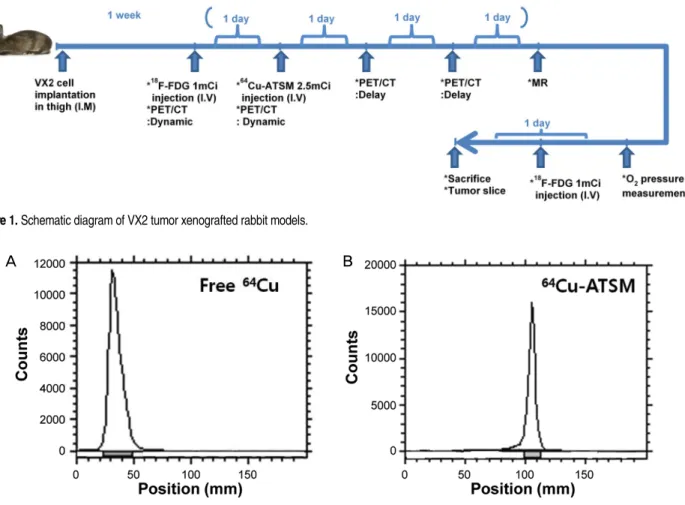
최종적으로 1 mL의 에탄올을 이용하여 카트리지에 흡착 된 순수 64Cu-ATSM만 획득한다. 이렇게 얻어진 64Cu-ATSM 의 품질관리로서 방사화학적 순도를 알아보기 위해 ra- dio-TLC를 시행하였다(Figure 2). 이동상(mobile phase)은 95% CH3CN/Water를 사용하였다. pH는 pH paper를 통해 확인하였다. 최종생산물인 에탄올 상태의 순수 64Cu-ATSM 은 생리식염수(0.9% saline)를 섞어 10% 에탄올 상태로 만든 다음, 최종적으로 0.22 μm의 멸균 필터를 통과시켜 멸균 용기로 옮긴다.
2. VX2 종양모델
VX2 암종(Seoul National University Hospital)을 이식해 놓은 토끼(New Zealand white rabbit, female, n=4)에서 종 양만 분리 후 Phosphate-Buffered Saline (PBS, GibcoBRL, Life Technologies, Gaithersburg, MD, USA)에 넣고 잘게 잘라준 후 1 mL를 마취시킨 새로운 토끼의 허벅지 근육 에 이식하여 종양을 키운다.
3. PET/CT와 MRI 영상
전체적인 실험일정은 Figure 1과 같이 진행하였다. 임 상용 PET/CT (Biograph, Siemens Medical System) 장비를 이용하여 VX2 암 종을 이식한 후 1주일된 토끼로 영상 을 획득하였다. 일주일을 한 과정으로 18F-FDG (1 mCi) 를 정맥주사로 투여하고 40분 후의 영상을 얻었고 하루 뒤에 64Cu-ATSM (2.5 mCi)를 18F-FDG와 같은 방법으로 정맥주사로 투여하고 40분 후의 영상을 얻었다. 그리고
64Cu-ATSM 영상은 주사 후 24시간과 48시간의 지연 영 상을 각각 얻었다. 그 후 3T MRI (magnetic resonance imaging, Magnetom Tim-Trio)에서 surface coil을 이용하여 T1 및 T2 강조 영상을 얻었다. T1 강조영상은 VIBE 시퀀 스를 사용하여 TR/TE=11.6 ms/3.37 ms, FA=10o, FOV=
100×90.6 mm, Matrix=290×320, AVE=2의 조건에서 획득 하였고, T2 강조영상은 turbo spin echo 시퀀스를 사용하여 TR/TE=3150 ms/38 ms, FA=120o, FOV=100×100 mm, Matrix
=320×320, AVE=2의 조건으로 획득하였다. 가돌리늄 조영증 강 MR 영상은 Omniscan (Gadodiamide, Gd-DTPA-BMA) 0.1 mmol/kg을 주입한 후에 T1 강조영상을 얻었다.
A
B
C
D
E
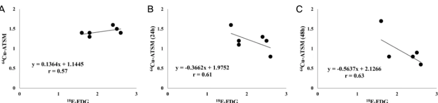
Figure 4. Analyse of ROI in PET imaging. Correlation between 18F-FDG and 64Cu-ATSM (A), 18F-FDG and 64Cu-ATSM _24 h (B), 18F-FDG and
4. 영상분석
획득한 영상으로 mean standardized uptake value (SUVmean) 값을 구해준 후 IRW(Inveon Research Workplace) 프로 그램을 이용하여 각 종양 내 동일 부분의 ROI를 구획 후, 수치화하여 결정계수(coefficient of determination, R2), 상관계수(coefficient of correlation, r)와 P-value를 구하였다.
5. 산소분압측정
토끼 허벅지의 종양에 임의로 4개의 점을 정한 후 1 cm간격으로 산소분압측정기(OXYLITE, Oxford Optronix Ltd, UK)에 연결한 O2 탐침(pO2 E-Serises Sensor, Oxford Optronix Ltd, UK)을 이용하여 종양조직 내의 깊이에 따 른 부분별 산소분압도를 측정하였다.
6. 자가방사기록법
영상획득에 이용된 토끼에 희생 1시간 전 18F-FDG 1 mCi를 정맥주사 후, 토끼의 VX2 종양을 적출하여 -8 0℃에서 1시간 얼린 후 냉동전신절삭기을 이용해 0.5 cm 간격으로 50 μm 냉동절편을 만들어 BAS image plate에 감광시켜 18F-FDG 영상을 획득하였다. 약 13시 간의 반감기를 가진 64Cu-ATSM의 특성을 이용하여 PET 영상 획득을 위해 2.5 mCi 주사하였던 같은 조직 에서 24시간 후에 감광하여 64Cu-ATSM의 종양 내 분포 를 확인하였다.
Results
1.
64Cu-ATSM의 생산
64CuCl2 와 H2ATSM 을 실온에서 10분간 반응 후 방사 성표지가 다 끝난 반응용액을 C18 Sep-Pak 카트리지에 통 과시켜 순수 64Cu-ATSM만 획득 시 방사화학 수율은 95%
로 나타났으며, 크로마토그라피를 확인한 결과 64Cu-ATSM 의 Rf 값은 0.9, 64CuCl2의 Rf 값은 0.0이었다(Figure 2).
2.
64Cu-ATSM과
18F-FDG- PET/CT 영상 비교
64Cu-ATSM 분포는 초기영상과 지연영상에서 섭취가 다르게 나타났다. 18F-FDG영상과 비교하여 보았을 때
64Cu-ATSM 24시간 이전의 초기영상은 유사한 패턴을 나 타냈다(Figure 3). 각 영상의 종양 내 6개 부분의 ROI 분 석에서 18F-FDG 는 초기 64Cu-ATSM 값과 양의 상관계수 (r = 0.57, P < 0.01)를 나타냈고, 24시간과 48시간 이후 의 64Cu-ATSM 값과는 음의 상관계수(r = -0.61, P < 0.01, r = -0.63, P < 0.01)를 확인할 수 있었다(Figure 4).
3.
64Cu-ATSM과
18F-FDG의 조직내 분포 비교
종양 내의 산소분압측정 결과는 64Cu-ATSM 24시간 이 후의 지연영상과 광범위하게 일치하였다(Figure 5). 자가 방사기록법을 이용한 실험에서, 64Cu-ATSM이 축적된 조 직은 주로 활성화된 세포로 이루어져 저산소 상태로 발전 될 예정의 종양 주위에서 관찰되었다. 18F-FDG가 축적된 조직은 64Cu-ATSM이 축적된 조직뿐만 아니라 이미 괴사 된 세포도 상당부분 관찰되었고 64Cu-ATSM이 축적된 부 분보다 더 광범위하게 분포되었다(Figure 6). 64Cu-ATSM 의 섭취가 높은 조직은 산소분압측정 결과에서도 조직
A B C
Figure 5. O2 pressure measurement in VX2 tumor. O2 probe measure- ment interval: 1 cm.
Figure 6. Double-tracer autoradiography with 18FDG and 64Cu-ATSM in VX2 tumor. Comparing 18FDG and 64Cu-ATSM in the same tissue.
내 산소포화도가 현저하게 낮게 나타난다는 것을 확인할 수 있었다.
Discussion
Cu-ATSM은 저산소 부분의 영상제로써 개발되었고, 심 장과 종양에서의 저산소 영상에 60Cu- 또는 62Cu-ATSM이 유용하다고 이전 논문들에 기술되었다(14,26,27). 종양학 에서, 64Cu-ATSM은 내부 방사선 치료의 잠재적인 의약품 으로 고려되고 있다(28). 저산소 환경의 종양 내 Cu-ATSM 축적의 특이성에 대한 연구가 필요하다.
따라서, 본 연구에서는 임상용 PET/CT를 이용하여 토 끼의 저산소 상태 종양 내 64Cu-ATSM과 18F-FDG의 분포 를 비교한 결과 저산소 종양 내 64Cu-ATSM의 섭취가 시 간이 경과함에 따라 18F-FDG의 섭취 패턴이 초기영상 (64Cu-ATSM 주사 직후)과 지연영상(24 또는 48시간)에서 다르게 나타나는 것을 확인하였다(Figure 3). 이 결과는 ROI분석에 따른 상관계수에서도 차이를 나타냈다(Figure 4). 토끼의 종양 내 산소분압을 측정한 결과로는 종양의 가장자리보다는 중심부위의 산소분압이 낮아지는 분포 를 확인할 수 있었고, 이 데이터를 토대로 종양의 가운데 부분으로 저산소 환경이 형성된 것을 추측할 수 있다 (Figure 5). 자가방사측정법으로는 64Cu-ATSM과 18F-FDG 가 동시에 주사된 토끼에서 적출한 조직을 절단해 각 방 사성의약품의 조직 내 분포를 비교하였는데, 대부분의 조 직에서 18F-FDG가 섭취된 조직 중 일부분에 64Cu-ATSM이 섭취된 것을 확인할 수 있었고, 64Cu-ATSM이 섭취된 부분 은 활성화된 세포가 대부분 분포하고 있는 것을 조직사 진으로 확인할 수 있었다(Figure 6). 또, 자가방사측정 데 이터의 64Cu-ATSM 섭취가 높은 부분은 PET영상의
64Cu-ATSM 섭취가 높은 부분과 일치할 뿐만 아니라 조직 내 산소분포가 낮은 부분과도 일치하는 것을 확인할 수 있 었다. 이 결과들을 토대로 18F-FDG는 종양에서 광범위하게 저산소 조직을 영상화하는 반면, 64Cu-ATSM이 저산소 조직 영상화에 탁월한 역할을 한다는 것을 입증하였다. 64Cu-ATSM
은 저산소 조직으로 발전 가능한 세포가 존재하는 종양 부분을 영상화하는 것에 18F-FDG보다 더 자세한 정보를 제공할 수 있다. 64Cu-ATSM은 또한 저산소 부분을 보다 특이적으로 영상화하여 저산소 상태이거나 저산소 상태 로 진행 가능한 영역을 표적화하여 종양 치료에서의 주 목표 중 하나인 영상으로 저산소 환경을 검출할 수 있는 유용한 수단이다.
Conclusion
64Cu-ATSM의 지연영상이 18F-FDG의 영상보다 특이적 으로 저산소 부분을 영상화하는 것으로 나타났고, 이를 종양 내 산소 분압과 자가방사기록법 실험을 통해서도 확인해 볼 수 있었다. 따라서 64Cu-ATSM의 섭취된 영상 을 통해 종양내의 저산소 조직의 정도 및 위치 등을 보 다 정확하게 평가할 수 있을 것이다.
Acknowledgments
이 연구는 미래창조과학부 한국원자력의학원 연구운 영비지원사업(No. 50441-2015)과 원자력연구개발사업 (NRF-2012M2A2A7013480)의 지원을 받아 수행하였음.
References
1. Brahimi-Horn C, Berra E, Pouyssegur J. Hypoxia: the tumor's gateway to progression along the angiogenic pathway. Trends Cell Biol 2001;11:S32-36.
2. Goonewardene TI, Sowter HM, Harris AL. Hypoxia-induced pathways in breast cancer. Microsc Res Tech 2002;59:41-48.
3. Chitneni SK, Palmer GM, Zalutsky MR, Dewhirst MW.
Molecular imaging of hypoxia. J Nucl Med 2011;52:165-168.
4. Padhani AR, Krohn KA, Lewis JS, Alber M. Imaging oxygen- ation of human tumours. Eur Radiol 2007;17:861-872.
5. Mees G, Dierckx R, Vangestel C, Van de Wiele C. Molecular imaging of hypoxia with radiolabelled agents. Eur J Nucl Med Mol Imaging 2009;36:1674-1686.
6. Lewis JS, Welch MJ. PET imaging of hypoxia. J Nucl Med 2001;
45:183-188.
7. Chapman JD, Franko AJ, Sharplin J. A marker for hypoxic cells in tumours with potential clinical applicability. Br J Cancer 1981;43:546-550.
8. Krohn KA, Link JM, Mason RP. Molecular imaging of hypoxia. J Nucl Med 2008;49(Suppl):129S-148S.
9. Dehdashti F, Grigsby PW, Mintun MA, Lewis JS, Siegel BA, Welch MJ. Assessing tumor hypoxia in cervical cancer by posi-
tron emission tomography with 60Cu-ATSM: relationship to therapeutic response a preliminary report. Int J Radiat Oncol Biol Phys 2003; 55:1233-1238.
10. Dehdashti F, Mintun MA, Lewis JS, Bradley J, Govindan R, Laforest R. In vivo assessment of tumor hypoxia in lung cancer with 60Cu-ATSM. Eur J Nucl Med Mol Imaging 2003;30:844-850.
11. Fujibayashi Y, Taniuchi H, Yonekura Y, Ohtani H, Konishi J, Yokoyama A. Copper-62-ATSM: a new hypoxia imaging agent with high membrane permeability and low redox potential. J Nucl Med 1997;38:1155-1160.
12. Lewis JS, McCarthy DW, McCarthy TJ, Fujibayashi Y, Welch MJ.
Evaluation of 64Cu-ATSM in vitro and in vivo in a hypoxic tu- mor model. J Nucl Med 1999;40:177-183.
13. Lewis JS, Sharp TL, Laforest R, Fujibayashi Y, Welch MJ. Tumor uptake of copper-diacetyl-bis(N4-methylthiosemicarbazone):
effect of changes in tissue oxygenation. J Nucl Med 2001;42:655-661.
14. Takahashi N, Fujibayashi Y, Yonekura Y, Welch MJ, Waki A, Tsuchida T, Sadato N, Sugimoto K, Itoh H. Evaluation of 62Cu labeled diacetyl-bis(N4- methylthiosemicarbazone) as a hy- poxic tissue tracer in patients with lung cancer. Ann Nucl Med 2000;14:323-328.
15. Tanaka T, Furukawa T, Fujieda S, Kasamatsu S, Yonekura Y, Fujibayashi Y. Double-tracer autoradiography with Cu-ATSM/
FDG and immunohistochemical interpretation in four different mouse implanted tumor models. Nucl Med Biol 2006;33:743-750.
16. Obata A, Kasamatsu S, McCarthy DW, Welch MJ, Saji H, Yonekura Y, Fujibayashi Y. Production of therapeutic quantities of 64Cu using a 12 MeV cyclotron. Nucl Med Biol 2003;30:535-539.
17. Burgman P, O'Donoghue JA, Lewis JS, Welch MJ, Humm JL, Ling CC. Cell line-dependent differences in uptake and retention of the hypoxia-selective nuclear imaging agent Cu-ATSM. Nucl Med Biol 2005;32:623-630.
18. Obata A, Kasamatsu S, Lewis JS, Furukawa T, Takamatsu S, Toyohara J, Asai T, Welch MJ, Adams SG, Saji H, Yonekura Y, Fujibayashi Y. Basic characterization of 64Cu-ATSM as a radio- therapy agent. Nucl Med Biol 2005;32:21-28.
19. Dehdashti F, Grigsby PW, Lewis JS, Laforest R, Siegel BA, Welch MJ. Assessing tumor hypoxia in cervical cancer by PET with
60Cu-labeled diacetyl-bis(N4-methylthiosemicarbazone). J Nucl Med 2008;49:201-205.
20. Grigsby PW, Malyapa RS, Higashikubo R, Schwarz JK, Welch MJ, Huettner PC. Comparison of molecular markers of hypoxia and imaging with 60Cu-ATSM in cancer of the uterine cervix.
Mol Imaging Biol 2007;9:278-283.
21. Dietz DW, Dehdashti F, Grigsby PW, Malyapa RS, Myerson RJ, Picus J, Ritter J, Lewis JS, Welch MJ, Siegel BA. Tumor hypoxia detected by positron emission tomography with 60Cu-ATSM as a predictor of response and survival in patients undergoing neo- adjuvant chemoradiotherapy for rectal carcinoma: a pilot study.
Dis Colon Rectum 2008;51: 1641-1648.
22. Obata A, Yoshimi E, Waki A, Lewis JS, Oyama N, Welch MJ, Saji H, Yonekura Y, Eujibayashi Y. Retention mechanism of hy-
poxia selective nuclear imaging/radio-therapeutic agent Cu-di- acetyl-bis(N4-methylthio- semicarbazone) (Cu-ATSM) in tu- mor cells. Ann Nucl Med 2001;15:499-504.
23. Dearling JL, Lewis JS, Mullen GE, Welch MJ, Blower PJ. Copper bis (thiosemicarbazone) complexes as hypoxia imaging agents:
structure activity relationships. J Biol Inorg Chem 2002;7:249-259.
24. Holland JP, Giansiracusa JH, Bell SG, Wong LL, Dilworth JR. In vitro kinetic studies on the mechanism of oxygen-dependent cellular uptake of copper radiopharmaceuticals. Phys Med Biol 2009;54:2103-2119.
25. Obata A, Yoshimoto M, Kasamatsu S, Naiki H, Takamatsu S, Kashikura K, Furukawa T, Lewis JS, Welch MJ, Saji H, Yonekura Y, Fujibayashi Y. Intra-tumoral distribution of 64Cu-ATSM: a comparison study with FDG. Nucl Med Biol 2003;30:529-534.
26. Chao KS, Bosch WR, Mutic S, Lewis JS, Dehdashti F, Mintun MA, Dempsey JF, Perez CA, Purdy JA, Welch MJ, A novel ap- proach to overcome hypoxic tumor resistance: Cu-ATSM-guid- ed intensity-modulated radiation therapy. Int J Radiat Oncol Biol Phys 2001;49:1171-1182.
27. Takahashi N, Fujibayashi Y, Yonekura Y, Welch MJ, Waki A, Tsuchida T, Sadato N, Sugimoto K, Nakano A, Lee JD, Itoh H, Copper-62 ATSM as a hypoxic tissue tracer in myocardial ischemia. Ann Nucl Med 2001;15:293-296.
28. Lewis J, Laforest R, Buettner T, Song S, Fujibayashi Y, Connett J, Welch MJ, Copper-64-diacetyl-bis(N4-methylthiosemicarbazone):
an agent for radiotherapy, Proc Natl Acad Sci USA 2001;98:
1206-1211.