Thioacetamide 유발 간 섬유화에 대한 모과의 효과
이진아1․이세희1․신미래1․노정숙2․노성수1
1대구한의대학교 한의과대학 본초약리학교실
2동명대학교 식품영양학과
The Effect of Chaenomelis Fructus Extract on Thioacetamide-Induced Liver Fibrosis
Jin A Lee
1, Se Hui Lee
1, Mi-Rae Shin
1, Jeong Sook Noh
2, and Seong-Soo Roh
11
Department of Herbology, College of Korean Medicine, Daegu Haany University
2
Department of Food Science and Nutrition, Tongmyong University
ABSTRACT This study was conducted to evaluate the effect of Chaenomelis Fructus (CF) extract on thioacetamide (TAA)-induced liver fibrosis in mice. The mice were divided into four groups for examination: Normal group (Normal, n=9), group with liver fibrosis administered distilled water (Control, n=9), group with liver fibrosis administered silymar- in 100 mg/kg (Silymarin, n=9), group with liver fibrosis administered CF 200 mg/kg (CF, n=9). Liver fibrosis was established in the mice via an injection of TAA (1 week 100 mg/kg B.W., 2∼3 weeks 200 mg/kg B.W., 4∼8 weeks 400 mg/kg B.W., three times a week, I.P) for 8 weeks and they were administered silymarin and CF extract (every day, P.O) with the TAA. After the autopsy, we analyzed the expression of inflammation-related proteins and fibrosis-re- lated factors in the liver tissue by the western blot test. The results of the experiment showed that the administration of CF regulated the expression of phosphorylated AMPK (p-AMPK) and Sirtuin 1 (SIRT1) and inhibited the expression of the nuclear factor kappa-light-chain-enhancer of activated B cells (NF-κB) pathway. In addition, the expressions of matrix metalloproteinase-2 (MMP-2), TIMP metallopeptidase inhibitor 1 (TIMP-1), ɑ-smooth muscle actin (ɑ-SMA), and collagen Ⅰ related to fibrosis were suppressed. These results suggest that the progression of liver fibrosis may be alleviated by regulating the AMPK/SIRT1/NF-κB pathway, as well as the expression of fibrosis-related factors.
Key words: Chaenomelis Fructus, liver fibrosis, thioacetamide
Received 1 April 2021; Revised 7 May 2021; Accepted 10 May 2021
Corresponding author: Seong-Soo Roh, College of Korean Medicine, Daegu Haany University, 136, Sincheondong-ro, Suseong-gu, Daegu 42158, Korea, E-mail: [email protected]
Author information: Jin A Lee (Graduate student), Se Hui Lee (Graduate student), Mi-Rae Shin (Professor), Jeong Sook Noh (Professor), Seong-Soo Roh (Professor)
Copyright ⓒ 2021 by The Korean Society of Food Science and Nutrition. All rights Reserved.
This is Open Access article distributed under the terms of the Creative Commons Attribution Non-Commercial License (https://creativecommons.org/licenses/
by-nc/4.0) which permits unrestricted non-commercial use, distribution, and reproduction in any medium, provided the original work is properly cited.
서 론
간(Liver)은 인체에서 가장 큰 장기 중 하나로 에너지 저 장, 영양소 대사, 해독, 순환 등의 중요한 역할을 가진다(Lee 등, 2006a). 현대인들은 과학기술의 급격한 발달로 화학 독 성물질이 증가한 환경에서 생활하고 있으며, 신약 등이 개발 되면서 여러 독성 물질에 대한 노출이 증가하고 있다(Park 등, 2015). 한국 40대 남성이 가장 높은 사망률을 보이는 질환은 간질환으로 알코올성 간질환, 간염, 간 섬유화 및 간 경변을 포함하며, 이는 간암의 대표적인 원인이 된다(Kim 등, 2019). 그중 간섬유화는 세포외기질(extracellar ma- trix) 단백질의 축적과 함께 손상이 일어나는 것이 특징이며,
섬유화 과정에서 핵심적인 역할을 하는 간 성상세포(hepat- ic stellate cells)는 바이러스, 독성 물질 등과 같은 자극에 의한 간세포 손상과 함께 활성화되어 콜라겐과 같은 세포외 기질의 생성을 촉진하고, 이 과정에서 세포외기질이 과도하 게 증식되어 간 섬유화로 진행된다(Bataller와 Brenner, 2005; Kim 등, 2012).
현재 간 섬유화 치료제 개발을 위해 주로 염증의 원인을 제거하는 방법, 간 성상세포의 활성화를 억제하여 교원질의 생성 억제 또는 파괴 증가를 유도하는 방향의 연구가 진행되 고 있으나 뚜렷한 치료제가 개발되어 있지 않은 실정이다 (Lee, 2006a).
모과(Chaenomelis Fructus)는 장미과(Rosaceae)에 속
한 모과나무(
Chaenomeles sinensis
Koehne)의 성숙한 과 실로 중국이 주 원산지이며, 원형 또는 타원형의 모양을 가 진다(Lee 등, 2007). 모과의 성분으로는 oleanolic acid, proanthocyanidin, saponin, flavonoid 등이 알려져 있으며 (Lee 등, 2020), 한방에서 모과는 간 기능 강화, 위장 강화 등의 효능을 가지고 있어 자양강장, 간염, 소화불량 등을 치 료한다고 알려져 있다(Lee 등, 2020). 그 외 많은 연구를 통 해 항산화 효과(Lee 등, 2007), 항염증 효과(Ryu 등, 2012), 항비만 효과(Kim 등, 2017) 등이 알려져 있으며, 에탄올에 의해 유발된 간 독성에서 모과 추출물이 간 조직 보호 효과 를 나타낸다고 밝혀졌다(Lee 등, 2006b). 또한, 선행 연구 결과, thioacetamide(TAA)로 유발된 급성 간 손상에서 모 과 추출물이 NF-κB의 활성을 억제함으로써 간 손상을 완화 시킨다는 것을 확인하였다(Lee 등, 2021).이에 본 연구에서는 급성 간 손상에서 염증 완화 효과를 나타냈던 모과 추출물이 TAA에 장기간 노출되어 간 섬유화 가 일어난 동물에서 어떠한 효과를 미치는지 확인함으로써 간 보호 효과를 가지는 새로운 기능성 소재로서의 활용 가능 성을 확인하였다.
재료 및 방법
시료 추출
본 실험에서 사용한 모과는 옹기한약국(Daegu, Korea) 에서 구입하였으며, 생약규격집에 맞추어 관능검사 후 약전 규격에 적합한 것만을 정선하여 사용하였다. 모과 200 g을 분쇄하여 증류수 2,000 mL를 첨가한 후 열탕 추출기를 이 용해 2시간 추출하였다. 얻어진 추출물은 감압 추출장치로 농축한 다음 동결 건조기를 이용해 완전 건조시켜 파우더(수 율, 13.3%; 수분함량, 9.1%M)를 얻었으며, -80°C에서 보 관하였다.
시약
본 실험에 사용된 Thioacetamide(TAA), potassium phosphate monobasic, potassium phosphate dibasic은 Sigma-Aldrich Co.(St. Louis, MO, USA)에서 구입하여 사 용하였다. Nitrocellulose membranes는 Amersham GE Healthcare(Little Chalfont, UK)에서 구입하였고, sirtuin 1(SIRT1), phosphorylation of nuclear factor-kappa B p65(NF-κBp65), inhibitor of nuclear factor kappa B al- pha(IκBα), phosphorylation inhibitor of nuclear factor kappa B alpha(p-IκBα), tumor necrosis factor-alpha (TNF-α), interleukin-6(IL-6), matrix metalloproteinase histone 2(MMP-2), tissue inhibitors of metallopro- teinases 1(TIMP-1), alpha-smooth muscle actin(α- SMA), collagen Ⅰ, histone, β-actin은 Santa Cruz Bio- technology(Dallas, TX, USA)로부터 구입하여 사용하였 다. AMP-activated protein kinase(AMPK), phospho-
AMP-activated protein kinase(p-AMPK)는 Cell Signal- ing Technology, Inc.(Beverly, MA, USA)에서 구입하여 사용하였으며, 2차 항체는 GeneTex, Inc.(Irvine, CA, USA) 에서 구입하여 사용하였다. Protease inhibitor mixture, ethylenediaminetetraacetic acid(EDTA)는 Wako Pure Chemical Industries, Ltd.(Osaka, Japan)에서 구입하여 사용 하였으며, 2′,7′-dichlorofluorescein diacetate(DCFH- DA)와 dihydrorhodamine 123(DHR123)은 Molecular Probes(Eugene, OR, USA)에서 ECL western blotting detection reagents는 GE Healthcare로부터 구입하여 사 용하였다. 단백질 정량을 위한 BCA protein assay kit은 Thermo Scientific(Waltham, MA, USA)에서 구입하였다.
실험동물
7주령의 웅성 C57BL/6(Dae Han Bio Link Co., Ltd., Chungbuk, Korea)를 구입하여 1주일 동안 실험실 환경에 적응시킨 후 실험을 진행하였다. 동물 사육실 조건은 con- ventional system으로 온도 22±2°C, 습도 50±5%, 명암주 기(light: dark cycle)는 12시간 주기로 조절하였고, 사료 (조단백질 18% 이상, 조지방 5.0% 이상, 조섬유 5.0% 이하, 조회분 8.0% 이하, 칼슘 1.0% 이상, 인 0.85% 이상, 칼륨 0.55% 이상, 나트륨 0.25% 이상, 마그네슘 0.15% 이상;
NIH-41, Zeigler Bros, Inc., Gardners, PA, USA)와 물을 충분히 공급하였다. 동물실험의 윤리적, 과학적 타당성 검토 및 효율적인 관리를 위하여 대구한의대학교 동물실험윤리 위원회(Institutional Animal Care and Use Committee:
IACUC)의 승인(승인번호: DHU2021-025)을 받았다.
간 섬유화 모델
실험군은 정상군(normal), DW를 투여한 대조군(control), silymarin 50 mg/kg 투여군(silymarin), 모과 추출물 200 mg/kg 투여군(CF) 총 4군으로 각각 9마리씩 분류하였다.
모든 동물은 일정한 시간에 1회/1일 체중을 측정하였으며, 정상군(normal)을 제외한 모든 동물(control, silymarin 및 CF)은 8주간 3회/1주 DW에 녹인 TAA(1주 100 mg/kg B.W., 2~3주 200 mg/kg B.W., 4~8주 400 mg/kg B.W.) 복강투여 및 DW에 녹인 해당 약물을 1회/1일 경구투여 하 였다(Thi Thanh Hai 등, 2018). 실험종료 후 마취하여 간 조직을 적출하였으며, 복대정맥에서 혈액을 채취하였다 (Table 1).
혈액 분석
실험동물의 복대정맥에서 채혈한 혈액을 4,000 rpm으로 10분간 원심분리하여 혈청을 얻었으며, 분리한 혈청을 이용 하여 GOT(glutamic oxaloacetic transaminase) 및 GPT (glutamic pyruvic transaminase) level을 측정하였다.
GOT와 GPT(Asan Pharmaceutical Co., Ltd., Seoul, Ko- rea)는 시약 세트를 구입하여 측정하였다.
Table 1. Body weight and death rate
Body weight (g)
Death rate (%)
Initial Final Gain
Normal Control Silymarin
CF
23.57±0.42 23.25±0.38 23.41±0.37 23.46±0.34
30.79±0.50 24.23±0.32***
24.19±0.30 24.27±0.47
7.26±0.46 0.98±0.27***
0.60±0.36 0.81±0.32
-
-
-
-
All date are expressed mean±SD (n=9). Normal, Normal group; Control, liver fibrosis-induced with distilled water group; Silymarin, liver fibrosis-induced with silymarin 50 mg/kg group; CF, liver fibrosis-induced with Chaenomelis Fructus 200 mg/kg group.
Significance: ***
P<0.001 vs. Normal group.
조직 Western blotting
간의 세포질을 얻기 위해 100 mM Tris-HCl(pH 7.4), 5 mM Tris-HCl(pH 7.5), 2 mM MgCl2, 15 mM CaCl2, 1.5 M sucrose, 0.1 M DTT, protease inhibitor cocktail 을 첨가한 buffer A를 넣고 tissue grinder(BioSpec Prod- ucts, Bartlesville, OK, USA)로 분쇄한 후 아이스 위에서 30분간 정치시켰다. 그 후, 10% NP-40 용액을 첨가하여 12,000 rpm으로 2분간 원심분리하여 세포질을 포함하고 있는 상층액을 분리하였다. 핵을 얻기 위해 10% NP-40가 더해진 buffer A에 두 번 헹구고, 100 µL의 buffer C(50 mM HEPES, 0.1 mM EDTA, 50 mM KCl, 0.3 mM NaCl, 1 mM DTT, 0.1 mM PMSF, 10% glycerol)를 첨가해 재부 유시킨 뒤 10분마다 vortex를 3번 하였다. 4°C에서 12,000 rpm으로 10분간 원심 분리한 후 핵을 포함하고 있는 상층액 을 얻어 -80°C에서 각각 냉동 보관하였다. 간 조직 세포질 에서 AMPK, p-AMPK, SIRT1, IκBα, p-IκBα, TNF-α, IL- 6, MMP-2, TIMP-1, α-SMA, collagen Ⅰ 및 β-actin과 핵에서 NF-κBp65와 histone 단백질 발현을 측정하기 위해 12 µg의 단백질을 8~14% SDS-polyacrylamide gel을 이 용하여 전기영동 후, acrylamide gel을 nitrocellulose membrane으로 이동시켰다. 준비된 membrane에 각각의 1차 antibody(1:1,000)를 처리하여 4°C에서 overnight 시 킨 다음 PBS-T로 6분마다 5회 세척하고, 각각 처리된 1차 antibody에 사용되는 2차 antibody(1:3,000)를 사용하여 상온에서 1시간 30분 반응시킨 후, PBS-T로 6분마다 5회 세척하였다. 그 후 enhanced chemiluminescence(ECL) 용 액에 노출시켜 Sensi-Q2000 Chemidoc(Lugen Sci Co., Ltd., Seoul, Korea)를 이용해 단백질 발현을 확인하였으 며, 해당 band를 ATTO Densitograph Software(ATTO Corporation, Tokyo, Japan)프로그램을 사용하여 band를 정량하였다. 각각의 단백질 수준을 정상군의 단백질 수준으 로 나눈 후 상대적 비로 나타내었다(fold of Normal).
조직학적 관찰
간 조직을 10% neutral buffered formalin에 고정시킨 다음 graded alcohol로 탈수시킨 후, 파라핀으로 포매하여 block을 제작한 다음 microtome으로 5 µm 두께의 조직 절 편을 제작하여 슬라이드를 weigert iron hematoxylin에서
7분간 염색하였으며, 세척 후 biebrich scarlet-acid fuch- sin에서 2분 동안 염색하였다. 그 후, phosphotungstic- phosphomolybdic acid에서 7분 동안 배양하고 aniline blue로 10분 동안 염색하였으며, 1% 아세트산에서 3분 동 안 고정하여 광학현미경으로 병변의 유무를 관찰하였다.
통계분석
In vivo
의 수치는 평균과 표준편차로 표시하였으며 SPSS program for windows version 25(SPSS Inc., Chicago, IL, USA)를 사용하여 one-way analysis of variance (ANOVA) test를 실시한 후, least-significant differ- ences(LSD) test로 사후검증을 실시하여 각 군의 평균 차이 에 대한 통계적 유의성을P
<0.05,P
<0.01,P
<0.001에서 검증하였다.결과 및 고찰
간 무게 및 GOT, GPT 측정
간 효소인 GOT와 GPT는 정상의 경우 소량이 혈청에 존 재하지만, 간세포가 손상을 받을 경우 다량의 효소가 혈액으 로 흘러 들어가 혈중 GOT 및 GPT의 농도가 증가한다고 알려져 있으며(Kim, 2008; Park 등, 2014), 혈중 GOT와 GPT의 농도가 증가하는 것은 간세포의 손상 및 사멸과 밀접 한 연관성이 있다. GOT와 GPT는 간염 등의 간질환에 의해 혈중으로 유리되어 농도가 높아지기에 간 손상의 판단지표 로 널리 이용되고 있다(Park 등, 2014).
간 무게 측정 결과, normal군의 간 무게는 0.88±0.022 g으로 나타났으며, control군에서는 0.99±0.024 g으로 12% 유의하게 증가하였고 CF군에서는 0.89±0.018 g으로 control군 대비 10% 유의하게 감소하였다. GOT 수치를 측 정한 결과, normal군 대비 control군에서 4배 이상 유의하 게 증가하였으며, CF군에서는 control군 대비 53% 유의하 게 감소하였다. GPT 또한 normal군 대비 control군에서 2 배 이상 유의하게 증가하였으며, control군 대비 CF군에서 26% 유의하게 감소하였다(Table 2). 모과의 성분 중 하나 인 oleanolic acid는 기존 연구에서 증가한 GPT의 활성을 감소시킨다고 알려져 있다(Jung 등, 2016). 본 연구 결과 TAA로 인해 증가한 GOT와 GPT 수치가 모과 추출물 투여
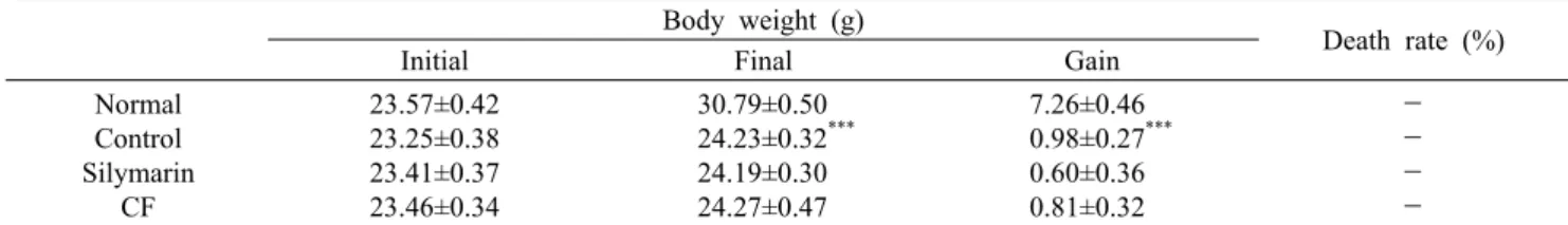
Fig. 1. Expression of AMPK/SIRT1 pathway in liver tissue. All date are expressed mean±SD (n=9). Normal, Normal group; Control,
liver fibrosis-induced with distilled water group; Silymarin, liver fibrosis-induced with silymarin 50 mg/kg group; CF, liver fibrosis-in- duced with Chaenomelis Fructus 200 mg/kg group. Significance: ###P<0.001 vs. Normal group,
*P<0.05,
**P<0.01 vs. Control group.
Table 2. Liver weight, serum GOT and GPT levels
Liver weight (g) GOT (IU/L) GPT (IU/L) NormalControl Silymarin
CF
0.88±0.022 0.99±0.024##
0.90±0.021**
0.89±0.018**
15.44±5.29 66.68±10.48###
37.08±3.51**
31.53±2.93**
9.58±1.60 21.79±2.74###
16.04±1.27* 16.13±0.23* All date are expressed mean±SD (n=9). Normal, Normal group;
Control, liver fibrosis-induced with distilled water group;
Silymarin, liver fibrosis-induced with silymarin 50 mg/kg group;
CF, liver fibrosis-induced with Chaenomelis Fructus 200 mg/kg group. Significance: ##
P<0.01,
###P<0.001 vs. Normal group,
*P<
0.05, **
P<0.01 vs. Control group.
를 통해 유의적으로 감소한 것을 확인하였으며, 이는 모과가 TAA로 인한 간 손상을 감소시킴으로써 간의 무게 및 혈중 GOT와 GPT의 수치를 감소시키는 것으로 판단된다.
Western blotting
AMPK/SIRT1 단백질 분석: AMP-activated protein kinase(AMPK)는 serine/threonine kinase로서 생체 내의 에너지 인식 및 항상성 조절을 담당하며, 세포 내의 에너지 를 소모할 경우 증가하는 AMP에 의해 활성화되어 ATP의 사용을 억제하는 인자로 잘 알려져 있다(Choi, 2012). 여러 연구를 통해 AMPK가 염증 반응을 조절하며, AMPK의 활성 화는 염증 전사인자인 NF-κB의 활성을 억제한다고 밝혀졌 다(Velagapudi 등, 2017; Goldfine과 Shoelson, 2017).
SIRT1 또한 AMPK에 의해 활성화되어 염증 반응에 관여하 여 항염증 작용을 한다고 알려져 있으며(Nguyen 등, 2019), Yang 등(2010)의 연구에서는 SIRT1이 NF-κB의 전사를 억제하여 염증 관련 인자의 발현을 조절한다고 알려졌다.
AMPK/SIRT1 단백질의 발현을 확인한 결과, normal군 대비 control군에서 p-AMPK의 발현이 50% 유의하게 감소 하였으며, SIRT1의 발현 또한 28% 유의하게 감소하였다.
반면, control군 대비 CF군에서 p-AMPK의 발현이 53%
유의하게 증가하였으며, SIRT1의 발현 또한 25% 유의하게 증가하였다(Fig. 1). 이러한 결과는 모과가 AMPK의 활성을 유도할 뿐 아니라 SIRT1의 발현을 증가시킴으로써 염증 반 응에 관여하여 염증을 완화할 것이라 판단된다.
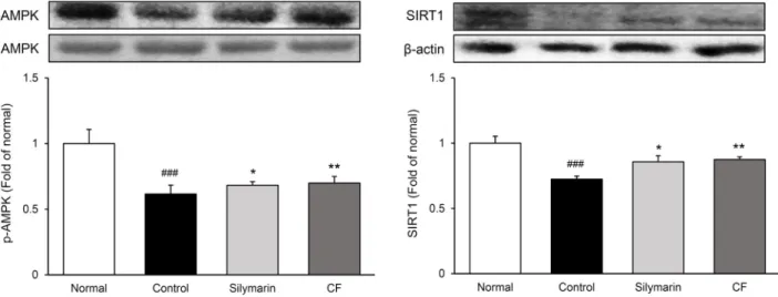
염증 관련 단백질 분석: 염증 전사인자로 알려진 NF-κB 는 IκBα에 의해 비활성화 상태로 세포질 내에 존재하다가 여러 자극에 의해 IκBα의 인산화가 일어나면서 핵 내로 이동 하여 염증반응을 활성화된다고 알려져 있다(Ngabire 등, 2018). NF-κB는 세포 외부의 자극에 대한 방어 및 면역세 포의 활성화를 조절하며 염증반응에 있어서 사이토카인 등 다양한 유전자들의 발현을 조절한다(Woo 등, 2018).
이에 본 실험에서 NF-κBp65와 p-IκBα의 발현을 확인한 결과, normal군 대비 control군에서 NF-κBp65의 발현이 70% 유의하게 증가하였으며, p-IκBα의 발현 또한 60% 이 상 유의하게 증가하였다. 반면, control군 대비 silymarin군 에서 NF-κBp65 31%, p-IκBα 15% 유의하게 감소하였으 며, CF군에서 NF-κBp65 35%, p-IκBα 23% 유의하게 감 소하여 양성대조군인 silymarin군보다 NF-κBp65의 활성 및 IκBα의 인산화를 뛰어나게 억제시켰다. TNF-α 및 IL-6 의 발현을 확인한 결과, normal군 대비 control군에서 TNF-α의 발현이 80% 유의하게 증가하였으며, IL-6의 발 현 또한 45% 이상 유의하게 증가하였다. 반면, control군 대비 CF군에서 TNF-α 20%, IL-6 18% 유의하게 감소하게 감소하였다(Fig. 2). 이전 연구에서 모과에 함유된 proan- thocyanidin이 NF-κB를 억제함으로써 간 손상을 완화한다 고 알려져 있으며(El-Shitany와 Eid, 2017; Lee 등, 2021), 본 연구에서도 마찬가지로 NF-κBp65의 활성을 억제함으 로써 NF-κB 관련 인자의 발현이 하향 조절되었다. 이는 모 과가 AMPK/SIRT1 경로를 통해 활성화된 NF-κBp65의 활 성 및 IκBα의 인산화를 억제함으로써 TNF-α와 IL-6 같은
Fig. 2. Expression of inflammation-related proteins in liver tissue. All date are expressed mean±SD (n=9). Normal, Normal group;
Control, liver fibrosis-induced with distilled water group; Silymarin, liver fibrosis-induced with silymarin 50 mg/kg group; CF, liver fibrosis-induced with Chaenomelis Fructus 200 mg/kg group. Significance: ##
P<0.01,
###P<0.001 vs. Normal group,
*P<0.05,
**
P<0.01 vs. Control group.
cytokine의 발현을 억제하는 것으로 판단된다.
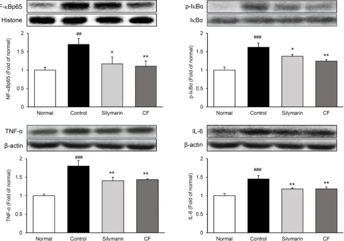
MMP-2 및 TIMP-1 단백질 분석: 인체 내에서 세포외 기질이 과도하게 생성되면 단백질분해효소인 matrix me- talloproteinase(MMP)에 의한 세포외기질의 분해 과정이 일어나며, MMP의 활성을 억제하는 tissue inhibitors of matrix metalloproteinase(TIMP) 또한 광범위하게 작용한 다(Lee, 2006b). Ⅳ형 collagenase인 MMP-2는 섬유증을 유발하는 세포외기질 분해에 작용하며, TIMP-1은 세포외 기질 분해를 억제함으로써 세포외기질의 과도한 침착을 유 도해 섬유증 진행에 관여한다(Xu 등, 2020). 이전 연구에서 모과의 oleanolic acid가 TIMP-1과 α-SMA의 발현 감소 및 간 성상 세포의 기능을 조절함으로써 간 섬유증을 완화한 다고 알려졌다(Wu 등, 2008).
본 실험 결과, normal군 대비 control군에서 MMP-2의 발현이 35% 유의하게 감소하였으며, control군 대비 CF군 에서 65% 유의하게 증가하였다. 반면, normal군 대비 con- trol군에서 TIMP-1의 발현이 33% 유의하게 증가하였으며, control군 대비 CF군에서 40% 감소하여 normal군과 비슷 한 수준을 나타냈다(Fig. 3). 이러한 결과는 모과가 세포외
기질 분해를 돕는 MMP-2 및 세포외기질 분해를 억제하는 TIMP-1의 발현을 조절함으로써 간 섬유화의 진행을 완화 한 것으로 판단된다.
α-SMA 및 collagen Ⅰ 단백질 분석: 간 손상 후 섬유화 단계에서는 세포외기질을 생산하는 간 성상세포가 활성화 됨으로써 형질이 전환되어 교원질(collagen Ⅰ)을 포함한 세포외기질의 생성 증가, α-SMA의 발현 증가, 세포증식 증 가 등이 발생한다(Kwon, 2006).
실험 결과, α-SMA의 발현이 normal군 대비 control군에 서 약 1.5배 유의하게 증가하였으며, control군 대비 CF군 에서 27% 유의하게 감소하였다. 또한, collagen Ⅰ의 발현 은 normal군 대비 control군에서 45% 유의하게 증가하였 으며, control군 대비 CF군에서 25% 유의하게 감소하였다 (Fig. 4). Wu 등(2008)의 연구와 같이 본 실험에서도 CF군 에서 α-SMA의 발현이 감소되었으며, collagen Ⅰ의 발현 또한 감소되었다. 이는 모과가 섬유화 단계에서 활성화되는 간 성상세포의 활성화를 억제시킴으로써 간 섬유화 진행을 완화시킨 것으로 판단된다.
Fig. 3. Expression of MMP-2 and TIMP-1 proteins in liver tissue. All date are expressed mean±SD (n=9). Normal, Normal group;
Control, liver fibrosis-induced with distilled water group; Silymarin, liver fibrosis-induced with silymarin 50 mg/kg group; CF, liver fibrosis-induced with Chaenomelis Fructus 200 mg/kg group. Significance: #
P<0.05,
###P<0.001 vs. Normal group,
**P<0.01
vs. Control group.Fig. 4. Expression of α-SMA and collagen Ⅰ proteins in liver tissue. All date are expressed mean±SD (n=9). Normal, Normal
group; Control, liver fibrosis-induced with distilled water group; Silymarin, liver fibrosis-induced with silymarin 50 mg/kg group;CF, liver fibrosis-induced with Chaenomelis Fructus 200 mg/kg group. Significance: ##
P<0.01,
###P<0.001 vs. Normal group,
*P<0.05,
**
P<0.01 vs. Control group.
조직학적 관찰
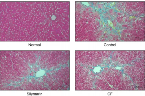
간 조직에서 Masson’s trichrome 염색을 실시한 결과, normal군에 비해 control군에서는 넓은 범위의 교원섬유의 침착을 확인하였으며, CF군에서는 control군에 비해 교원 섬유의 침착 및 조직학적 섬유화 변화가 완화된 것을 확인하 였다(Fig. 5).
요 약
본 실험에서는 TAA(thioacetamide)로 유발한 간 섬유화 동 물 모델에 모과 추출물이 미치는 효과를 확인하기 위하여 8주간 3회/주 TAA 복강투여 및 1회/1일 경구투여 하였다.
모과 추출물을 경구투여 하였으며 실험 종료 후 간 조직을
적출하였다. 간 조직 western blotting을 통해 염증 관련 단백질의 발현을 확인한 결과, 모과 투여군에서 p-AMPK, SIRT의 발현이 유의하게 증가하였으며, 염증 전사인자 NF- κB, IκBα 및 염증성 사이토카인 TNF-α, IL-6의 발현이 유 의하게 감소하였다. 또한, 섬유화와 관련된 MMP-2, TIMP- 1, α-SMA 및 collagen Ⅰ과 같은 세포외기질 관련 인자가 유의적으로 감소하였다. Masson’s trichrome staining으로 간 조직을 관찰한 결과, 모과 투여군에서는 대조군에 비하여 교원섬유의 침착 및 조직학적 섬유화 변화가 완화된 것을 확 인하였다. 이상의 결과를 종합해보면 모과 추출물은 AMPK/
SIRT1/NF-κB의 발현을 조절할 뿐만 아니라 섬유화 관련 단백질의 발현을 억제함으로써 간 섬유화의 진행을 완화하 는 것으로 생각된다.
Fig. 5. Liver histological examination
through Masson’s trichrome staining (original magnification ×200). Normal, Normal group; Control, liver fibrosis- induced with distilled water group;Silymarin, liver fibrosis-induced with silymarin 50 mg/kg group; CF, liver fibrosis-induced with Chaenomelis Fruc- tus 200 mg/kg group.
감사의 글
이 논문은 2021년도 정부(과학기술정보통신부)의 재원으로 한국연구재단의 지원(No. 2018R1A5A2025272)을 받아 수행된 연구입니다.
REFERENCES
Bataller R, Brenner DA. Liver fibrosis. J Clin Invest. 2005. 115:
209-218.
Choi HC. Effects of AMP-activated protein kinase activating compounds and its mechanism. Yeungnam Univ J Med. 2012.
29:77-82.
El-Shitany NA, Eid B. Proanthocyanidin protects against cispla- tin-induced oxidative liver damage through inhibition of in- flammation and NF-κβ/TLR-4 pathway. Environ Toxicol.
2017. 32:1952-1963.
Goldfine AB, Shoelson SE. Therapeutic approaches targeting inflammation for diabetes and associated cardiovascular risk.
J Clin Invest. 2017. 127:83-93.
Jung S, Lee S, Ko KS. Effects of oleanolic acid and hederagenin on acute alcohol-induced hepatotoxicity in mice. J Korean Soc Food Sci Nutr. 2016. 45:307-312.
Kim DH, Kwon B, Kim SJ, Kim HJ, Jeong SI, Yu KY, et al.
Anti-obese effects and signaling mechanisms of Chaenomeles
sinensis extracts in 3T3-L1 preadipocytes and obese mice fed
a high-fat diet. Herbal Formula Science. 2017. 25:457-469.Kim SJ, Kim SY, Kim JA, Park IS, Yu KY, Chung CH, et al. Inhibitory effect of Cirsium japonicum root or flower ex- tract on hepatic stellate cells activation. Kor J Pharmacogn.
2012. 43:27-31.
Kim YJ. Interpretation of liver function tests. Korean J Gas- troenterol. 2008. 51:219-224.
Kim YS, Kim SK, Kwon DA, Kim HK, Lee HS. Hepatoprotec- tive effect of Chrysanthemum zawadskii extract (CZE) in ex- perimentally induced liver damage model in vitro and in vivo.
J Korean Soc Food Sci Nutr. 2019. 48:189-197.
Kwon OS. Cellular and molecular mechanisms of liver fibrosis.
Proceedings of Single Topic Symposium of Korean Associa-
tion for the Study of the Liver. 2006 Sep 1. Incheon, Korea.
p 18-28.
Lee EG, Kim KB, Jeong JM. Hepatoprotective effects of poly herbal formulation (Hepa-1000) on t-BHP-induced toxicity in human hepatoma cells. J Korean Soc Food Sci Nutr. 2006a.
35:1121-1126.
Lee JA, Shin MR, Lee JH, Park HJ, Roh SS. The effect of Chaenomelis Fructus on thioacetamide-induced acute hepatic injury. J Korean Soc Food Sci Nutr. 2021. 50:322-329.
Lee JH, Im SY, Lee WR. Protocatechuic acid content and phys- iological activities of Chaenomeles sinensis extracts prepared with different methods. Kor J Pharmacogn. 2020. 51:199-206.
Lee KS. Hepatic fibrogenesis. Korean J Gastroenterol. 2006a.
48:297-305.
Lee YM, Lee JJ, Shin HD, Lee MY. Protective effects of Chae-
nomeles sinensis koehne extract on ethanol-induced liver dam-
age in rat. J Korean Soc Food Sci Nutr. 2006b. 35:1336-1342.Lee YM, Shin HD, Lee JJ, Lee MY. Antioxidative effect of
Chaenomelis fructus ethanol extract. Korean J Food Preserv.
2007. 14:177-182.
Lee YS. Antifibrotic therapy in chronic liver disease: past and future prospectives. Single Topic Symposium of Korean Association for the Study of the Liver. 2006b Sep 1. Incheon, Korea. p 41-55.
Ngabire D, Seong YA, Patil MP, Niyonizigiye I, Seo YB, Kim GD. Anti-inflammatory effects of Aster incisus through the inhibition of NF-κB, MAPK, and Akt pathways in LPS-sti- mulated RAW 264.7 macrophages. Mediators Inflamm. 2018.
2018:4675204. https://doi.org/10.1155/2018/4675204 Nguyen PA, Won JS, Rahman MK, Bae EJ, Cho MK. Modula-
tion of Sirt1/NF-κB interaction of evogliptin is attributed to inhibition of vascular inflammatory response leading to at- tenuation of atherosclerotic plaque formation. Biochem Phar- macol. 2019. 168:452-464.
Park KH, Yoon H, Han BS, Lee JB, Jeong MH, Cho N, et al.
Effects of aqueous Azadirachta indica extract on hepatotox- icity in rats. Korean J Environ Agric. 2014. 33:395-402.
Park YM, Kim JA, Kim CH, Lim JH, Seo EW. Effect of cultured
Acer tegmentosum cell extract against hepatic injury induced
by D-galactosamine in SD-rats. Korean J Plant Res. 2015.28:551-560.
Ryu HW, Kim YS, Lim EM. The antiinflammatory effects of
Chaenomelis fructus herba water extract on mouse RAW
264.7 cell. J Korean Obstet Gynecol. 2012. 25(3):1-15.Thi Thanh Hai N, Thuy LTT, Shiota A, Kadono C, Daikoku A, et al. Selective overexpression of cytoglobin in stellate cells attenuates thioacetamide-induced liver fibrosis in mice.
Sci Rep. 2018. 8:17860. https://doi.org/10.1038/s41598-018- 6215-4
Velagapudi R, El-Bakoush A, Lepiarz I, Ogunrinade F, Olajide OA. AMPK and SIRT1 activation contribute to inhibition of neuroinflammation by thymoquinone in BV2 microglia. Mol Cell Biochem. 2017. 435:149-162.
Woo HS, Lee SM, Heo JD, Lee MS, Kim YS, Kim DW. Anti-in- flammatory activity of extracts of Hovenia dulcis on lipopoly-
saccharides-stimulated RAW264.7 cells. Korean J Plant Res.
2018. 30:466-477.
Wu LM, Wu XX, Sun Y, Kong XW, Zhang YH, Xu Q. A novel synthetic oleanolic acid derivative (CPU-Ⅱ2) attenuates liver fibrosis in mice through regulating the function of hepatic stellate cells. J Biomed Sci. 2008. 15:251-259.
Xu S, Mao Y, Wu J, Feng J, Li J, Wu L, et al. TGF-β/Smad and JAK/STAT pathways are involved in the anti-fibrotic ef- fects of propylene glycol alginate sodium sulphate on hepatic fibrosis. J Cell Mol Med. 2020. 24:5224-5237.
Yang XD, Tajkhorshid E, Chen LF. Functional interplay between acetylation and methylation of the RelA subunit of NF-κB.
Mol Cell Biol. 2010. 30:2170-2180.