신한약복합물 HT005의 성장기 흰쥐 장골길이 성장에 미치는 효과
김미연1,2, 마르쿠스 라미레즈1, 이동헌1,2, 임동욱1, 최호영1, 김호철1,2*1)
1: 경희대학교 한의과대학 본초학교실 2: 뉴메드 한의과학기술연구소
New Multi-herb Mixture, HT005, Induces Longitudinal Bone Growth in Male Rats
Mi-Yeon Kim1,2, Marcus J. Ramirez1, Donghun Lee1,2, Dong Wook Lim1, Ho-Young Choi1, Hocheol Kim1,2*
1: Department of Herbal Pharmacology, College of Oriental Medicine, Kyung Hee University 2: Korea Institute of Science and Technology for Eastern Medicine (KISTEM), NeuMed Inc
ABSTRACT
Objectives : It was investigated new herbal prescription HT005 supported by traditional Korean medicine has a activity on the longitudinal bone growth of rats. HT005 were consisted of the root of Angelica sinensis, the fruit of Alpinia oxyphylla, the root of Astragalus membranaceus, the stem of Eleutherococcus senticosus, and the root of Dioscorea japonica, and Poria cocos.
Method : To investigate the effect on longitudinal bone growth in adolescent male Sprague-Dawley rats, the longitudinal length of growth plate was measured directly by the tetracycline fluorescent labeling method and chondrocyte staining method. HT005 administered orally for four days, and the tetracycline was injected twice on the growth plate of the animals for fluorescent dying. The rate of longitudinal bone growth was measured the length between the tetracycline bands which fixed on the 3rd day and 5th day after injection. Cresyl violet was also used to stain the chondrocytes in the growth plate. The length of growth plate after administration was compared. Expression of IGF-1 and BMP-2 in the growth plate was investigated by immunohistochemistry.
Results : HT005 showed a significant longitudinal bone growth which was 301.0±6.1 μm/day at the dose of 100 mg/kg and 283.8±1.25 μm/day (p< 0.001)at the dose of 10 mg/kg of HT005, compared to control group by the tetracycline fluorescent labeling method. HT005 showed a significant chondrocyte length on growth plate which was measured 797.19±3.31 μm (p< 0.001) at the dose of 100 mg/kg and 720.14±2.19 (p< 0.001) μm at the dose of 10 mg/kg compared to the control group by cresyl-violet staining method. Both the number and intensity of BMP-2 and IGF-1 positive cells were increased in the hypertrophic zone of growth plate. There was a significant correlation between BMP-2 and IGF-1 expression and heights of chondrocytes in growth plate.
Conclusions : Treatment of HT005 on Sprague-Dawley rats markedly increased the longitudinal bone growth. Therefore, HT005 may be an alternative herbal source to growth hormone as it promotes bone growth in children afflicted by growth retardation.
Key words : HT005, longitudinal bone growth, growth plate height, IGF-1, BMP-2
* Corresponding author : Hocheol Kim, Department of Herbology, College of Oriental Medicine, Kyung Hee University, 1 Hoegi-dong Dongdaemun-gu Seoul Republic of Korea
․ Tel : 82-2-961-0419 ․ E-mail : [email protected]
․ Acceptance:2010년 2월 24일 ․ Adjustment:2010년 3월 19일 ․ Adoption:2010년 3월 22일
Introduction
To develop new prescription for child growth based on the traditional medicine, we had reviewed Dongeuibogam by Huh-Jun and other report. From these search, it was previously reported ‘Jaoga-Yukmiwon’ prescription has bone growth effect1). One herb among the constituents of the Jaoga-Yukmiwon, Eleutherococcus senticosus, is used to treat the weakness of children’s muscles and bones2) and also promotes the longitudinal bone growth in adolescent male rats1) and increases bone mineral density of the mouse tibia3). Also it has been often used to treat heart diseases and stroke as well as tonify qi, strengthen muscle and bone, tranquilize, and dispel wind dampness in traditional Korean medicine4). Through the further research, we have found that Astragalus membranaceus inhibits the bone loss of the tibia and lumbar5) and accelerates the proliferation and promotion of bone marrow stromal cells6). Also, A.
membranaceus, which is used to reinforce ‘Qi’ (vital energy) in traditional medicine can strengthen the superficial resistance4) and allow the growth hormone in the pituitary culture7). Therefore, we prepared new herbal prescription, namely HT005, and studied the bone growth effect.
HT005 was consisted of six medicinal herbs, the root of Angelica sinensis, the fruit of Alpinia oxyphylla, the root of Astragalus membranaceus, the stem of Eleutherococcus senticosus, and the root of Dioscorea japonica, and Poria cocos.
Long bone growth takes place at the growth plate by endochondral ossification, a two-step process in which cartilage is first formed and then remodeled into bone.
Growth plate chondrocyte proliferation and hypertrophy lead to formation of new cartilage, chondrogenesis. At the same time, the growth plate is invaded from the bony metaphysic by blood vessels and bone cell precursors, which remodel the cartilage into bone. Such remodeling appears to be triggered by apoptosis or cell death of the hypertrophic chondrocytes adjacent to the metaphyseal bone. This coordinated processes of chondrogenesis and ossification lead to long bone growth.
In this study, new herbal prescription, HT005 was investigated the bone growth effect by tetracycline fluorescent dying and chondrocyte proliferation in the epiphyseal plate of Sprague Dawley (SD) rats and studied the expression of BMP-2 and IGF-1by immunohistochemistry.
Materials and methods
1. Plant materialThe dried roots of A. sinensis, A. membranaceus and D. japonica, the dried fruits of A. oxyphylla (20g), the dried stem of E. senticosus and P. cocos were purchased at Kyungdong oriental drug market in Seoul, Korea. They were identified by Professor Dr. Ho-Young Choi and the Voucher Specimens (#HP114, #HP123,
#HP064, #HP234, #HP060, #HP065) were deposited in the Department of Herbal Pharmacology, College of Oriental Medicine, Kyung Hee University, Seoul, Korea.
2. Preparation of HT005
The dried roots of A. Sinensis(40 g), A. membranaceus (40 g) and D. japonica (20 g), the dried fruits of A.
oxyphylla (20 g), the dried stem of E. senticosus (40 g), P. cocos (20 g) were mixed and extracted twice with distilled water for 3 h by reflux heater. The filtrates were evaporated by rotary evaporator and lyophilized by a freeze-dryer (Operon TM, Seoul, Korea).
The powder was stored at -20℃ until use. The yield of the freeze-dried product of HT005 was about 12%.
3. Animals
To investigate the effects of the HT005 treatment on longitudinal bone growth, three week-old male Sprague-Dawley rats, weighing 60±10 g each, were used (Samtako Co., Osan, Korea). Animals were housed under controlled temperature (23±2℃), relative humidity (55±10%) and lighting (07 : 00-19 : 00 h) conditions, with food and water made available adlibitum. After 1 week of acclimatization, the rats were exposed to the herbal mixture.
4. Short-term measurement of longitudinal bone growth
To investigate the short-term effect on longitudinal bone growth, tetracycline was used as a fluorescence marker to label the bone line on the surface of the tibia8). Tetracycline plays the role of fluorescent dye under ultraviolet illumination. The longitudinal bone growth rates were assessed by measuring the length between the fluorescent line formed by tetracycline and the epiphyseal end line of the growth plate8). On the
third and fifth days of the experimentation process, all rats were injected i.p. with tetracycline (10 mg/kg, Sigma Chemicals Co, USA) for measurements of bone growth. On the sixth day, all animals were totally anesthetized using ether and then sacrificed9). Animals were randomly allocated into four groups according to weight. The control group was given daily intraperitoneal (i.p.) injections of saline. The human growth hormone treated group was given daily subcutaneous (s.c.) injections of recombinant human growth hormone (HGH, 20 μg/kg, Green Cross Co., Korea). The HT005 treated group (10 and 100 mg/kg, p.o.) were given daily the extract of HT005 for five consecutive days in each case.
Recombinant human growth hormone, 20 μg/kg (HGH, Green Cross Co, Korea) was subcutaneously injected once daily. This treatment was maintained for 5 consecutive days in each case.
On the third and fifth days of the experimentation process, all rats were injected i.p. with tetracycline (10 mg/kg, Sigma Chemicals Co, USA) for measurements of bone growth. On the sixth day, all animals were totally sacrificed and then the tibias were dissected free of soft tissue9). The dissected tibias were fixed in 4% paraformaldehyde for 24 h, and undergone decalcification by immersion in 10% ethylene diamine tetra acetic acid solution for 24 h (Sigma Chemicals Co, USA). After dehydration by immersion in 30% sucrose for one day10), Each bone sample was sectioned longitudinally at a thickness of 40 μm with a sliding microtome (HM440, Zeiss, Germany)11), which were then also used for measuring bone growth and immunohistochemical analysis. Measurements and calculations of bone growth were obtained, by use of a computer program (Optimas 6.5, Optimas Corp., Bothell, USA)8). Focus was placed between the two fluorescent bands formed by chelating of tetracycline and calcium on the epiphyseal plate. Corresponding to each injection of tetracycline, which was visible by using a fluorescence microscope8). The mean values were from three different sections within the fluorescent band gap.
5. Measurement of height of growth plate
Cresyl violet was used to stain the chondrocytes in the growth plate of samples. Each sample was sectioned longitudinally at a thickness of 40 μm with a sliding microtome as described previously and slides were prepared. The 0.5% cresyl violet solution used for the sections was made as follows. Cresyl violet (2.5
mg; Sigma Chemical Co, St Louis, USA), distilled water (300 ml), 1 M sodium acetate (30 ml of 13.6 g/
92 ml), and 1 M acetic acid (170 ml of 28.95 ml glacial acetic acid / 500 ml distilled water) were mixed for 7 days on magnetic stirrer. The slides were then dipped into distilled water for 5 min, into cresyl violet for 5 min, 50% ethanol for 5 min, 75% ethanol for 5 min, 90% ethanol for 5 min, 100% ethanol for 5 min and xylene for 5 min mounted with permount solution and cover glass. The heights of resting zone, proliferative zone and hypertrophic zone in growth plate were measured in three different areas per sample, for each of the three sections and then an average value was calculated.
6. Immunohistochemistry of BMP-2 and IGF-1
For the detection of BMP-2 and IGF-1 in the growth plate, antigen-specific immunohistochemistry was performed as previously described11). Momentarily, decalcified tissue sections were washed twice in 0.1 M PBS and washed twice in 10 & hydrogen peroxide for 10 minutes. Then washed twice with 0.1 M PBS, the 40 um sections were treated and incubated with 0.3%
triton-X at 37℃ for 15 min. After washing the sections twice in 0.1 M PBS, endogenous peroxidase was blocked with 3% H2O2, the sections were blocked in 0.5% bovine serum albumin (BSA, in 0.1 M PBS) for 30 minutes. The sections were then incubated with goat Bone Morphogenetic Protein-2 primary anti body and rabbit Insulin-like Growth Factor-1primary antibody (1 : 500, Santa Cruz Biotechnology, CA) over night at room temperature in a humid chamber. After 24 h, sections were then washed three times with 0.5%
bovine serum albuminin 0.1 M phosphate buffered saline, and then incubated with the biotinylated anti-goat secondary antibody (1 : 200, Vector Laboratories, Burlingame, CA) and biotinylated anti-rabbit secondary antibody (Jackson Immuno Research Laboratories, USA) (1 : 200) for 60 m respectively. After being washed twice with 0.1 M phosphate buffered saline for 15 minutes, the sections were incubated with avidin-biotin-peroxidase complex (1 : 100, Vectastain ABC Kit, Vector Laboratories, Burlingame, CA) for 1 h at room temperature. After another washing with 0.1 M phosphate buffered saline, the sections were stained and reacted with 0.05% 3,3’
diaminobenzidine solution containing hydrogen peroxide in phosphate buffered saline. The reaction was stopped
by washing with phosphate buffered saline, the slides were then dehydrated with the use of 50%, 75%, 95%, 100% ethanol and xylene in order. The sections were then mounted on glass slides with permount medium solution (Fisher Scientific, PA). Micrographs of sections were taken and the labeling index was calculated according to the method reported by Wilsman12).
7. Statistical analysis
All results were calculated and expressed as mean ± SEM. All differences between groups were analyzed by one-way analysis of variance (ANOVA) followed by Tukey’ multiple comparison test. Values of p< 0.05 were considered to indicate statistical significance.
Result
1. Short-term effect of longitudinal bone growth rate
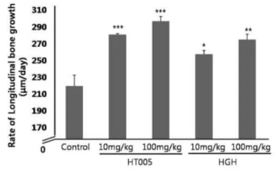
Bone growth effect of HT005 was assessed by taking measurements of the gap between the two bands formed by tetracycline at three different locations to obtain an average number. The two fluorescence bands (one located above and one below) corresponding to each given injection (3rd and 5th day) of tetracycline. Tetracycline was used to label the site of mineralization, whereas it plays the function of fluorescent dye under ultraviolet illumination (Fig. 1 A~E).
At the dose of 100 mg/kg, HT005 caused a significant acceleration of longitudinal bone growth which was 301.0±6.1 μm/day (38.07%, p< 0.001, Fig.
1C) compared to control group, which was 218.0±14. 1μ m/day (p< 0.001). At the dose of 10 mg/kg, HT005 caused a significant acceleration of longitudinal bone growth of 283.8±1.25 μm/day (21.86%, p<0.001) compared to control group. At the dose of 10 mg/kg, HGH caused an acceleration of longitudinal bone growth of 258.7±4.91 μm/day (18.67%, p< 0.05) compared to control group (p< 0.05). the dose of 100 mg/kg, HGH caused an acceleration of longitudinal bone growth which was 277.55±6.61μm/day (27.29%, p<0.01) compared to the control group (Fig. 2).
HT005 showed a dose-dependent and significant increase in the longitudinal bone growth compared to the control group.
Fig. 1. Fluorescence photomicrograph of the longitudinal section of the proximal tibia
Control (A), HT005 10 mg/kg and 100mg/kg (B & C), HGH 10 and 100 mg/kg (D & E) Scale bar 250 μm.
Fig. 2. Longitudinal bone growth of each group in adolescent rats, each value is the mean±SEM of nine animals
Control group : (saline, i.p.), HT005 : (10 and 100 mg/kg, p.o.) and HGH : (10 and 100 mg/kg, i.p.). Significantly different from the control, *** p < 0.001, ** p < 0.01, * p < 0.05.
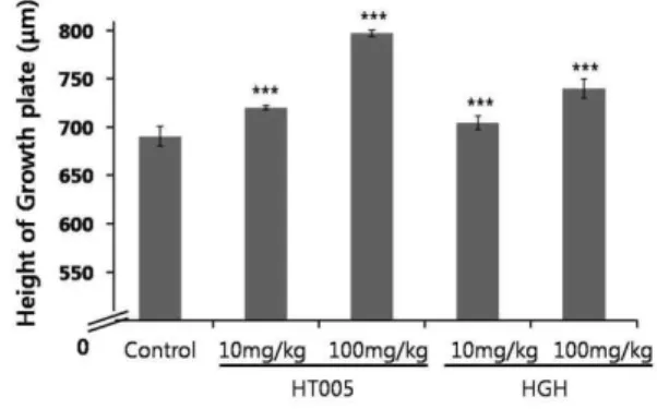
2. Effects on growth plate thickness
Longitudinal bone growth depends on a complex synchronization of the rate of proliferation, advancement and development of hypertrophy, which results in the longitudinal expansion and progression of the growth plate. The heights of the total growth plates were measured at three different locations within the growth plate for each sample and animal in each group by histological study.
At the dose of 100 mg/kg, HT005 caused a significant acceleration of the growth plate by 797.19 μ m (15.42%, p<0.001 ; Fig. 3C) compared to the control group, which was 690.67 μm (p<0.001 ; Fig. 3A). At the dose of 10 mg/kg, HT005 caused the significant difference of the growth plate, which was 720.14±2.19 μm (4.27%, p<0.001 ; Fig. 3B) compared to the control group.
In HGH group, the overall growth plate thickness was approximately 740.14 at 100 mg/kg (7.16%, p<0.001) and 704.4 at 10 mg/kg (1.99%, not significant).
HGH and HT005 treated group displayed a greater
growth plate thickness than the control group. Following administration of HT005, overall growth plate thickness increased significantly and dose dependently (Fig. 4).
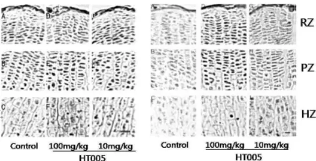
Fig. 3. Histological sections of proximal tibial growth plates viewed by light microscopy containing cresyl violet-stained chondrocytes in the three separate
(RZ : resting; PZ : proliferative, HZ : hypertrophic) zones of the growth plate in untreated, male 3-week-old adolescent rats (A), rats orally administered with 10 and 100 mg/kg of HT005 (B & C), rats injected s.c. with 10 and 100 mg/kg of HGH (D & E). Scale bar = 50 μm.
Fig. 4. Height of growth plate of each group in adolescent rats
Each value is the mean±SEM of nine animals.
Control group : (saline, i,p.). HT005 : (10 and 100 mg/kg, p.o.) and HGH : (10 and 100 mg/kg, s.c.). Significantly different from the control,* p < 0.05, ** p < 0.01, *** p < 0.001.
3. Effects on each zonal height
The heights of three principal layers (the resting, proliferative and hypertrophic zones) in the growth plate were measured at three different locations within the growth plate for each sample and animal in each group by histological study (Fig. 3A~E). The three principle layers of growth plate can be divided by its particular shapes. Chondrocytes in resting zone are irregularly scattered in a bed of cartilage matrix (compressed and packed), whereas chondrocytes in proliferative zone are arranged in columns parallel to the long axis of the bone. Chondrocytes in hypertrophic zone eventually become spherical, greatly enlarged, and then degenerated13).
The resting zone heights of HGH and HT005 group
were 11.30 μm and 16.40 μm at 10 mg/kg, and those of HGH and HT005 group were 12.23 μm and 17.00 μm at 100 mg/kg, respectfully. HT005 and HGH did affect the heights of resting zone significantly compared to control (Table 1).
The proliferative zone heights of HGH and HT005 group were 305.11 μm and 237.84 μm at 10 mg/kg, and Those of HGH and HT005 group were 265.51 μm and 286.70 μm at 100 mg/kg, respectfully. HT005 100 mg/kg and HGH 10 mg/kg group showed a significant effect on the heights of proliferative zone (Table 1).
The hypertrophic zone heights of HGH and HT005 group were 472.05 μm and 517.58 μm at 10 mg/kg and those of HGH and HT005 group were 479.89 μm and 573.88 μm at 100 mg/kg, respectfully. HT005 100 mg/kg group had a significant increase of the hypertrophic zone (Table 1).
Finally, the height of resting, proliferative, and hypertrophic zones increased by 30.16%, 16.34%, and 13.13% at 10 mg/kg and 34.92%, 40.26%, and 25.69%
respectively at 100 mg/kg of HT005.
Every zonal height of HT005 was statistically significant and dose-dependently increased compared to control.
Table 1. Each zonal height on growth plate of each group Group Zonal height (mean, μm ± SEM)
Resting zone Proliferative zone Hypertropic zone Control 12.60 ± 1.50 204.44 ± 2.64 456.55 ± 12.30 HT005 10
mg/kg 16.40 ± 1.40* 237.84 ± 19.50 517.58 ± 5.20 100mg/kg 17.00 ± 1.34 286.70 ± 11.10** 573.88 ± 16.30* HGH 10
mg/kg 11.30 ± 1.17 305.11 ± 8.40** 472.05 ± 26.70 100mg/kg 12.23 ± 0.73* 265.51 ± 6.68 479.89 ± 14.06 Control group : (saline, i.p.), HT005 : (10 and 100 mg/kg, p.o.) and HGH : (10 and 100 mg/kg, i.p.). Significantly different from the control, * p < 0.05, ** p < 0.01.
4. Effect on BMP-2 and IGF-1 expression
Immunohistochemical studies were performed to evaluate the expression of BMP-2 and IGF-1 in the three major principle zones of the growth plate. BMP-2 as well as IGF-1 staining showed the highest change in the cytoplasm of the hypertrophic zone chondrocytes.
Treatment of HT005 markedly increased the BMP-2 and IGF-1 expression in hypertrophic zone chondrocytes of growth plate (Fig. 5). Both the number and intensity of BMP-2 and IGF-1 positive cells were increased in
Fig. 5. Immunohistochemical localization of BMP-2 (L) and IGF-1 (R) in the growth plate
Representative saline treated control group was shown at the first column, the second and the third one HT005 (100 and 10 mg/kg) treated groups. The first row is the resting zone (RZ); the second row is the proliferative zone (PZ); the third row is the hypertrophic zone (HZ). Scale bar = 50 μm.
the hypertrophic zone. In contrast, staining of BMP-2 was lighter in the proliferative zone chondrocytes. It was supported there BMP-2 and IGF-1 expression and chondrogenesis effect of HT005 were correlated.
Discussion
In this study, HT005 showed a significant longitudinal bone growth compared to control group. In the immuno- histochemical study, BMP-2 expressed markedly in the proliferative zone and IGF-1 expressed markedly in the hypertrophic zone chondrocytes in HT005 treated group compared to the controls.
The growth plate is a highly organized cartilage structure entrapped between the epiphyseal and metaphyseal bone at the distal ends of the long bones. The thickness of growth plate or the length of chondrocytes zone in growth plate is meaningful in evaluating the longitudinal bone growth rate directly14).
The growth plate can be divided into distinctive histological zones of chondrocytes at different stages of differentiation, beginning with the resting zone, extending through the proliferative and hypertrophic zone15). The heights of resting, proliferative and hypertrophic region of growth plate by cresyl violet staining were measured.
HT005 treated group increased the heights of the all the zone and especially that of proliferative zone were increased significantly and dose-dependently compared to controls. It is suggesting that HT005 could promote bone growth by enhancing the chondrogenesis of growth plate. The resting zone, the uppermost region of growth plate, is composed of only a few of chondrocytes. The proliferative zone, characterized by flattened chondrocytes, is the driving force behind bone
elongation. After several mitoses, the chondrocytes were transformed to the hypertrophic zone, in which the cell division has ceased and enlarged greatly and finally transformed to the bone matrix which becomes calcified16).
The changes in the growth plate organization induced by HT005 may be related to transformation in the production of certain cytokines, including both BMP-2 and IGF-1.
The BMP is a member of TGF-β family and acts as growth and differentiation factors. IGF-1 is a growth factor structurally related to insulin and in turn induces subsequent cellular activities, particularly on bone growth. While TGF-β is unable to initiate the entire osteo-induction cascade by itself, BMP-2 uniquely exhibits ectopic bone formation property17,18). Especially, BMP-2 plays an important role in the development of epiphyseal growth plate19). Even though IGF-1 does not have the same functions of BMP-2, which has autocrine and paracrine activities in addition to the initially observed endocrine activities on bone. The IGF-1 receptor, like the insulin receptor, has intrinsic tyrosine kinase activity20). Of the various forms of BMP, BMP-2 is known as a positive regulator of growth plate chondrogenesis. BMP-2 stimulates longitudinal bone growth by stimulating growth plate chondrocyte proliferation and hypertrophy140. Mechanism of IGF-1 action in promoting the statural growth involves
‘insulin-like’ anabolic effects, supporting the extraordinary biosynthetic activity, somatic growth and matrix production that characterize hypertrophic chondrocytes21). HT005 showed a significant longitudinal bone growth which was 301.0±6.1 μm/day at the dose of 100 mg/kg (p<0.001) and 283.8±1.25 μm/day (p<0.001) at the dose of 10 mg/kg of HT005, compared to control group. In the immunohistochemical study, BMP-2 expressed markedly in the proliferative zone and IGF-1 expressed markedly in the hypertrophic zone chondrocytes in HT005 treated group compared to the controls.
These results suggest us that HT005 triggers BMP-2 and IGF-1 generation in chondrocytes zone, consequently plays a significant role in longitudinal bone growth and growth plate chondrogenesis.
Further studies are needed to identify the active components of HT005 that can provide confirmation of growth promoting and to better understanding the mechanisms that take part in this process. It is also necessary to study clinically on bone growth in children afflicted by growth retardation to be an alternative to growth hormone.
Acknowledgements
This work was supported by the Second Stage of Brain Korea 21 project in 2009 and by the Korea Science and Engineering Foundation (KOSEF) grant funded by the Korea government (MEST) (No. 2009- 0063466)
References
1. Leem K, Park SY, Lee DH et al. Effects of Jaoga-Yukmiwon(R), a Korean herbal medicine, on chondrocyte proliferation and longitudinal bone growth in adolescent male rats. Phytother Res.
2003 ; 17 : 1113-6.
2. Kim EH, Kim YJ, Lee HJ et al. Acupuncture increases cell proliferation in dentate gyrus after transient global ischemia in gerbils. Neurosci Lett.001 ; 297 : 21-4.
3. Yang DS, Cha MH, Kang BJ, Oh SW, Kim YE, Yoon Y. A study on the longitudinal bone growth of growth-stimulating material with eleutherococcus senticosus. Food Sci Technol 2003 ; 35 : 702-7.
4. Kim H. Textbook of herbal pharmacology. Seoul : Jipmoondang. 2001.
5. Kim C, Ha H, Lee JH, Kim JS, Song K, Park SW. Herbal extract prevents bone loss in ovariectomized rats. Arch Pharm Res. 2003 ; 26 : 917-24.
6. Liu HJ, Wang XP, Lin J et al. The effect of icariin and astragalosid I on the proliferation and differentiation of bone marrow stromal cells.
Zhong Yao Cai. 2006 ; 29 : 1062-5.
7. Kim C, Ha H, Kim JS, Kim YT, Kwon SC, Park SW. Induction of growth hormone by the roots of Astragalus membranaceus in pituitary cell culture.
Arch Pharm Res. 2003 ; 26 : 34-9.
8. Hansson LI, Menander-Sellman K, Stenstrom A, Thorngren KG. Rate of normal longitudinal bone growth in the rat. Calcif Tissue Res. 1972 ; 10 : 238-51.
9. Schipani E, Ryan HE, Didrickson S, Kobayashi T, Knight M, Johnson RS. Hypoxia in cartilage:
HIF-1alpha is essential for chondrocyte growth arrest and survival. Genes Dev. 2001 ; 15 : 2865-76.
10. Sun W, Gould TW, Vinsant S, Prevette D, Oppenheim RW. Neuromuscular development after
the prevention of naturally occurring neuronal death by Bax deletion. J Neurosci. 2003 ; 23 : 7298-310.
11. Leem K, Park SY, Lee DH, Kim H. Lovastatin increases longitudinal bone growth and bone morphogenetic protein-2 levels in the growth plate of Sprague-Dawley rats. Eur J Pediatr. 2002 ; 161 : 406-7. E
12. Wilsman NJ, Farnum CE, Green EM, Lieferman EM, Clayton MK. Cell cycle analysis of proliferative zone chondrocytes in growth plates elongating at different rates. J Orthop Res. 1996 ; 14 : 562-572.
13. Hunziker EB. Mechanism of longitudinal bone growth and its regulation by growth plate chondrocytes. Microsc Res Tech. 1994 ; 28 : 505-19.
14. De LF, Barnes KM, Uyeda JA et al. Regulation of growth plate chondrogenesis by bone morphogenetic protein-2. Endocrinology. 2001 ; 142 : 430-6.
15. Robson H. Bone growth mechanisms and the effects of cytotoxic drugs. Arch Dis Child. 1999 ; 81 : 360-4.
16. Jee SS. The Skeletal Tissues. In: Leon Weiss, ed.
Histology: Cell and Tissue Biology. New York : Elsevier Science Publishing Company. 1983 : 228-31.
17. Yazaki Y, Matsunaga S, Onishi T et al.
Immunohistochemical localization of bone morphogenetic proteins and the receptors in epiphyseal growth plate. Anticancer Res. 1998 ; 18 : 2339-44.
18. Zerath E, Holy X, Mouillon JM et al.
TGF-beta2 prevents the impaired chondrocyte proliferation induced by unloading in growth plates of young rats. Life Sci 1997 ; 61 : 2397-406.
19. Wozney JM, Rosen V. Bone morphogenetic protein and bone morphogenetic protein gene family in bone formation and repair. Clin Orthop Relat Res.
1998 ; 26-37.
20. Huang Y, Kim SO, Yang N, Jiang J, Frank SJ.
Physical and functional interaction of growth hormone and insulin-like growth factor-I signaling elements. Mol Endocrinol. 2004 ; 18 : 1471-85.
21. Wang J, Zhou J, Cheng CM, Kopchick JJ, Bondy CA. Evidence supporting dual, IGF-I- independent and IGF-I-dependent, roles for GH in promoting longitudinal bone growth. J Endocrinol. 2004 ; 180 : 247-55.