접수일자 : 2010년 2월 4일 완료일자 : 2010년 7월 10일
감사의 글 : 본 연구는 2009년 한라대학교 교비 연구 비 지원을 받아 수행되었습니다.
유방조영 영상의 대비개선을 위한 형체기반 호모몰픽필터 Morphology-Based Homomorphic Filter for Contrast Enhancement
of Mammographic Images
황희수 Heesoo Hwang 한라대학교 전기전자과
School of Electrical Engineering, Halla University, Wonju, Gangwon, 220-712, Korea 요 약
본 논문은 유방 조영 영상의 대비를 향상시키기 위해 새로운 호모몰픽 필터(MBHF)를 제안한다. 제안된 필터는 영상을 형 체적으로 다수 분할한 후 각각의 형체 즉, 서브-밴드에 대해 그 구성요소와 이득이 차분진화를 통해 최적의 값을 갖도록 설계된다. 결과 분석을 통해 제안된 방법이 영상 대비를 향상시키는 것을 보이며 웨이블렛 기반의 호모몰픽 필터와 성능이 비교된다. 성능평가 기준으로는 인간의 시각적 인식을 고려한 WPSNR(Weighted Peak Signal to Noise Ratio)을 사용한다.
키워드 : 형체기반 필터, 호모몰픽 필터, 유방 조영, 이미지 대비, 차분진화 Abstract
In this paper, a new MBHF(Morphology-Based Homomorphic filter) is presented to enhance contrast in mammographic images. The MBH filtering is performed based on the morphological sub-bands, in which an image is morphologically decomposed. The filter is designed to have optimal gain and structuring element in each sub-band through differential evolution. Experimental results show that the proposed method improves the contrast in mammographic images such that an evaluation criterion, WPSNR(Weighted Peak Signal to Noise Ratio) which takes into account human visual system is increased compared with a wavelet-based Homomorphic filter.
Key Words : Morphological filter, homomorphic filter, mammography, contrast enhancement, differential evolution
1. Introduction
The contrast enhancement among adjacent regions or features in mammographic images is aimed at support- ing activities such as disease diagnosis and monitoring, and surgical planning. The mammography remains one of the diagnostic imaging modalities where a breast tu- mor is hard to be detected. The major reason for the poor visualization of small malignant tumor masses is due to the small differences in the x-ray attenuation between normal and malignant glandular tissue [1]. The poor visibility of small lesions has also been a problem in mammography. To improve the visibility of breast tumors for a human observer, an image processing technique is required to decrease the noise and the in- terference from the structures that may be similar to
tumors [2].
The homomorphic filtering techniques have shown that they are capable of compressing the dynamic range and enhancing the contrast simultaneously [2,3]. They concentrate on reinforcing the details of the image to be enhanced in terms of the frequency domain. However, these linear approaches have drawback of not well solving problems involving geometrical components in the image. MGA(Multiscale Geometric Analysis) based methods have been applied to image enhancement and show good effectiveness [10] but require iterative com- putation for subband decomposition and its parameters.
A mathematical morphology is widely used to enhance or detect the geometrical structure of the image object [4]. The mathematical morphology can decompose an image into different sub-bands through a multi-reso- lution analysis, where each sub-band contains the ob- jects of a specific size. As a nonlinear methodology, the mathematical morphology is a new and powerful tech- nique to solve the above-mentioned problems.
Concerning the feature extraction from the local in- formation, another issue is to design the mapping gain.
The conventional mapping gain used in the wavelet
sub-bands for the noise suppression and the contrast enhancement is experimentally determined [2,3]. Since a feature of image object is very difficult to express mathematically, finding an optimal mapping gain for the feature extraction and a structuring element for the morphological filter is difficult and time-consuming task by trial and error. A GA(Genetic Algorithm)-based op- timization of the structuring element is described [5].
Although many GA versions have been recently pro- posed [6], they are time-consuming since GAs require inherently large population and long evolution in order to prevent the wrong and early convergence of GA. For the fast convergence differential evolution scheme was proposed [7], and has been applied to image processing areas [8,9].
For the contrast enhancement this paper presents a MBH filtering method. The proposed method decom- poses an image into morphological sub-bands, and then performs the homomorphic filtering using the morpho- logical sub-bands. The optimal filter parameters, which are mapping gains and structuring elements for the morphological sub-bands are searched by differential evolution scheme. Experimental results are given to verify the effectiveness of the proposed method.
2. Morphology-Based Homomorphic Filter
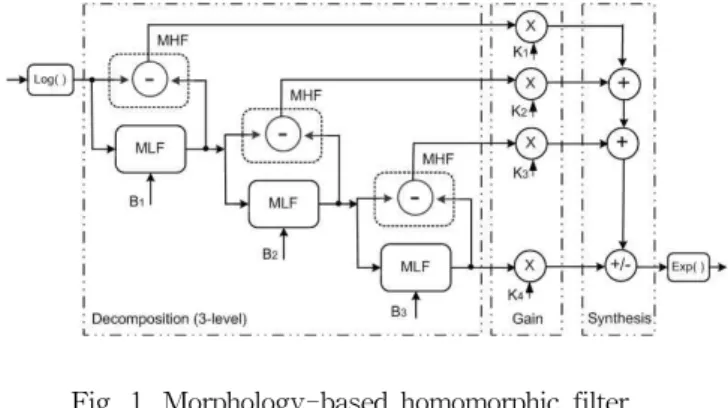
The MBHF of 3-level is shown in Fig. 1. The loga- rithm of the image is decomposed into several sub-bands through the morphological low and high pass filters with the different sized-structuring elements. The left and the right dotted boxes in Fig.1 indicate the part of the image decomposition and that of the re- construction, respectively.
Fig. 1. Morphology-based homomorphic filter structure(3-level)
MLF : Morphological low-pass filter MHF : Morphological high-pass filter
: Structuring element
Let be the logarithm of the original image. The morphological low and high pass filters uses the closing operator(●) for the dark feature enhancement. The op-
erator is defined as in (1).
● (1) Here denotes two dimensional variable and de- notes i-thlevel( ≥ ). The size of the structuring ele- ment should be increased in the subsequent decom- position level, i.e. ≤ .
According to the extensity of closing, the following relationship holds: ≥ . For 3-level an image is decomposed into three high passed sub-bands, ,
, and one low passed sub-band, . The feature-enhanced image can be obtained from the re- construction process: the summation of the weighted sub-bands, , , , and then exponential operation. Similarly, the morphological low and high pass filters using opening operator(○) for the bright feature enhancement are defined as in (2).
○ (2) Since the morphological filter can analyze the geo- metrical features of an image by locally comparing it with the structuring element, each high passed sub-band contains the objects of specific size and shape which are smaller than those of the structuring element.
The mapping gain and the structuring element should be determined to enhance the interest feature. In the homomorphic and wavelet-based homomorphic filter the gains , for the low and high frequency com- ponents are experimentally decided by the guideline of
and [2,3]. The experimental decision on the gains requires a repetitive task. The method of finding an optimal gain and the structuring element through the evolutionary search will be described in the next section.
3. Design of Morphology-Based Homomorphic Filter
Since mammographic images have bright and dark details, both bright and dark features need to be enhanced. To do this the following approach is introduced. Two MBH blocks of Fig. 1 which improves dark details are combined in series as in Fig. 2. The first MBHF receives the reversal of original image and enhances the dark details of the reversal, i.e. the bright details of the original image. The reversal of its output image is inputted to the second MBHF, which enhances the dark details of the reversal, i.e. the dark details of the original image.
To search the optimal gains and structuring elements in the proposed MBHF, differential evolution scheme is employed. Differential evolution scheme is a simple, ef- fective and powerful evolutionary algorithm, which has
Fig. 2. Series connected MBHFs for contrast enhancement
been utilized for a stochastic optimization to minimize or maximize a fitness function that can model the ob- jectives of the problem with constraints [8]. As a fit- ness function of (3) is utilized.
×
(3) Here is the -th row and -th column pixel value of the output image , and is that of the target image . and are the total numbers of row and column in the images.
Our goal is to minimize the dissimilarity between the output image × pixels obtained from the mor- phology-based homomorphic filter and the correspond- ing synthetic target image × pixels. Under the conditions that the shape of the structuring element is symmetric disk-like and ≤ , the optimal size
of the structuring element and the optimal gain
of each sub-band are searched through the differ- ential evolution process. In order to enhance the dark features of blood vessels, the synthetic input and target images sized of 100×130 pixels containing the patterns of blood vessels as in Fig. 3 are constructed. The only blood vessel areas of the target image get darker than those of the input image.
(a) Input image
(b) Target image
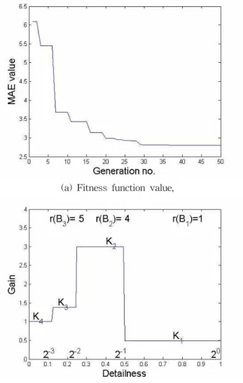
Fig. 3. Training images for differential evolution The used differential evolution control parameters are as follows: population size = 5, maximum generation number = 50, differential amplification factor = 0.5, and
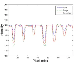
crossover rate = 0.5. The evolution result for the target image was shown in Fig. 4. The results demonstrated that the optimal gains and structuring elements turned out to be practical with fast convergence and effective to find the optimal solution.
4. Experimental Results
To evaluate the performance of the proposed method, WPSNRs of six different case images [12] are com- pared and given in Table 1, in which WPSNR [11] is an evaluating criterion which takes into account human visual system, and WHF Wavelet-based Homomorphic Filter [2].
The processing results of the mammographic image are shown in Fig. 5: the original image [12], and the images processed by the histogram equalization, WHF method, and the proposed method with the optimal pa- rameters of Fig. 4 (b). It is noticeable that the contrast of the image is improved and blood vessels are more clearly seen, on the other hand the images processed by the histogram equalization and WHF are too bright and considerably noisy. The proposed method seems to be very useful in the mammographic image enhancement.
(a) Fitness function value,
(b) Optimal gains(’s) and the sizes of structuring element(’s)
(c) A line sample
Fig. 4. Differential evolution result
Table 1. Comparison of contrast improvement
Images
Criterion: WPSNR Proposed Histogram
equalization
WHF[2]
(3-level decomposition:
=6, =4, =2, and
=1) mdb010 41.50dB 11.59dB 40.15dB mdb015 41.35dB 11.28dB 39.21dB mdb117 40.95dB 13.17dB 37.97dB mdb132 38.32dB 26.47dB 36.05dB mdb148 38.18dB 18.73dB 36.66dB mdb209 40.18zdB 13.70dB 37.69dB
(a) Original image
(b) Histogram equalization
(c) WHF[2]
(d) Proposed
Fig. 5. Results of contrast enhancement in mammographic image
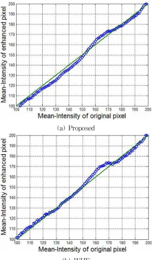
The effectiveness of the proposed method is verified by the intensity mapping results shown in Fig. 6. Fig.
6(a) shows that not only the bright pixels of the in- tensity between 60 and 170 but also the dark pixels of the intensity between 120 and 150 are enhanced. These bright and the dark details correspond to blood vessels and dark background around them, respectively. After the proposed filtering the blood vessels are more light- ened and their background more darkened. Fig. 6(b)
shows that the only bright details are enhanced.
(a) Proposed
(b) WHF
Fig. 6. Intensity mapping results
The absolute pixel values of the difference between the enhanced and original images are displayed in Fig.
7, in which the proposed method enhances the details of blood vessels better than WHF method. WHF method enhances noisy components comparatively.
(a) Proposed
(b) WHF
Fig. 7. Comparison of enhanced components
5. Conclusion
In this paper a filtering method is presented for the contrast enhancement in mammographic images. The method uses two MBHFs connected in series, in which MBHF combines the morphological sub-band decom- position and the homomorphic enhancement features.
The morphological sub-bands with the optimal gains are merged to reconstruct an enhanced image. The op- timal gain and structuring element of each sub-band are searched through differential evolution. Experimental results show that the proposed method has improved the contrast of the interest feature in mammographic image. Therefore, the method is considered to be of great use in supporting activities such as breast tumor diagnosis and its surgical planning.
References
[1] P. C. Johns and M. J. Yaffe, “X-ray character- ization of normal and neoplastic breast tissue,”
Physics in Medicine and Biology, vol. 32, no. 6, pp. 675-695, 1987.
[2] J. H. Yoon, and Y. M. Y. M., “Enhancement of the contrast in mammographic images using the homomorphic filter method,” IEICE Trans. Inf. &
Syst., vol.85-D, no.1, pp. 298-303, 2002.
[3] H. I. Ashiba, K. H. Awadallah, S. M.
El-Halfway, and F. E. El-Samie, “Homomorphic enhancement of infrared images using the addi- tive wavelet transform,” Progress In Electromagnetics Research, vol. 1, pp. 123-130, 2008.
[4] P. Maragos, “Morphological filtering for image enhancement and feature detection,” in ‘The Image and Video Processing Handbook’, Elsevier Academic Press, pp. 135-156, 2005.
[5] N. Harvey, and S. Marshall, “The use of genetic algorithms in morphological filter design,”
Elsevier Signal Processing: Image Communication, vol. 8, no. 1, pp. 55-72, 1996.
[6] M. Paulinas, and A. Usinska, “A survey of ge- netic algorithms applications for image enhance- ment and segmentation,” Inform. Tech. &
Control, vol. 36, no. 3, pp. 278-284, 2007.
[7] R. Storn, “Differential evolution - a simple and efficient heuristic strategy for global optimization over continuous spaces,” Journal of Global Optimization, vol. 11, no. 4, pp. 341-359, 1997.
[8] P. Thomas, and D. Vernon, “Image registration by differential evolution,” Proc. Int. Conf.
Machine Vision & Image Processing, pp.
221-225, 1997.
[9] S. Rahnamayan, H. R. Tizhoosh, and M. M. A.
Salama, “Image thresholding using differential evolution,” Proc. Int. Conf. Image Processing, Computer Vision & Pattern Recognition, pp.
244-249, 2006.
[10] X. Zhang and H. Xie, "Mammograms enhance- ment and denoising using generalized gaussian mixture model in nonsubsampled contourlet transform domain," Journal of Multimedia, vol. 4, no. 6, pp. 389-396, 2009.
[11] F. Kammoun and M. Salim Bouhlel, "A percep- tual image coding method of high compression rate," Int. J. of Signal Processing, vol.1, no. 1, pp. 46-50, 2005.
[12] J Suckling et. al.,The Mammographic Image Analysis Society Digital Mammogram Database Exerpta Medica. International Congress Series 1069, pp. 375-378, 1994. http://peipa.essex.
ac.uk/info/mias.html
저 자 소 개
Heesoo Hwang
received the B.S., M.S. and Ph.D.
degrees in Electrical Engineering from Yonsei University in 1986, 1988 and 1993, respectively. He is currently an associate Professor in the School of Electrical Engineering at Halla University, Korea. His research interests include data based modeling, prediction, and diagnosis.