of January 13, 2016.
This information is current as
Macrophages, and Pulmonary Inflammation Expression, Mediator Release from
Aerosolized Syk Antisense Suppresses Syk
L. Jones, A. Dean Befus and Alan D. Schreiber
Pyoung-Han Hwang, Jong-Gu Park, Warren Finlay, Richard Chen, Nadir Hirji, Fiona L. Wills, Mark Gilchrist,
Grant R. Stenton, Moo-Kyung Kim, Osamu Nohara, Chin-Fu
http://www.jimmunol.org/content/164/7/3790 doi: 10.4049/jimmunol.164.7.3790
2000; 164:3790-3797; ; J Immunol
References
http://www.jimmunol.org/content/164/7/3790.full#ref-list-1
, 18 of which you can access for free at:
cites 33 articles This article
Subscriptions
http://jimmunol.org/subscriptions
is online at:
The Journal of Immunology Information about subscribing to
Permissions
http://www.aai.org/ji/copyright.html Submit copyright permission requests at:
Email Alerts
http://jimmunol.org/cgi/alerts/etoc
Receive free email-alerts when new articles cite this article. Sign up at:
Print ISSN: 0022-1767 Online ISSN: 1550-6606.
Immunologists All rights reserved.
Copyright © 2000 by The American Association of 9650 Rockville Pike, Bethesda, MD 20814-3994.
The American Association of Immunologists, Inc.,
is published twice each month by The Journal of Immunology
at Keimyung Univ Med Lib on January 13, 2016 http://www.jimmunol.org/ Downloaded from at Keimyung Univ Med Lib on January 13, 2016 http://www.jimmunol.org/ Downloaded from
Mediator Release from Macrophages, and Pulmonary Inflammation 1
Grant R. Stenton,* Moo-Kyung Kim,
‡Osamu Nohara,* Chin-Fu Chen,
‡Nadir Hirji,*
Fiona L. Wills,* Mark Gilchrist,* Pyoung-Han Hwang,
§Jong-Gu Park,
¶Warren Finlay,
†Richard L. Jones,* A. Dean Befus,
2* and Alan D. Schreiber
‡Syk protein tyrosine kinase (PTK) is involved in signaling in leukocytes. In macrophages, Fc ␥-receptor cross-linking induces Syk PTK phosphorylation and activation, resulting in Syk-dependent events required for phagocytosis and mediator release. We hypothesized that Syk antisense oligodeoxynucleotides (ASO) delivered by aerosol to rat lungs in vivo would depress Syk PTK expression, mediator release from alveolar macrophages, and Syk-dependent pulmonary inflammation. RT-PCR and RT-in situ PCR demonstrated that aerosolized Syk ASO administration reduced Syk mRNA expression from alveolar macrophages com- pared with cells isolated from sham-treated rats. Western blot analysis confirmed that Syk PTK expression was reduced after Syk ASO treatment. Compared with sham-treated rats (scrambled oligodeoxynucleotide), Syk ASO treatment suppressed Fc ␥-recep- tor-mediated nitric oxide (86.0 ⴞ 8.3%) and TNF (73.1 ⴞ 3.1%) production by alveolar macrophages stimulated with IgG-anti- IgG complexes. In contrast, Fc ␥-receptor-induced IL-1 release was unaffected by Syk ASO treatment. Additionally, Syk ASO suppressed Ag-induced pulmonary inflammation, suggesting that Syk ASO may prove useful as an anti-inflammatory therapy in disorders such as asthma. The Journal of Immunology, 2000, 164: 3790 –3797.
S timulation of macrophage Fc ␥R leads to downstream sig- naling events, gene transcription, mediator release, and phagocytosis. In macrophages, cross-linking of Fc ␥R also results in the activation of Src and Syk protein tyrosine kinases (PTK).3These protein tyrosine kinases associate with specific rec- ognition sequences, immunoreceptor tyrosine-based activation motifs, present in the intracellular domains of Fc ␥R (1–5).
In addition to being expressed in macrophages (6, 7), Syk (p72
Syk) PTK is expressed in eosinophils (8), T cells (9 –11), B cells (10, 11), neutrophils (12), and mast cells (13–15). Recently, Matsuda et al. (16) observed that in vitro treatment of human pe- ripheral blood monocytes with Syk antisense oligodeoxynucleoti- des (ASO) inhibited Syk mRNA and protein expression compared with cells treated with scrambled oligodeoxynucleotides (ODN).
This inhibition correlated with the suppression of Fc ␥R-mediated
phagocytosis and indicated that Syk PTK plays a critical role in Fc ␥R-mediated cellular signaling and function in monocytes and macrophages. These data suggest that Syk ASO may be effective in vivo in inhibiting Syk PTK expression and cellular function in several cell populations including eosinophils, mast cells, and macrophages.
Alveolar macrophages are the most abundant cell type in the airways and are important in host defense and the inflammatory response. Alveolar macrophages rapidly internalize liposomes and are likely to be exposed to Syk ASO after aerosolization (17).
Therefore, we studied the effects of Syk ASO liposome com- plexes delivered in vivo by aerosolization on a rat model of pul- monary inflammation. In these studies we also examined the ef- fects of in vivo aerosolized Syk ASO liposome complexes on the expression of Syk PTK and on the function of alveolar macrophages.
Materials and Methods
Reagents
Syk ASO and the scrambled ODN, respectively, were prepared by the Nucleic Acid Facility of the Department of Chemistry at the University of Pennsylvania Cancer Center and the Core DNA Services at University of Calgary. 1,2-Dioleoyl-3-trimethylammonium-propane (DOTAP) and dio- leoylphosphatidyl-ethanol-amine (DOPE) phospholipids were purchased from Avanti Polar Lipids (Alabaster, AL). DOTAP liposomal transfection reagent for in vitro studies was purchased from Boehringer Mannheim (Indianapolis, IN). RPMI 1640, Eagle’s MEM (EMEM), FBS, penicillin, and streptomycin were purchased from Life Technologies (Grand Island, NY). Rat IgG2a ( ) and mouse monoclonal anti-rat IgG2a (used to form IgG-anti-IgG complexes) were purchased from Serotec (Toronto, Ontario, Canada). Rabbit anti-murine Syk polyclonal Ab was purchased from Up- state Biotechnology (Lake Placid, NY), and the isotype control, polyclonal rabbit IgG, was purchased from Serotec. F(ab ⬘)
2goat anti-rabbit HRP was supplied by The Jackson Laboratory (Bar Harbor, ME). MTT, PMSF, apro- tinin, p-tosyl arginine methyl ester, and leupeptin were supplied by Sigma (St. Louis, MO). Rat recombinant TNF and rat IL-1 ELISA kits were purchased from R&D Systems (Minneapolis, MN). Chemiluminescence reagent was purchased from DuPont NEN (Boston, MA).
Departments of *Medicine and†Mechanical Engineering, University of Alberta, Ed- monton, Alberta, Canada;‡University of Pennsylvania School of Medicine, Philadel- phia, PA 19104;§Department of Pediatrics, Chonbuk National University, Chonju, Chonbuk, Korea; and ¶Institute for Medical Sciences, Dongsan Medical Centre, Taegu, Korea
Received for publication April 27, 1999. Accepted for publication January 13, 2000.
The costs of publication of this article were defrayed in part by the payment of page charges. This article must therefore be hereby marked advertisement in accordance with 18 U.S.C. Section 1734 solely to indicate this fact.
1This work was supported by the Alberta Lung Association, the Medical Research Council of Canada (A.D.B.), the National Institutes of Health (Grant HL-27068 to A.D.S.), and a Postdoctoral Fellowship from the Canadian Lung Association/Medical Research Council of Canada/GlaxoWellcome (to G.R.S.).
2Address correspondence and reprint requests to Dr. A. Dean Befus, Astra Chair in Asthma Research, Glaxo-Heritage Asthma Research Laboratories, Department of Medicine, University of Alberta, Edmonton, Alberta, T6G 2S2, Canada. E-mail ad- dress: [email protected]
3Abbreviations used in this paper: PTK, protein tyrosine kinase; ASO, antisense oligodeoxynucleotide; ODN, oligodeoxynucleotides; EMEM, Eagle’s MEM;
DOTAP, 1,2-dioleoyl-3-trimethylammonium-propane; DOPE, dioleoylphosphatidyl- ethanol-amine; HDC, histidine decarboxylase; BAL, bronchoalveolar lavage.
Copyright © 2000 by The American Association of Immunologists 0022-1767/00/$02.00
at Keimyung Univ Med Lib on January 13, 2016 http://www.jimmunol.org/ Downloaded from
Animals
Male Sprague Dawley rats were used throughout this study. They were housed in the University of Alberta animal housing facility in filter-top cages to minimize unwanted infection. They were exposed to 12-h light/
dark cycles and were given food and water ad libitum. All rats were in- fected by s.c. injection of 0.5 ml PBS containing 3000 larvae (L3 stage) of
Nippostrongylus brasiliensis and were studied 4 wk later (18). The wormsare expelled by the host within 2–3 wk, and the inflammation in the lungs and the intestine subsides, leaving rats sensitized for re-exposure experi- ments. This work was approved by the University of Alberta Animal Ethics Committee in accordance with guidelines of the Canadian Council for Animal Care.
Syk ASO and scrambled ODN
A stem-loop rat Syk ASO was designed as described by Matsuda et al. (16) for human Syk ASO. The 60 base ASO consisted of the following se- quence: 5 ⬘-GCCGCGGTTGCCCGCCATGTCTGATTTGATTCTTGAG ATTTGGTAGTATCCCTCCGCGGC-3 ⬘. The scrambled ODN, also a 60- basesequence, has the same content of A, T, G, and C, and consisted of the following sequence: 5 ⬘-GCCGCGGTTCCTGAGTGTATCTGATTCGTA CGATTCTATGGTCGTCGTCTCATCCGCGGC-3 ⬘. The stem-loop ODN and the scrambled ODN have three phosphorothioate backbone modifica- tions, which increases their resistance to nuclease degradation. The stem- loop ASO is designed to interact with Syk mRNA at three sites, increasing its efficiency (16).
Liposome preparation
Liposomes were prepared using a method modified from Legendre and Szoka (19). One milligram DOTAP in chloroform and 1 mg DOPE in chloroform were mixed, and the chloroform was evaporated, leaving a film of 1:1 DOTAP:DOPE. Two milliliters of sterile saline were added, result- ing in liposome formation composed of equal parts of DOTAP and DOPE.
The liposomes varied in size and were dispersed in an ultrasonic bath (for
⬃1 min) until they were ⬃200 nm in diameter. The diameters of the li- posomes suspended in saline were determined by dynamic light scattering using a Brookhaven (BI90) particle sizer (Brookhaven Instruments, Holtsville, NY).
Incubation of liposomes with Syk ASO
Because DOTAP:DOPE liposomes are cationic, they form complexes with negatively charged ODN (20). Complexes were formed by incubating a 2.5:1 ratio of the liposomes (1.25 mg) with either Syk ASO (0.5 mg) or the scrambled ODN (0.5 mg) for 45 min at room temperature in sterile saline (final volume, 9 ml). The ratio of liposome:Syk ASO was optimized as described below.
In vitro studies of Syk ASO
To examine rat Syk ASO in vitro, we studied the effects on Syk expression in the rat basophilic cell line RBL-2H3. RBL-2H3 cells were grown in EMEM supplemented with 17% FBS, 100 U penicillin, and 100 g/ml streptomycin, and 4 mM glutamine at 37°C in 5% CO
2. A total of 1 ⫻ 10
6RBL-2H3 cells were seeded in each well of a 24-well plate. Twenty-four hours later, ASO/liposome complexes were added using complexes formed between Syk ASO and DOTAP transfection agent. In vitro, RBL-2H3 cells readily take up Syk ASO when it is complexed with DOTAP liposomes.
With RBL-2H3 cells in vitro, there is no need to include in the liposome formulation the lipid DOPE, which was used in vivo to enhance Syk ASO delivery. Each addition consisted of 2 l DOTAP (1 mg/ml stock) and 1 l Syk ASO (1 mg/ml stock), which were allowed to form complexes in EMEM (75 l total volume). The ASO-liposome complexes (75 l) were then added to each well containing 175 l culture medium without serum and the cells were incubated at 37°C for 24 h. A second volume of ASO- liposome complexes (75 l) was then added, the culture medium was ad- justed to 5% FBS (final volume, 1.0 ml), and the RBL-2H3 cells were incubated for 1 day before study. After treatment with liposome, scrambled ODN-liposome complexes, or Syk ASO-liposome complexes, cells were used for RT-PCR analysis of Syk mRNA expression and Western blot analysis of Syk protein expression.
After control and ASO-liposome treatment, a histamine release assay was also performed by cross-linking the RBL-2H3 IgE receptor Fc⑀RI using J17 (murine anti-rat Fc ⑀RI Ab) and measuring the resulting hista- mine release by enzyme immunoassay kit (Immunotech, Marseille, France) as described below. Twenty-four-well plates containing 1 ⫻ 10
5RBL-2H3 per well in 1.0 ml EMEM were incubated overnight at 37°C. The cells were washed once with PBS and incubated on ice with 1.0 ml PAGCM (hista- mine release buffer) and 100 l of the anti-rat Fc⑀RI Ab for 30 min at 4°C.
After one wash with PBS, the RBL-2H3 cells were incubated as follows:
1.0 ml PAGCM alone (negative control), 1.0 ml PAGCM containing 10 l calcium ionophore (50 g/ml stock) (positive control), or 1.0 ml PAGCM containing 10 l murine anti-rat Fc⑀RI Ab (1 mg/ml) for 30 min at 37°C.
The PAGCM containing histamine was removed from the cells and as- sayed by enzyme immunoassay. Standards were included to establish a dose curve for histamine.
Aerosolized administration of Syk ASO and harvesting of alveolar macrophages
To restrict their movement, animals were confined in plexiglass animal chambers, which were then placed in small plastic boxes with lids for the duration of aerosolization. Nine milliliters of liposome, scrambled ODN/
liposome, or Syk ASO/liposome complexes were administered by nebuli- zation for ⬃30 min using a Sidestream nebulizer, model 1200A durable (Medic-Aid, Pagham, U.K.). Twenty-four hours later the procedure was repeated, and another 24 h later the animals were anaesthetized with an i.p.
injection of 0.5 ml Rompun (20 mg/ml) and 0.5 ml Ketalean (Ketamine, 100 mg/ml) and then sacrificed by severing of the abdominal aorta.
The trachea of each rat was cannulated with polyethylene tubing (1.14-mm diameter; Becton Dickinson, Sparks, MD) attached to a needle, and 5 ml of ice-cold PBS was massaged into the lungs 12 times as de- scribed previously (21). The PBS was aspirated into ice-cold polypro- pylene tubes. The cells were kept on ice until they were washed by cen- trifugation at 150 ⫻ g for 20 min and resuspended in PBS, yielding ⱖ95%
alveolar macrophages as determined by staining of cytospins with May- Gru¨nwald-Geimsa stain. Cell viability was ⬎95% as determined by trypan blue exclusion. The isolated macrophages were then studied 1) for RNA isolation and RT-PCR analysis of Syk mRNA expression, 2) by Western blot analysis of Syk protein expression, or 3) by analysis of mediator re- lease.
Optimization of aerosol Syk ASO delivery
To confirm that the aerosolization procedure delivered particles to the lower airways, methylene blue was aerosolized. On frozen tissue sections of lung, areas in and around the bronchioles were stained blue, confirming that the nebulizer delivered dye to the lower airways. In addition, the neb- ulizer produced methylene blue droplets of 5.1 ⫾ 0.4 m, which are small enough to enter the lower airways, further validating the aerosolization system. Nebulized Syk ASO/liposome complexes had droplet sizes of 4.1 ⫾ 0.1 m, similar to those of methylene blue, suggesting that ODN/
liposome droplets were small enough to penetrate the lower airways. Par- ticle sizes were measured using Doppler anemometry (Dantec Electronics, Mahwah, NJ) at the exit of the nebulizer.
Optimization of liposome/Syk ASO ratio and concentration Because i.p. delivery controls the precise dose of Syk ASO administered and because this may not be the case with aerosolization, we used data from i.p. treated rats to optimize liposome:Syk ASO ratio and concentra- tion. We examined the ability of varying liposome:Syk ASO ratios and concentrations to inhibit IgE-mediated (Syk-dependent) histamine release from purified peritoneal mast cells (22). The optimum ratio and Syk ASO concentration was identified as having the strongest inhibition of IgE-me- diated histamine release. Liposome:Syk ASO (2.5:1) delivered i.p. (results not shown) was found to be the optimum ratio, as reported earlier by Zelphati and Szoka (23), for studies of maximal cellular uptake of antisense in vitro. In a similar manner, the optimum concentration of Syk ASO administered i.p. was determined to be 0.25 mg ASO/animal/day.
However, because a large proportion of aerosolized Syk ASO is not inhaled by the rats in our aerosolization delivery system, we increased the aerosol dose of Syk ASO or scrambled ODN to 0.5 mg/animal/day for all experiments using aerosol delivery and maintained the 2.5:1 liposome:Syk ASO ratio.
RNA isolation and solution-phase RT-PCR
Total RNA was isolated from alveolar macrophages and RBL-2H3 using TRIzol reagent (Life Technologies, Burlington, Canada) and quantified using an OD
260/280ratio, frozen at ⫺70°C until RT-PCR analysis of Syk RNA expression. One rat per treatment group provided sufficient alveolar macrophages to yield at least 1 g total RNA. RNA was reverse tran- scribed by SuperScript RNase (Life Technologies, Grand Island, NY) us- ing a PTC-100 programmable thermal controller (MJ Research, Cam- bridge, MA) according to the manufacturer’s protocols. PCR was modified from the Life Technologies, (Burlington, Canada) Taq DNA polymerase protocol, with changes in the concentration of dNTP (1.23 mM) and 10 ⫻ PCR buffer (67 mM Tris (pH 8.8), 1.5 mM MgCl
2, 16.6 mM (NH
4)
2SO
4, 3791 The Journal of Immunology
at Keimyung Univ Med Lib on January 13, 2016 http://www.jimmunol.org/ Downloaded from
and 10 mM BME) in a total volume of 20 l. Semiquantitative PCR was initially performed using increasing mRNA and cDNA concentrations to determine the appropriate optimal PCR cycle numbers. After preliminary study of PCR cycle numbers, 27 cycles were used (95°C for 45 s, 62°C for 45 s, and 72°C for 2 min). Twenty-seven PCR cylcles were performed with synthesized cDNA as templates using two primers, Rat-5 ⬘ Syk, 5⬘-TTTG- GCAACATCACCCGG-3 ⬘; and Rat-3⬘ Syk, 5⬘-ACTTATGATGGCTT- GCTC-3 ⬘. Products were run on a 2% agarose gel and were stained with ethidium bromide, producing a 400-bp Syk product. Controls included his- tidine decarboxylase primers Rat-5 ⬘ histidine decarboxylase (HDC), 5⬘- GGAGCCCAGTGAATACCATG-3 ⬘; and Rat-3⬘ HDC, 5⬘-TGCAGAG- GACTGTCAGCGAA-3 ⬘ (460-bp HDC product), for in vivo-treated alveolar macrophages and in vitro-treated RBL-2H3.
RT-in situ PCR
RT-in situ PCR was modified and performed as previously described (24).
The macrophages were fixed for 16 h in 10% buffered neutral formalde- hyde (BDH, Toronto, Ontario, Canada) at 22°C, washed twice with diethyl pyrocarbonate-treated water, and then placed on silane-coated glass slides (Perkin-Elmer, Norwalk, CT), each with three spots for test, positive, and negative controls. The cells were allowed to dry overnight, were digested in 10,000 U/ml pepsin (Boehringer Mannheim, Mannheim, Germany) in 0.01 M HCl for 1 h at 22°C, and then were treated with RNase-free DNase I (2,000 U/ml; Boehringer Mannheim, Indianapolis, IN) at 37°C for 24 h.
The test specimens were treated with a RT solution containing 1 M an- tisense primer or 25 g/ml oligo(dT)
12–18primer (Life Technologies, Bur- lington, Canada) with Moloney murine leukemia virus RT (Life Technol- ogies, Burlington, Canada) for 3 h at 37°C. Amplification of the cDNA was accomplished using a hot start method (24) with a PCR solution containing 4.5 mM MgCl
2; 80 M each of dATP, dCTP, dGTP, and dTTP; 0.8 M of each primer; 16 M of digoxigenin-11-dUTP (Boehringer Mannheim, Indianapolis, IN), and 120 U/ml Taq polymerase (Life Technologies, Bur- lington, Canada) using a GeneAmp In Situ PCR System 1000 (Perkin- Elmer). Cycling conditions were 5 min at 94°C and 30 cycles of 94°C for 1 min, 64°C (-actin), 51°C (Syk) for 1 min, and 72°C for 1.5 min. The digoxigenin-11-dUTP-labeled PCR product was detected after incubation with alkaline phosphatase anti-digoxigenin conjugate (Boehringer Mann- heim, Indianapolis, IN) (3.75 U/ml in 0.1 M Tris-HCl and 0.15 M NaCl (pH 7.5)) for 30 min at 22°C and after development in 4-nitro blue tetra- zolium chloride/5-bromo-4-chloro-3-indolyl phosphate (Boehringer Mann- heim, Indianapolis, IN) substrate solution. Test spots with DNase digestion and the RT step showed target mRNA expression in the cytoplasm. Posi- tive controls without DNase treatment to monitor the length of protease digestion showed nuclear DNA priming, and negative controls, in which the cells were treated with DNase and the RT step was eliminated, showed that no priming of genomic DNA was detectable. The primers used for RT-in situ PCR Syk detection were the same as for solution-phase RT-PCR as described in the previous section. -Actin controls were performed for RT-in situ PCR using the following primers: Rat -actin 5⬘ primer, 5⬘- GTGGGGCGCCCCAGGCACCA-3 ⬘; and 3⬘ primer; 5⬘-GTCCTTAAT- GTCACGCACGATTTC-3 ⬘ (526-bp -actin product).
Western blot analysis of Syk protein in RBL-2H3 cells
Cultured RBL-2H3 cells were treated with liposome alone (2 g), 2 g of control scrambled ODN and liposome, or Syk ASO and liposome at 1 ⫻ 10
5cells/ml as described above. Cell lysates were prepared from 5 ⫻ 10
6cells by boiling in Laemmli sample buffer (2% SDS, 10% glycerol, 100 mM DTT, and 60 mM Tris (pH 6.8)) for 5 min. Protein was loaded from lysates of equal cell numbers, separated by SDS-PAGE (12.5% polyacryl- amide gel), and transferred to a nitrocellulose membrane in sample buffer (25 mM Tris, 190 mM glycine, and 20% methanol). The membrane was then incubated overnight at 4°C with 1 g/ml rabbit anti-murine Syk poly- clonal Ab before incubation (1.5 h at room temperature) with goat anti- rabbit HRP. Bands on the membrane were visualized with chemilumines- cence reagent according to the manufacturer’s protocol. After detection of Syk protein, the Abs were removed by incubating the membrane in a strip- ping buffer containing 100 mM 2-ME, 2% SDS, and 62.5 mM Tris-HCL (pH 6.7) for 30 min at 50°C with occasional agitation. The membrane was then reprobed with an anti-actin Ab (Actin I-19; Santa Cruz Biotechnology, Santa Cruz, CA). Bands on the membrane were visualized with chemilu- minescence reagent.
Western blot analysis of Syk protein in alveolar macrophages Alveolar macrophages from sham- and Syk ASO-treated animals were lysed for 1 h at 4°C with 1% Nonidet P-40 in the presence of the following protease inhibitors: 100 g/ml PMSF, 5 g/ml aprotinin, 5 g/ml p-tosyl arginine methyl ester (serine protease inhibitors), and 5 g/ml of leupeptin
(thiol protease inhibitor). Protein extracted from 0.5 ⫻ 10
6cells was sep- arated on 10% SDS-PAGE and blotted onto Hybond-C Super membrane (Amersham, Oakville, Ontario, Canada). The membrane was blocked for 1.5 h in 3% skim milk and 5% goat serum at room temperature. The membrane was then incubated overnight at 4°C with 1 g/ml rabbit anti- murine Syk polyclonal Ab before incubation (1.5 h at room temperature) with goat anti-rabbit HRP. Isotype controls were performed using purified polyclonal rabbit IgG as the primary Ab. Bands on the membrane were visualized with chemiluminescence reagent according to the manufactur- er’s protocol.
Release and measurement of NO from alveolar macrophages Rat alveolar macrophages (2 ⫻ 10
5cells/well) suspended in complete RPMI 1640 medium (5% FBS) were allowed to rest for 1 h in a humidified incubator (37°C and 5% CO
2) before stimulation for 24 h with IgG-anti- IgG complexes to induce NO production. IgG-anti-IgG complexes were prepared by incubating (37°C for 10 min) 100 g/ml IgG2a with 20, 50, and 100 g/ml mouse monoclonal anti-rat IgG2a. These complexes were resuspended and 20 l was added to cells to make a final volume of 200
l and final concentrations of 2, 5, and 10 g/ml mouse anti-rat IgG2a.
Because alveolar macrophage NO is rapidly converted to nitrite (NO
2⫺), the Greiss method of NO
2⫺detection was used to determine NO produc- tion. Cell-free supernatants were mixed with equal volumes of a 1:1 mix- ture of 1% sulfanilamide:0.1% N-(1-napthyl) ethylenediamine dihydro- chloride dissolved in 2.5% H
3PO
4and incubated for 10 min at room temperature (25). NO
2⫺concentration was determined at 540 nm with a Molecular Devices Vmax kinetic microplate reader (Molecular Devices, Menlo Park, CA). NaNO
2(0.85–100 M) was used to establish a standard curve for every experiment.
Release and measurement of TNF and IL-1  from alveolar macrophages
Purified alveolar macrophages (2 ⫻ 10
5cells/well) in complete RPMI 1640 medium were added to each well of a 96-well plate and were allowed to rest for 1 h in a humidified incubator (37°C and 5% CO
2). The cells were then stimulated with IgG-anti-IgG complexes for 6 h and 24 h to induce TNF and IL-1  production, respectively, as described for NO release ex- periments. The bioactivity of TNF was tested for cytotoxicity toward
FIGURE 1.Effects of in vitro-administered Syk ASO on Syk mRNA levels. A, Examination of Syk expression by RT-PCR using rat Syk prim- ers. Lane 1, DNA ladder; lane 2, cells treated with Syk ASO/liposome complexes; lane 3, cells treated with scrambled ODN/liposome complexes;
lane 4, cells treated with liposome; lane 5, no treatment; lane 6, positive
control (human Syk plasmid). The Syk product is 400 bp. Syk expression is inhibited by Syk ASO treatment. B, Examination of HDC expression by RT-PCR using rat HDC primers. Lane 1, DNA ladder; lane 2, cells treated with Syk ASO/liposome complexes; lane 3, cells treated with scrambled ODN/liposome complexes; lane 4, cells treated with liposome. The HDC product is 460 bp. HDC expression is not affected by Syk ASO treatment.
at Keimyung Univ Med Lib on January 13, 2016 http://www.jimmunol.org/ Downloaded from
WEHI 164.13 using the MTT assay previously described (26). Briefly, 50
l/well of rat recombinant TNF standard or samples were added into flat- bottom Linbro 96-well plates. Each sample was tested in duplicate. Eight two-fold serial dilutions starting from 100 pg/ml were used to establish the standard curve. Fifty microliters/well of 1 ⫻ 10
5WEHI 164.13 cells/ml in RPMI medium supplemented with 10% FBS and 50 M 2-ME was added and incubated for 20 h. Then, 10 l/well of MTT (5 mg/ml) was added and further incubated for 3 h. Isopropanol-HCl (150 l) was used to dissolve the purple formazan precipitates. The plate was read on a Vmax kinetic microplate reader (Molecular Devices) at 570 nm. Abs (IgG-anti-IgG) did not interfere with the TNF assay.
IL-1 released from macrophages was measured in cell-free superna- tants using ELISA instructions provided by R&D Systems.
Pulmonary inflammation
A rat model of pulmonary inflammation was induced by i.v. injection via the tail vein of 100 l of saline containing 50 worm equivalents of
N. brasiliensis Ag per sensitized rat as described previously (21). Sham-challenged animals received 100 l saline alone. Eight hours after Ag challenge, animals were sacrificed and cells from the bronchoalveolar spaces were harvested by lavage as described above. Total bronchoalveolar lavage (BAL) cell counts and cell differentials, expressed as a percentage of total cells, were obtained.
Statistical analysis
Data are expressed as mean ⫾ SEM for n separate experiments. After ANOVA, statistical analysis was performed using a two-tailed Student’s
t test.Results
Effects of Syk ASO in vitro
We first determined the effects of Syk ASO in vitro using the cell line RBL-2H3. Syk ASO substantially reduced the level of Syk
mRNA in RBL-2H3 compared with controls (Fig. 1A). In contrast, the expression of HDC mRNA was not affected by Syk ASO treat- ment (Fig. 1B).
We also examined the effect of Syk ASO on Syk protein ex- pression in RBL-2H3 using Western blot analysis. Cell lysates were probed with anti-Syk Abs to detect Syk protein expression.
Syk ASO treatment suppressed Syk protein expression, whereas sham or scrambled ODN treatment had no effect (Fig. 2). In ad- dition, Syk ASO treatment of RBL-2H3 significantly reduced his- tamine release from 1.9 ⫾ 0.3 ng/10
5cells to 0.5 ⫾ 0.2 ng/10
5cells (74% inhibition) after Fc ⑀RI cross-linking (n ⫽ 3; data not shown).
Effects of in vivo Syk ASO treatment on Syk mRNA and Syk protein expression by alveolar macrophages
Treatment of rats in vivo on days 1 and 2 with Syk ASO/liposome complexes inhibited alveolar macrophage Syk mRNA expression, as determined by solution-phase RT-PCR, compared with sham treatment (liposome alone or scrambled ODN/liposome com- plexes) (Fig. 3A). Syk ASO treatment had no effect on control HDC mRNA expression (Fig. 3B).
Similar results were obtained using RT-in situ PCR to detect Syk mRNA expression (Fig. 4). Fig. 4 demonstrates that the in situ
FIGURE 2.Detection of Syk tyrosine kinase expression in RBL-2H3.
Western blot RBL-2H3 lysates (5 ⫻ 10
6cells/lane) after treatment on days 1 and 2 with complexes of liposome (lane 1), scrambled ODN/liposome (lane 2), or Syk ASO/liposome (lanes 3 and 4). p72
Syk(Syk) was detected using rabbit anti-murine Syk polyclonal Ab. A representative of three in- dependent experiments is shown. The molecular mass markers are shown, and the arrow indicates the position of the 72-kDa Syk protein.
FIGURE 3.
Effects of in vivo-administered Syk ASO on Syk mRNA levels. A, RT-PCR analysis of Syk mRNA expression by alveolar macro- phages after no treatment (lane 6) or aerosolized liposome (lanes 2 and 7), scrambled ODN/liposome complex (lanes 3 and 8), or Syk ASO/liposome complex (lanes 4 and 9) treatment in vivo. Human Syk plasmid positive control (lanes 1 and 10) and DNA ladder (lane 5) are shown. Rats were treated with aerosol preparations on days 1 and 2, and alveolar macro- phages were harvested 24 h later. Results are representative of three inde- pendent experiments. The Syk product is 400 bp. B, RT-PCR analysis of HDC mRNA expression by alveolar macrophages after aerosolized lipo- some (lane 1), scrambled ODN/liposome complex (lane 2), or Syk ASO/
liposome complex (lane 3) treatment in vivo. DNA ladder is shown in lane 4.
Rats were treated with aerosol preparations on days 1 and 2, and alveolar macrophages were harvested 24 h later. Results are representative of three independent experiments. The HDC product is 460 bp.
3793 The Journal of Immunology
at Keimyung Univ Med Lib on January 13, 2016 http://www.jimmunol.org/ Downloaded from
Syk mRNA signal is detected in the cytoplasm of alveolar mac- rophages (Fig. 4A1). In Fig. 4A1, the nuclei were unstained, re- flecting the removal of genomic DNA by DNase digestion. In con- trast, the absence of DNase digestion (Fig. 4A2) leads to nuclear staining in the positive control, demonstrating that the PCR step is successful. In the negative control (Fig. 4A3), when the cells were treated with DNase and the RT step was eliminated, the cells were unstained, indicating that the product detected in the cytoplasm of cells in the test spot (Fig. 4A1) was from cDNA and was not priming from contaminating genomic DNA.
Fig. 4B demonstrates the detection of in situ Syk and -actin mRNA. Positive and negative controls were also performed as described above (results not shown). Treatment of rats on days 1 and 2 with Syk ASO/liposome complexes suppressed alveolar macrophage Syk mRNA expression (Fig. 4B3) compared with sham treatment (liposome alone, Fig. 4B1,; or scrambled ODN/
liposome complexes, Fig. 4B2). Similar staining intensity in Fig. 4, B4–B6, indicates that -actin mRNA expression from alveolar macrophages after Syk ASO treatment (Fig. 4B6) was not signif- icantly different from that of macrophages isolated from sham- treated animals (Fig. 4, B4 and B5).
In a similar manner, Western blot analysis confirmed that the same 2-day Syk ASO/liposome complex treatment inhibited Syk PTK expression in macrophage cell lysates compared with cell lysates from sham-treated animals (Fig. 5). Purified Syk protein, supplied by the manufacturer of the rabbit anti-murine Syk Ab and isolated from Jurkat cells, was used as a positive control for West- ern blot analysis of Syk protein. Bands of 72 kDa were visible by Western blot analysis of cell lysates from sham-treated animals. In contrast, the 72-kDa Syk protein band was markedly reduced in cell lysates from Syk ASO-treated animals. Isotype controls for the anti-Syk Ab showed no detectable bands at 72 kDa (data not shown). Thus, RT-PCR and Western blot analyses clearly indicate
that Syk ASO treatment suppresses Syk PTK mRNA and protein expression.
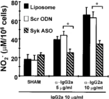
Effects of in vivo Syk ASO treatment on Fc ␥R-mediated NO release from alveolar macrophages
Alveolar macrophages were stimulated with IgG-anti-IgG com- plexes containing 5 or 10 g/ml anti-IgG2a and 10 g/ml IgG2a
FIGURE 4. A, RT-in situ PCR analysis of Syk mRNA expression (controls). 1, Test spot. Detection of Syk mRNA in the cytoplasm. Cells undergoDNase digestion to ensure there is no amplification of genomic Syk DNA, hence the nuclei are negative for Syk expression. Reverse transcription (RT) is performed to convert Syk mRNA to Syk cDNA, and the cDNA is amplified by PCR. 2, Positive control. Cells are treated as in 1, except that there is no DNase digestion step. Therefore, amplification of genomic Syk DNA occurs, and the nuclei are positive for Syk expression. 3, Negative control. DNase treatment and PCR steps are performed without the cDNA-forming RT step. Hence, no genomic or cytoplasmic amplification is observed. B, RT-in situ PCR analysis of Syk (1-3) and -actin (4-6) mRNA expression by alveolar macrophages after aerosolized liposome (1 and 4), scrambled ODN/liposome complex (2 and 5), or Syk ASO/liposome complex treatment (3 and 6). Rats were treated with aerosol preparations on days 1 and 2, and alveolar macrophages ( ⱖ95% purity) were harvested 24 h later. Syk ASO/liposome complexes inhibited Syk mRNA expression (3) but not -actin mRNA expression (6). Positive and negative controls, as described for A, were performed for alveolar macrophages from each treatment group (data not shown).
Results are representative of two independent experiments.
FIGURE 5.
Detection of Syk tyrosine kinase expression. Western blot of alveolar macrophage lysates ( ⬎95% purity; 0.5 ⫻ 10
6cells/lane) after aerosolized liposome, scrambled ODN/liposome complex (Scr ODN), or Syk ASO/liposome complex treatment. Rats were treated with aerosol preparations on days 1 and 2, and alveolar macrophages were harvested 24 h later. A positive control for Syk (Syk ⫹ve) was performed using purified Syk protein supplied by the manufacturer of the rabbit anti-murine Syk Ab. p72
Syk(Syk) was detected using rabbit anti-murine Syk polyclonal Ab. Isotype control (rabbit IgG) was negative for Syk (data not shown). A representative of three independent experiments is shown. The molecular mass markers are shown, and the arrow indicates the position of the 72-kDa Syk protein.
at Keimyung Univ Med Lib on January 13, 2016 http://www.jimmunol.org/ Downloaded from
for 24 h, which resulted in the release of significant levels of NO (measured as [NO
2⫺]). IgG2a in the absence of anti-IgG2a (sham) failed to induce NO release above spontaneous levels (Fig. 6).
When rats were treated with Syk ASO/liposome complexes (com- pared with control treatment with scrambled ODN/liposome com- plexes) once daily for 2 days before alveolar macrophage isolation and stimulation (10 g/ml IgG-anti-IgG2a), NO release was in- hibited by 73.1 ⫾ 3.1% when spontaneous NO release was sub- tracted from Fc ␥R-mediated NO release (Fig. 6; n ⫽ 4). There was no significant difference in Fc ␥R-mediated NO release from alve- olar macrophages after treatment with scrambled ODN/liposome complex or liposome alone.
Effects of in vivo Syk ASO treatment on Fc ␥R-mediated TNF and IL-1  release from alveolar macrophages
The effects of Syk ASO treatment on cytokine release from alve- olar macrophages were determined by measuring TNF and IL-1  production (Figs. 7 and 8). After liposome, scrambled ODN/lipo-
some complex, or Syk ASO/liposome complex treatment on days 1 and 2, alveolar macrophages were isolated by BAL and were stimulated with IgG-anti-IgG2a complexes for 6 h (TNF) and 24 h (IL-1 ).
Although IgG-anti-IgG2a complexes induced significant TNF and IL-1  release, IgG2a in the absence of anti-IgG2a (Figs. 7 and 8; sham) failed to induce TNF or IL-1  release above spontaneous levels. Syk ASO/liposome complex treatment compared with con- trol treatment with scrambled ODN/liposome complexes signifi- cantly inhibited Fc ␥R-mediated (10 g/ml IgG-anti-IgG2a) TNF release by 86.0 ⫾ 8.3% when the spontaneous TNF release was subtracted from the Fc ␥R-mediated TNF release (Fig. 7; n ⫽ 4–5).
There was no significant difference in Fc ␥R-mediated TNF release from alveolar macrophages after treatment with scrambled ODN/
liposome complexes or liposome alone.
In contrast to the effects on NO and TNF release, in vivo Syk ASO/liposome complex treatment did not significantly inhibit IgG-anti-IgG2a complex-induced IL-1  release compared with treatment with scrambled ODN/liposome complexes or liposome alone (Fig. 8; n ⫽ 4).
Effects of in vivo Syk ASO treatment on pulmonary inflammation Using an established model of pulmonary inflammation, we stud- ied the effects of in vivo Syk ASO treatment on pulmonary in- flammation as measured by BAL cell number and differential. In these experiments, rats were treated once daily for 2 days with Syk ASO/liposome complexes, scrambled ODN/liposome complexes, or liposome alone before i.v. saline (sham) or Ag challenge (Fig.
9; n ⫽ 5). In the liposome treatment group, we observed a signif- icant increase in the total BAL cell number 8 h after Ag challenge (4.3 ⫾ 0.3 ⫻ 10
6) compared with 8 h after sham challenge (2.3 ⫾ 0.4 ⫻ 10
6). Similarly, the total BAL cell number from scrambled ODN/liposome complex-treated rats 8 h after Ag challenge was increased (4.9 ⫾ 1.0 ⫻ 10
6) compared with 8 h after sham chal- lenge (2.2 ⫾ 0.3 ⫻ 10
6). By contrast, no significant increase in BAL cell number was observed 8 h after Ag challenge (2.6 ⫾ 0.4 ⫻ 10
6) compared with sham challenge (2.0 ⫾ 0.7 ⫻ 10
6) in the Syk ASO/liposome complex treatment group.
The proportions of different cell populations in BAL were ex- amined after sham or Ag challenge (8 h). The majority of cells present in all groups were macrophages (range, 91–97%), although
FIGURE 6.Fc␥R-mediated NO release from alveolar macrophages af-
ter aerosolized liposome, scrambled ODN (Scr ODN), or Syk ASO. Rats were treated with aerosol preparations on days 1 and 2, and alveolar mac- rophages were harvested 24 h later. NO released 24 h after stimulation is represented by [NO
2⫺] in micromolar/10
6cells (mean ⫾ SEM; n ⫽ 4).
ⴱ, p ⬍ 0.05, Scr ODN vs Syk ASO. ␣-IgG2a represents anti-IgG2a.
“SHAM” represents macrophages treated with IgG2a (10 g/ml) only.
Spontaneous NO release from untreated macrophages isolated from lipo- some alone and scrambled ODN/liposome complex- and Syk ASO/lipo- some complex-treated animals was 24.8 ⫾ 4.6, 18.0 ⫾ 5.6, and 26.7 ⫾ 1.1
M/10
6cells, respectively. Data shown have not been corrected for spon- taneous NO release.
FIGURE 7.
Fc␥R-mediated TNF release from alveolar macrophages af- ter aerosolized liposome, scrambled ODN (Scr ODN), or Syk ASO. Rats were treated with aerosol preparations on days 1 and 2, and alveolar mac- rophages were harvested 24 h later. TNF released 6 h after stimulation is represented as pg/10
6cells (mean ⫾ SEM; n ⫽ 4–5). ⴱ, p ⬍ 0.05, Scr ODN vs Syk ASO. ␣-IgG2a represents anti-IgG2a. “SHAM” represents macro- phages treated with IgG2a (10 g/ml) only. Spontaneous TNF release from untreated macrophages isolated from liposome alone and scrambled ODN/
liposome complex- and Syk ASO/liposome complex-treated animals was 10.8 ⫾ 3.6, 8.4 ⫾ 3.1, and 7.9 ⫾ 2.4 pg/10
6cells, respectively. Data shown have not been corrected for spontaneous TNF release.
FIGURE 8.
Fc␥R-mediated IL-1 release from alveolar macrophages after aerosolized liposome, scrambled ODN (Scr ODN), or Syk ASO. Rats were treated with aerosol preparations on days 1 and 2, and alveolar mac- rophages were harvested 24 h later. IL-1 released 24 h after stimulation is represented as pg/10
6cells (mean ⫾ SEM; n ⫽ 4). ⴱ, p ⬍ 0.05, Scr ODN vs Syk ASO. ␣-IgG2a represents anti-IgG2a. “SHAM” represents macro- phages treated with IgG2a (10 g/ml) only. Spontaneous IL-1 release from untreated macrophages isolated from liposome alone and scrambled ODN/liposome complex- and Syk ASO/liposome complex-treated animals was 1.5 ⫾ 0.4, 1.5 ⫾ 0.4, and 1.8 ⫾ 0.3 pg/10
6cells, respectively. Data shown have not been corrected for spontaneous IL-1 release.
3795 The Journal of Immunology
at Keimyung Univ Med Lib on January 13, 2016 http://www.jimmunol.org/ Downloaded from
eosinophils (0.9 – 4.8%), neutrophils (0.2–2.9%), and lymphocytes (0.6 –1.7%) were observed in BAL from all treatment groups (Table I). Although Ag challenge induced an increase in absolute numbers of lymphocytes, neutrophils, and eosinophils, the relative abundance of each cell type did not change significantly as the macrophage numbers increased proportionately at 8 h after Ag challenge (Table I).
Discussion
Treatment with ASO has the potential to reduce mRNA levels, inhibit translation, and reduce protein levels. Two primary mech- anisms of action of ASO have been observed (27, 28). First, the ASO forms a DNA/RNA duplex that prevents mRNA translation.
Second, the DNA/RNA duplex formation induces RNase H acti- vation, which degrades the DNA/RNA duplex, thus destroying mRNA. Destruction of the mRNA results in reduced levels of functional protein, which can inhibit cell function.
ASO are anionic molecules that cross cell membranes poorly.
Cationic liposomes such as DOTAP therefore have been used to enhance delivery of ASO to target cells (23, 29, 30). For example, the CFTR gene has been complexed with DOTAP and successfully administered to and expressed in the nasal epithelium of humans with cystic fibrosis (30). Zelphati and Szoka (23) demonstrated that optimal delivery occurs when the liposome contains an addi- tional lipid such as DOPE. Furthermore, when a cationic liposome composed of 1:1 DOPE:DOTAP is used, the ideal ratio of lipo- some:ASO was 2.5:1. Our studies confirm this observation and use DOTAP-DOPE liposomes complexed with Syk ASO at a ratio of 2.5:1 for in vivo studies. For in vitro studies we used liposomes made of DOTAP alone complexed with Syk ASO at a ratio of 2:1.
We observed that in vitro delivery of Syk ASO to the rat mast cell line RBL-2H3 suppressed Syk mRNA and protein expression compared with control-treated cells. Furthermore, Syk ASO sup- pressed the Syk-dependent Fc ⑀RI-mediated histamine release from
RBL-2H3 cells. This Syk ASO-mediated suppression of histamine release supports the observation by Zhang et al. (31) that Syk tyrosine kinase is required for optimal Fc ⑀RI-dependent histamine release from mast cells.
Next, we studied the use of Syk ASO in vivo by delivering it to rat lungs by aerosolization. It is well-known that optimal Fc ␥R stimulation of macrophages is Syk PTK-dependent (6, 16). Similar to the in vitro effects on Syk PTK expression, we observed that aerosolized Syk ASO suppressed alveolar macrophage Syk mRNA and protein expression as well as immune complex-mediated NO and TNF release. However Fc ␥R-mediated IL-1 release from al- veolar macrophages was unaffected.
A previous report indicates that the administration of ASO di- rected at other targets in the lung may also be effective. Ag-in- duced bronchoconstriction in a rabbit model of asthma was inhib- ited after treatment with an ASO directed against the adenosine receptor (32). Our studies are the first to target the important in- tracellular signaling molecule Syk PTK in vivo.
It is of interest that there was no inhibition of Fc ␥R-mediated IL-1  release by alveolar macrophages after Syk ASO treatment.
We previously observed a similar differential regulation of TNF and IL-1  release after CD8␣ ligation of alveolar macrophages and demonstrated that CD8 ␣-mediated IL-1 release but not TNF release was inhibited by the protein kinase C inhibitor Ro 31-8220 (33). Thus, different intracellular pathways may be utilized for TNF and IL-1  release, a result further suggested by our obser- vations with Syk ASO treatment. Alternatively, IL-1  synthesis and release may require lower levels of active Syk PTK than NO and TNF release do.
Having observed that in vivo delivery of Syk ASO suppressed alveolar macrophage function, we studied the effects of Syk ASO in an in vivo model of systemic anaphylaxis and focused on pul- monary inflammation. It has been established that when rats pre- viously sensitized to N. brasiliensis are challenged by i.v. injection of Ag isolated from this parasite, anaphylactic shock occurs, one aspect of which is pulmonary inflammation. One method for as- sessing pulmonary inflammation is to determine the Ag-mediated changes in BAL cell number and cell types found in the BAL. We observed that Syk ASO treatment suppressed the Ag-mediated (8 h) increase in BAL cell number, indicating that Syk ASO has anti-inflammatory properties in the lung. However, the proportions of different cell populations in BAL did not change 8 h after Ag challenge. In future studies, we will examine the effects of Syk antisense on changes in cell populations found in BAL at different times after Ag challenge. We will also determine whether aero- solized Syk ASO selectively inhibits pulmonary inflammation or if it affects other components of the systemic anaphylaxis.
These data extend the observations of Matsuda et al. (16) and pioneer the use of aerosolized Syk ASO in vivo to inhibit Syk mRNA and protein expression as well as the Syk-dependent acti- vation of alveolar macrophages. Syk ASO may prove useful in the
FIGURE 9.Syk ASO inhibits Ag-induced pulmonary inflammation.
Rats were treated with aerosolized liposome, scrambled ODN, or Syk ASO on days 1 and 2, and 24 h later the rats were challenged with 100 l saline or Ag delivered i.v. Eight hours postchallenge alveolar macrophages were harvested. BAL cell number (mean ⫾ SEM; n ⫽ 5) served as an indicator of pulmonary inflammation. ⴱ, p ⬍ 0.05.
Table I. Cell composition (%) in BAL fluids from rats 8 h after Ag or saline challenge
aCell Type Lip-Sal (%) Lip-Ag (%) Scr ODN-Sal (%) Scr ODN-Ag (%) Syk ASO-Sal (%) Syk ASO-Ag (%)
Macrophage 94.2 ⫾ 2.2 94.1 ⫾ 2.8 96.3 ⫾ 1.5 91.0 ⫾ 2.6 96.9 ⫾ 0.5 95.8 ⫾ 1.5
Lymphocyte 0.7 ⫾ 0.4 0.7 ⫾ 0.3 0.6 ⫾ 0.4 1.3 ⫾ 0.6 1.1 ⫾ 0.5 1.7 ⫾ 0.5
Neutrophil 1.5 ⫾ 0.6 1.3 ⫾ 1.2 1.3 ⫾ 0.6 2.9 ⫾ 1.6 0.2 ⫾ 0.1 1.1 ⫾ 1.0
Eosinophil 3.1 ⫾ 2.0 3.9 ⫾ 1.8 0.9 ⫾ 0.4 4.8 ⫾ 2.3 1.2 ⫾ 0.5 1.0 ⫾ 0.3
Epithelial 0.5 ⫾ 0.4 0.1 ⫾ 0.1 0.8 ⫾ 0.8 0.1 ⫾ 0.1 0.6 ⫾ 0.4 0.3 ⫾ 0.3
aRats were treated with aerosolized liposome (Lip), scrambled ODN (Scr ODN), or Syk ASO on days 1 and 2, and 24 h later were challenged with 100l saline (Sal) or antigen (Ag) delivered intravenously. Eight hours postchallenge, BAL was performed. The percentages (mean⫾ SEM) of macrophages, lymphocytes, neutrophils, eosinophils, and epithelial cells recovered from BAL are shown (n⫽ 5).
at Keimyung Univ Med Lib on January 13, 2016 http://www.jimmunol.org/ Downloaded from
study of allergic disorders because cells such as eosinophils and mast cells express several Syk-dependent pathways of activation (8, 13–15). Thus, Syk ASO-mediated Syk suppression may prove useful in the development of therapy for inflammatory diseases of the airways, such as asthma and allergic disorders, in addition to being potentially beneficial at other sites of inflammation.
Acknowledgments
We thank Dr. T. Allen for helping to make the liposomes for this study. We are also thankful to Dr. T. J. Lin for helping us develop the Greiss assay for the quantification of NO
2⫺concentration and to Helena Orszanska for measuring the diameter of the aerosol droplets.
References
1. Indik, Z. K., J. G. Park, X. Q. Pan, and A. D. Schreiber. 1995. Induction of phagocytosis by a protein tyrosine kinase. Blood 85:1175.
2. Kiefer, F., J. Brumell, N. Al-Alawi, S. Latour, A. Cheng, A. Veillette, S. Grinstein, and T. Pawson. 1998. The Syk protein tyrosine kinase is essential for Fc␥ receptor signaling in macrophages and neutrophils. Mol. Cell. Biol. 18:
4209.
3. Crowley, M. T., P. S. Costello, C. J. Fitzer-Attas, M. Turner, F. Meng, C. Lowell, V. L. J. Tybulewicz, and A. L. DeFranco. 1997. A critical role for Syk in signal transduction and phagocytosis mediated by Fc␥ receptors on macrophages.
J. Exp. Med. 186:1027.
4. Bolen, J. B. 1995. Protein tyrosine kinases in the initiation of antigen receptor signaling. Curr. Opin. Immunol. 7:306.
5. Lin, C. T., Z. Shen, P. Boros, and J. C. Unkeless. 1993. Fc receptor-mediated signal transduction. J. Clin. Immunol. 14:1.
6. Darby, C., R. L. Geahlen, and A. D. Schreiber. 1994. Stimulation of macrophage Fc␥RIIIA activates the receptor-associated protein tyrosine kinase Syk and in- duces phosphorylation of multiple proteins including p95Vav and p62/GAP-as- sociated protein. J. Immunol. 152:5429.
7. Greenberg, S., P. Chang, and S. C. Silverstein. 1994. Tyrosine phosphorylation of the␥ subunit of Fc␥ receptors, p72syk, and paxillin during Fc receptor-mediated phagocytosis in macrophages. J. Biol. Chem. 269:3897.
8. Yousefi, S., D. C. Hoessli, K. Blaser, G. B. Mills, and H. U. Simon. 1996.
Requirement of Lyn and Syk tyrosine kinases for the prevention of apoptosis by cytokines in human eosinophils. J. Exp. Med. 183:1407.
9. Hunter, S., M. Kamoun, and A. D. Schreiber. 1994. Transfection of an Fc␥ receptor cDNA induces T cells to become phagocytic. Proc. Natl. Acad. Sci. USA 91:10232.
10. Weiss, A., and D. R. Littman. 1994. Signal transduction by lymphocyte antigen receptors. Cell 76:263.
11. Chan, A. C., D. Desai, and A. Weiss. 1994. Role of protein tyrosine kinases and protein tyrosine phosphatases in T cell antigen receptor signalling. Ann. Rev.
Immunol. 12:555.
12. Yan, S. R., M. Huang, and G. Berton. 1997. Activation of the p72syktyrosine kinase and formation of protein complexes containing p72sykand Src family kinases in neutrophils spreading over fibrinogen. J. Immunol. 158:1902.
13. Benhamou, M., J. S. Gutkind, K. C. Robbins, and R. P. Siraganian. 1990. Ty- rosine phosphorylation coupled to IgE receptor-mediated signal transduction and histamine release. Proc. Natl. Acad. Sci. USA 87:5327.
14. Hutchcroft, J. E., R. L. Geahlen, G. G. Deanin, and J. M. Oliver. 1992. Fc⑀RI- mediated tyrosine phosphorylation and activation of 72 kDa protein tyrosine ki-
nase, PTK72, in RBL-2H3 rat tumour mast cells. Proc. Natl. Acad. Sci. USA 89:9107.
15. Benhamou, M., N. J. P. Ryba, H. Kihara, H. Nishikata, and R. P. Siraganian.
1993. Protein-tyrosine kinase p72sykin high affinity IgE receptor signaling.
J. Biol. Chem. 268:23318.
16. Matsuda, M., J. G. Park, D. C. Wang, S. Hunter, P. Chien, and A. D. Schreiber.
1996. Abrogation of the Fc␥ receptor IIA-mediated phagocytic signal by stem- loop Syk antisense oligonucleotides. Mol. Biol. Cell 7:1095.
17. De Haan, A., G. Groen, J. Prop, N. Van Rooijen, and J. Wilschut. 1996. Mucosal immunoadjuvant activity of liposomes: role of alveolar macrophages. Immunol- ogy 89:488.
18. Befus, A. D., N. Johnston, and J. Bienenstock. 1979. Nippostrongylus brasilien- sis: mast cell and histamine levels in tissues of infected and normal rats. Exp.
Parasitol. 48:1.
19. Legendre, J. Y., and F. C. Szoka. 1992. Delivery of plasmid DNA into mamma- lian cell lines using pH-sensitive liposomes: comparison with cationic liposomes.
Pharm. Res. 9:1235.
20. Felgner, P. L., T. R. Gadek, M. Holm, R. Roman, H. W. Chan, M. Wenz, J. P. Northrop, G. M. Ringhold, and M. Danielson. 1987. Lipofection: a highly efficient, lipid-mediated DNA-transfection procedure. Proc. Natl. Acad. Sci. USA 84:7413.
21. Ramaswamy, K., R. Mathison, L. Carter, D. Kirk, F. Green, J. S. Davison, and D. Befus. 1990. Marked anti-inflammatory effects of decentralization of the su- perior cervical ganglia. J. Exp. Med. 172:1819.
22. Shanahan, F., J. A. Denburg, J. Fox, J. Bienenstock, and A. D. Befus. 1985. Mast cell heterogeneity: effects of neuroenteric peptides on histamine release. J. Im- munol. 135:1331.
23. Zelphati, O., and F. C. Szoka. 1996. Intracellular distribution and mechanism of delivery of oligonucleotides mediated by cationic lipids. Pharm. Res. 13:1367.
24. Nuovo, G. J. 1995. PCR in situ hybridization: protocols and applications. Ge- nome Res. 4:s123.
25. Lin, T. J., N. Hirji, O. Nohara, G. R. Stenton, M. Gilchrist, and A. D. Befus. 1998.
Mast cells express novel CD8 molecules that selectively modulate mediator se- cretion. J. Immunol. 161:6265.
26. Espevik, T., and J. Nissen-Meyer. 1986. A highly sensitive cell line, WEHI 164 clone 13, for measuring cytotoxic factor/TNF from human monocytes. J. Immu- nol. Methods 95:99.
27. Crooke, S. T. 1996. Progress in antisense therapeutics. Med. Res. Rev. 16:319.
28. Askari, F. K., and M. McDonnell. 1996. Antisense-oligonucleotide therapy.
N. Engl. J. Med. 334:316.
29. Alton, E. W. F. W., P. G. Middleton, N. J. Caplen, S. N. Smith, D. M. Steel, F. M. Munkonge, P. K. Jeffery, D. M. Geddes, S. L. Hart, R. Williamson, et al.
1993. Non-invasive liposome-mediated gene delivery can correct the ion trans- port defect in cystic fibrosis mutant mice. Nat. Genet. 5:135.
30. Porteous, D. J., J. R. Dorin, G. McLachlan, H. Davidson-Smith, H. Davidson, B. J. Stevenson, A. D. Carothers, W. A. Wallace, S. Moralee, C. Hoenes, et al.
1997. Evidence for safety and efficacy of DOTAP cationic liposomes mediated CFTR gene transfer to the nasal epithelium of patients with cystic fibrosis. Gene Ther. 4:210.
31. Zhang, J., E. H. Berenstein, R. L. Evans, and R. P. Siraganian. 1996. Transfection of Syk protein tyrosine kinase reconstitutes high affinity IgE receptor-mediated degranulation in a Syk-negative variant of rat basophilic leukemia RBL-2H3 cells. J. Exp. Med. 184:71.
32. Nyce, J. W., and W. J. Metzger. 1997. DNA antisense therapy for asthma in an animal model. Nature 385:721.
33. Lin, T. J., N Hirji, G. R. Stenton, M. Gilchrist, B. J. Grill, A. D. Schreiber, and A. D. Befus. 2000. Activation of macrophage CD8: pharmacological studies of TNF and IL-1 production. J Immunol. 164:1783.