일산화탄소 중독이 뇌내 아미노산 신경전달물질 함량변화에 미치는 영향
정민정
•박 송 자
* •이선희
•윤재순
이 화 여 자 대학교 약학대학
•K IST 도핑콘 트 롤 센 타 (Received August 13, 1990)
The Effect of Carbon Monoxide Intoxication on the Changes in Contents of Amino Acid Neurotransmitter of Rat Brain
Min Jung Jung, Son Ja Park*, Sun Hee Lee and Jae Soon Yun
College of Pharmacy, Eivha Women’s University, Seoul 120-750, Korea
*KIST, Doping Control (Jenter
Abstract— To study influence of carbonmonoxide (CO) poisoning on the content of amino acid neurotransmitter in brain, male rat was exposed to CO 5000 ppm for 30 minutes (60-75% HbCO). Aspartic acid and glutamic acid level in the cerebral ccrtex and aspartic acid level in the striatum were significantly decreased. GAB A level in the cerebral cortex was significantly increased after the 30 and 60 minutes of CO intoxication. Taurine level in both the cerebral cortex and the striatum was increased although nonsig
nificant. Consequently, the CO-induced hypoxia brain showed lower level of excitatory neurotransmitter, aspartic acid and glutamic acid and higher level of inhibitory neurotransmitter, GABA and taurine. These results suggest that the change in content of amino acid neurotransmitter in the rat brain may be concern
ed with several CO poisoning symptoms.
Keywords □ Amino acid neurotransmitter, aspartic acid, glutamic acid, taurine, 7-aminobutyric acid, CO intoxication.
일 산화탄소
(CO)는
sulfur oxide, hydrocarbon등과 더불어
5대 공기 오염물질 중의 하나이다
Cᄋ는 자동차 배기가스
,공장배출연기 특히 우리나
라의 경우는
CO를 가장 많이 발생하는 연탄을 가
정연료로 사용하고 있어 그 피해가 심각한 문제로
대두되고 있다
. CO는 체내에 흡입되면 쉽게 일산
화탄소헤모글로빈
(HbCO)을 형성하여 조직으로의 산소운반을 감소시키고 산소헤모글로빈
(Hb02)으로
4터 산소해리도 저해하여 결국 저산소증이 유발된 다
. 2,C
ᄋ는 뇌에는 특히 예민하여 0}주 낮은 농도의
CO
를 흡입해도 심한 중추신경병변을 나타내어
3>치 매
,우울,판단력장애,지각장애
,파킨슨씨병,언어
장애
,장기혼수 등 다양한 신경정신계 중상과 지연 성후유증이 나타나고4 이 지연성후유중은
C O증독 시의 저산소증에서만 나타난다고 하였다
. 4)이같은 중독증상 발현에 대한 정확한 원인은 밝혀지지 않았 으나 파킨슨씨병
,정신분열중 및 우울중 등과 같은 신경정신 질환이 중추신경계의 신경전달물질의 변화 에 기인한다는 것이 알려져 이러한 질환에 신경전달 물질이나 그 수용체에 작용하는 약물을 투여하였을
때 치료되는 것으로 보아
, CO중독시에도 이러한
신경중상을 나타내므로 뇌내 신경전달물질의 변동이 있을 것으로 예상된다
. Newby,6) Macmillan등
3>은
CO중독시에 뇌내
dopamine의 함량변화를 보 고하였다
.또한 여러 원인의 저산소증 상태에서 뇌
323
324_____________________________________ 정민정 • 박송자 ■ 이선희 • 윤재순
내
acetylcholine과 아미노산 신경전달물질의 함량 이 감소하고 이 때,행동장애가 나타난다는 결과도 보고되었다
. 7'8)따라서
C0중독으로 인한 저산소증에서도 아미노
산 신경전달물질의 함량변화가 있으리라 생각되어
C0에 가장 예민하다고 알려진 대뇌피질과 선조체 에서
9>아미노산 신경전달물질인
aspartic acid, glutamic acid, taurine및
y-aminobutyric acid의 함량변화에 미치는
Cᄋ의 영향을 연구하고자 하 였다
.실험재료 및 방법
측정용 시약一
L-Aspartic acid, L-glutamic acid, y-Aminobutyric acid (GABA), Taurine, 3- Mercaptopropionic acid, phenylisothiocyanate는
Sigma Chem.Co.에서 구입하였고
Triethylamine은
Aldrich Chem. Co.에서 과염소산
(HC1ᄋ
J은
Junal Chem. Co.에서 구입하였다
.아미노산 분석 에 사용되 는 용매 인
Acetonitrile, Methanol은
HPLC grade
를 사용하였으며 용매제조에 쓰이는
물은
Milli-Q grade의 물을 사용하였다
.실험기기一중독
chamber내의
CO농도의 측정 은
Cᄋ 검지관과 검지기
(Gastec. Co. Japan)로 하 였으며 원심분리기는
Beckman model J2-21을 사용하였다
. UV/visible spectrophotometer는
Shimazu UV-240을 사용하였고 아미노산의
phenylisothiocyanate
유도체화 반응은
PICO. TAG (Millipore)롤 사용하였다
.아미노산은
HP1090M liquid chromatograph (Hewlett-Packard)로 분 석 하 였 고
DR5 solvent delivery system, autosampler, autoinjector, DIODE-ARRAY detector, HP9000~3000 computer, HP9133 disc drive, HP7475 plotter, thinkjet printer및
PH hypersil octadecylsilane column (4.6 mm * 10 cm, particle size 5 /zm)을 사용하였다
.실험동물一체중
300~400g의
Wistar계 웅성 흰 쥐를 사용하였으며
,실험전까지 물과 고형사료를 자 유로이 공급하였다
.시육실내 온도는
23±2°C로 유 지하였고 명암은
12시간씩 자동조절 장치로 조절하 였다
.일산화탄소 중독증 모델一일산화탄소 중독은
180/
용적의
stainless steel chamber에
CO:N2
:
0 2 = 1:
79:
20(% )의 혼합
gas롤
2//min의 기속으로
30분간 홀려주어 행하였으며 이 때
chamber내 농도는 약
5000 ppm으로 유지되었다
.정상대조군은
CO gas대신 압축공기를 같은 조건으 로 홀려주었다
.시료 추출一저산소중군은
CO 5000 ppm으로
30분간 노출
,중독시켜 중독즉시와
30분
, 1시간,
a시 간 경과 후에 각각 단두 치사시켰으며 정상대조군은 압축공기를
30분간 홀려준 후 단두 치사시켰다
.사 후의
y-amino-butyric acid (GABA)증가를 막기 위해 단두시키기
2분 전에 꼬리 정맥에
1.5mM/kg의
3-mercaptopropionic acid를 주사하였다
.단 두한 후 즉각
dry ice에 넣고
10분간 방치 한 후 뇌 를 꺼내어
0.9%생리 식염액으로 세척하고
0~4°C에서 대뇌피질과 선조체조직에 빙냉한
0.4M HC104수용액을 각각
4m/,
lm /가하여
10분간 고 정시켜 단백질을 제거한 후
homogenizer로
30초 간 균질화시켰다
.이 균질액을
21,000 *용로
4°C에 서
10분간 원심분리하여 상등액을 취하고 침전에
0.4M HC104 2.5m/, 0.5m/ 를 각각 가하고 재균 질화하여 같은 조건에서
10분간 원심분리하여 얻은 상등액을
2M KOH로
pH 2.5로 맞추어 과잉의
HC104를 중화시킨 후 다시
21,000 *표로
4°C에서
10분간 원심분리하여 얻어진 상등액을 아미노산 정 량용 시료로 사용하였다
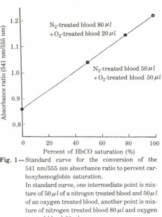
.일산화탄소 헤모글로빈량 측정ᅳ
CO중독용
chamber
에서 꺼낸 즉시 안정맥총에서 채혈하여
Tietz
등법11>에 따라
HbCO포화도를 측정하였다
. Hb02와
HbCO룰 함유하는 혈액의 알칼리 희석액 은 가시영역에서
double bond로 비슷한 흡수극대 롤 나타내는
Na2S2ᄋ
4처치시
Hb02는 탈산소 헤모 글로빈으로 변화하는 반면
HbCO는 이 시약에 영 향을 받지 않으므로
541 nm와
555nm에서 나타나 는
Hb02와
HbCO각각의 흡수극대율을 측정하여 그 비율로
HbCᄋ량을 표준곡선에서 구하였다
.표 준곡선은 흰쥐의 안정맥총에서 채혈한 혈액을
in vitro 로순수한
CO및 ᄋ2로 각각
15분 포화시 켜
100%와
0%의
HbCO를 만들어 표준곡선의 양 끝 점을 구하였다
.물리적으로 녹아 있는
CO를 구축 시키기 위해
N2gas로 처리하였다
.이 질소처치시 료와 산소처치시료를 각각
8 : 2및
5 : 5로 혼합하므
J. Pharm. Soc. Korea
N2-treated blood 80
^ I
+ 0 2-treated blood20^1
N2-treated blood
50Ml h
02-treated blood 50 /iI
0 20 40 60 80 100
Percent of HbCO saturation (%) Fig. 1 — Standard curve for the conversion of the
541 nm/555 nm absorbance ratio to percent car- boxyhemoglobin saturation.
In standard curve, one intermediate point is mix
ture of 50 of a nitrogen treated blood and 50/i
I
of an oxygen treated blood, another point is mixture of nitrogen treated blood
SO/jil
and oxygen treated blood20 u I.
Table I一Chromatographic gradient conditions.
Time (min) Percent of solution A
Percent of Solution B
Initial 100 0
15 95 5
16 70 30
18 60 40
21 50 50
Solution A: Phosphate buffer (0.01 M K3P 04 and 0.02 M KH2P04 in distilled water, pH 6.7-6.8)
Solution B: acetonitrile .
aspartic acid, glutam ic acid, tau rin e
및
y-aminobutyric acid (GABA)의 각 아미노산 표
준품의 농도에 따른
peak면적을 검량하여 작성하
였다
.통계 분석ᅳ모든 실험값은 평균土표준오차로 표시 하였으며
Student t-test에 의해 유의성을 검정하 여
p<0.05일 때 유의성 이 있다고 판정하였다
.결 과
로서 표준곡선의 중간점을 구하였다
(Fig.l).아미노산 정량ᅳ대뇌피질과 선조체를
0.4M HC1CX용액으로 제단백,균질화하여 얻은 시료
50 m/ 씩을 각각 취하여 반응용기에 넣고
PICᄋ
. TAG work station에서 감압 건조시켰다
. 12)완전히 건 조시 킨 후
methanol, H20, triethylamine, phenylisothiocyanate
를
7 : 1: 1: 1로 혼합한 유도체 시약
20# /룰 가하여
5초 동안 섞어주고
20분 동안 실온에서 방치 하여 반응시키고
PICO. TAG에서 감압 건조시켰다
. Na2HP04 710mg을
H20 1/ 에 용해시켜
10% H3P04로
pH 7.4로 조절하고 그 용 액
950m/ 에
acetonitrile 50m/ 를 가하여 혼합하 고 여과한 용액
250서 를 가하여 시료를 완전히 용 해시키 이를
HPLC를 이용한 아미노산 분석용 시 료로 사용하였다
.아미노산 분석을 위한 이동상은
pH 6.7—6.8의 인 산 염 완 중 액 과
acetonitrile을
gradient elution하였고
(Table I ) lm //m in의 속,
5# /의 주입용량으로
254nm에서 측정하였다
.아미노산의 정량은 외부 표준물질로
peak면적을 검량한 표준곡선을 이용하여 구하였다
.표준곡선은
일산화탄소 헤모글로빈량 변화
ᅳ혈액 중의
HbCO
포화도를 측정하기 위하여 정상 흰쥐에서 채
혈한 혈액을 순수한
CO와 ᄋ
2로 포화시켜 표준곡선 을 작성하였다
(Fig.l). CO 5000ppm으로
30분 중 독시킨 직후의
HbCO포화도는
72% 였고 지연호흡 에 의하여 회복시킬 때
3시간 후에 정상으로 회복되 었다
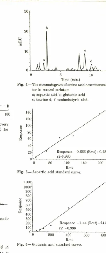
(Fig. 2).정상뇌 중 아미노산 신경전달물질 함량변화ᅳ대뇌 피질과 선조체에서 얻은 시료를 역상
chromatography
에 의해 분리하여 아미노산을 분석하였으며
정상대조군의 대뇌피질과 선조체 중의 아미노산을 분리한
chromatogram을
Fig. 3,4에 표시하였다
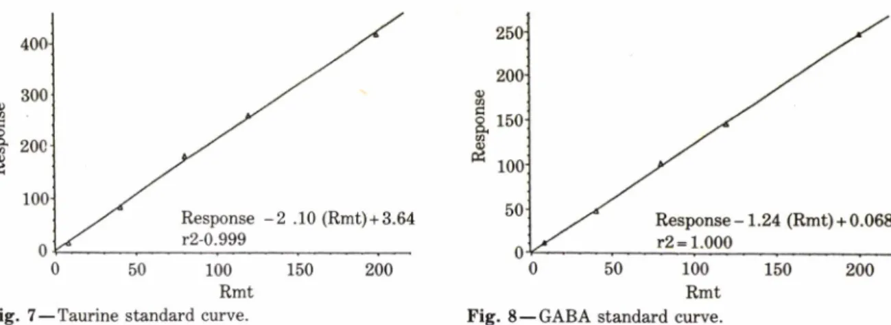
.아미노산은
Fig. 5~8에 나타낸
aspartic acid, glutamic acid, taurine, y-aminobutyric acid(GABA)
의 표준곡선을 이용하여 함량을 구하였다
.정상대조군의 대뇌피질과 선조체 중 아미노산 신경 전달물질의 함량은 각각
aspartic acid가
5.57,
4.24/^mole/g뇌조직이고
, glutamic acid가
16.67,16.04//mole/g
뇌조직이고
taurine이
5.12,6.35umole/g
뇌 조직 이 며
G A B A가
2.49,3.60(uni
lo UD lo/
u nl
T
—I 행
Q
) 0
- 43
O3J
<uo l
^l cl . ol acl yl
Vol. 34,No. 5, 1990
326 정 민 정 • 박 송 자 • 이 선 희 . 윤재순
1100
100040. ᄉᄉ
20- Response -0.666 (Rmt) +0.283
^
r2-0.980o-v^ --- ---
0 50 100 150 200
Rmt Fig. 5 — Aspartic acid standard curve.
0 30 60 90 120 150 180
Time after CO intoxication (min.) Fig. 2 — Carboxyhemoglobin percent during the recovery
time after intoxication in 5000 ppm CO for 30 min.
0 5 10
Time (min.)
Fig. 4 — The chromatogram of amino acid neurotransmit
ter in control striatum.
0 200 400 600 800
Rmt Fig. 6 — Glutamic acid standard curve.
물질의 함량변화一
C05000ppm으로
30분간 중독시 킨 후 중독 즉시
30, 60및
180분 경과시의 대뇌피 질의 아미노산 신경전달!■질의 함량을 측정한 결과
U '거
' r « ' '•ᄀ
* •—5 10
Time (min.)
Fig. 3 — The chromatogram of amino acid neurotransmit
ter in control cortex,
a; aspartic acid b; glutamic acid c; taurine d;
y
-aminobutyric aicd./^mole/g
뇌조직으로
aspartic acid는 대뇌피질 조 직에 더 많이 함유되어 있고
taurine과
GABA는
선조체 조직 중에 더 많이 함유되어 있다
(TableII).
일산화탄소 중독 대뇌피질 중 아미노산 신경전달
a; aspartic acid b; glutamic acid c; taurine d; 7 -aminobutyric aicd.
80| 30-
o
o
o
o
o
o
o
o
o c o
o
o
o
o
o
o
o
o 9 8 7 6 5 4 3 2 1
OJs u o ds o ) 05
0
o
o
o
2086
1
O) 1
suods9
t f
o- o- o-o
o 5
4
3
2
1
§
日
o
o 2
1
{%)
.sq q bfl
o u w l
^ l^o cl .I C TJ o tMo
J
/
Pharm. Soc. Korea
0
Response -2 .10 (Rmt) + 3.64 r2-0.999
0 50 100 150
Rmt Fig. 7一 Taurine standard curve.
100 Rmt Fig. 8•一GAB A standard curve.
Table II 一 The contents of amino acid neurotransmitter in cerebral cortex and striatum of normal rats Contents (/imol/g brain tissue)
Brain region Aspartic acid Glutamic acid Taurine GABA
Cerebral cortex Striatum
5.57 + 0.28 (6) 4.24 + 0.41 (6)
16.57 + 0.48 (6) 16.04 + 0.63 (6)
5.12 + 0.25 (7) 6.35 + 0.27 (7)
2.49 + 0.30 (7) 3.60 + 0.61 (6) Values are means土 S.E.M. of the number of animals in parentheses.
Table III — The contents of amino acid neurotransmitter in cerebral cortex after CO intoxication.
Contents (^/mol/g brain tissue)
Group Time after
intoxication (min) Aspartic acid Glutamic acid Taurine GABA Control - 5.57 ±0.28 (6) 16.67 ±0.48 (6) 5.12 ±0.25 (7) 2.49 土 0.30 (7) CO intoxication 0 1.13±0.06** (7) 4.94 士 0.21** (7) 5.04 ±0.12 (7) 3.32 ±0.20 (5) 30 1.27±0.11** (4) 4.32士0.18** (5) 6.18 士 0.45 (5) 3.66 ±0.27* (5) 60 1.16±0.03** (4) 4.53 士 0.36** (4) 4.74 ±0.39 (4) 3.73 ±0.38* (4) 180 2.48±0.11** (4) 7.29 ±0.43** (4) 4.98 ±0.48 (4) 2.52 ±0.14 (3) Values are means士 S.E.M. of the number of animals in parentheses.
Significantly different from the control values.
*p<0.05, **p<0.01
는
Table UI과 같고
,정상대조군에 대한 증감률은
Fig. 9, 10과 같 다
.정 상 뇌 에 서
aspartic acid, glutamic acid는
5.57, 16.67//mole/g뇌조직이었 으나
Cᄋ에서
30분 중독시킨 후의 저산소성 뇌에 서는
1.13, 4.94//mole/g뇌조직으로 각각
79.7, 70.3% 의 현저한 감소를 나타내었으며
(p<0.01) taurine, GABA함량은
CO중독으로
5.04,
3.32^mole/g
뇌조직으로
GABA의 경우
33.3%중가 되었지만 유의성은 없었고
taurine은 거의 변하지
않았다
.중독 후
30분 경과시에는
aspartic acid, glutamic acid의 함링은 중독 직후의 저산소성 뇌와 비교하여 커다란 변화를 나타내지 않았으며
GABA, taurine은 대조군에 비하여 각각
46.9%, 20.7%중가되었으나
taurine의 경우 유의성은 없 었다
.증독 후
1시간 경과시에는 중독 후
30분 경과 시에 비하여
aspartic acid glutamic acid는 큰 변 화를 보이지 않았으며
taurine은 대조군에 비하여
7.4%감소하였으나 유의성은 없었다
.중독 후
3시
asuodsa
t f
300
않
g I 20C a;100
Vol. 34, No. 5,1990
328 정 민 정 • 박 송 자 • 이 선 희 • 윤재순
Time after CO intoxication (min.) Fig. 9 一 The effects of recovery after CO intoxication on
the contents of aspartic acid and glutamic acid in rat cerebral cortex.
Each point is expressed as ratio to the change in contents of aspartic acid and glutamic acid in control group.
Vertical lines indicate土 S.E.M.
Significant difference compared with control values.
**p<0.01 • Aspartic acid ■ Glutamic aicd.
간 경과시에
aspartic acid, glutamic acid는 중독 직후에 비하여 증가는 되었으나 정상뇌에 비하여 각 각
55.4, 56.2%감소로 정상뇌의 함량에는 도달하지 못하였고
taurine, GABA함량은 정상뇌와 거의 유
Time after CO intoxication (min.) Fig. 10 — The effects of recovery after CO intoxication on
the contents of taurine and GABA in rat cerebral cortex.
Each point is expressed as ratio to the change in contents of taurine and GABA in control group.
Vertical lines indicate土S.E.M.
Significant difference compared with control values.
*p<0.05 • Taurine ■ GABA
사하게 회복되었다
.일산화탄소 중독 선조체 중 아미노산 신경전달물 질의 함량변화一
CO 5000 ppm으로
30분 중독시킨
Table VI 一 The contents of amino acid neurotransmitter in striatum after CO intoxication.
Contents
(ji
mol/g brain tissue)Group Time after
intoxication (min) Aspartic acid Glutamic acid Taurine GABA Control - 4.24 ±0.41 (6) 16.04 ±0.63 (6) 6.35 ±0.27 (7) 3.60 ±0.61 (7) CO intoxication 0 2.84±0.19** (7) 14.59 ±0.26* (6) 6.50土0.10 (7) 3.89 ±0.14 (6) 30 2.73 ±0.24* (5) 15.38 ±1.13 (5) 7.25 ±0.52 (3) 3.00 士 0.25 ⑷
60 3.44 ±0.75 (5) 16.92 ±0.68 (4) 6.57 土 0.28 (3) 2.81 ±0.31 (3) 180 4.27 ±0.23 (4) 17.12±1.02 (4) 6.45 ±0.08 (3) 3.50 ±0.32 (3) Values are means土S.E.M. of the number of animals in parentheses.
Significantly different from the control values.
*p<0.05, **p<0.01
-100
-30 0 30 60 180
Time after CO intoxication (min.) Fig. 11 —— The effects of recovery after CO intoxication Oil
the contents of aspartic acid and glutamic acid in rat striatum.
Each point is expressed as ratio to the change in contents of aspartic acid and glutamic acid in control group.
Vertical lines indicate± S.E.M.
Significant difference compared with control value
*p<0.05, **p<0.01
•ᅳ• Aspartic acid ■ Glutamic acid
후 중독 즉시
, 30,60및
180분 경과시 선조체에서의 아미노산 신경전달물질의 함량을 측정한 결과를
Table IV
에 표시하였고,정상대조군에 대한 중감률
은
Fig. 11,12에 표시하였다
.정상뇌에서
aspartic acid, glutamic acid는
4.24,
16.04 ;miole/g뇌조 직이었으나
30분 중독 후에는
2.84,14.59/miole/g뇌조직으로 정상뇌에 비해
35.0,
9.0% 의 감소를 나 타내었으며 정상뇌에서
taurine, GABA는
6.35,
3.60//mole/g뇌조직 인데 비하여
CO 30분 중독 후 의 저산소성뇌에서는
6.50,
3.89/imole/g뇌조직으 로 약간 중가되었으나 유의성은 없었다
.중독 후
30분경과시에
asparticacid는
2.73/miole/g뇌조직으 로 대조군에 비해
35.6%감소하였다
(p< 0.05).Glutamic acid
는
CO중독
30분 경과 후에
15.38 //mole/g뇌조직으로 정상뇌에 비해
4.1% 의 감소 에 그쳤다
. Taurine은
14.1%중가
, GABA는
16.6%감소하였으나 유의성은 없었다
.중독 후
1시
■100
■co->
-30 0 30 60
Time after CO intoxication (min.) Fig. 12 — The effects of recovery after CO intoxication on
the contents of taurine and GABA in rat striatum.
Each point is expressed as ratio to the change in contents of aspartic acid and glutamic acid in control group.
Open symbols represent control values.
• Taurine ■ GABA
간 경과시에는 중독 후
30분 경과시와 비교하여
aspartic acid, glutamic acid는 증가되 어 회복되 는 경향을 나타내었고
taurine, GABA는 감소되었 지만 유의성은 없었다
.중독 후
3시간 경과시에는
aspartic acid, glutamic acid, taurine, GABA는 대조군에 비하여 각각
0.7,
6.7, 1.5,
2.7% 의 근소한 증감률을 나타내어 모두 정상뇌와 거의 유사하게 회 복됨을 보였다
.고 찰
CO 5000 ppm
에서
30분간 중독시켰을 때
CO는 적혈구내 혈색소와의 친화력 이 산소보다
210배나 크 기 때문에 대기 중의
C O가 흡입되면 쉽게
H b과
결합하여
HbCO를 형성하여 조직으로의 산소운반
을 감소시킬 뿐만 아니라 이미 결합되어 있는
Hb02로부터의 산소해리도 저해되어 결국 심한 저 산소증이 유발되어
2> Fe2+이나
Cu2+과의 친화력도 강하여 이 같은 금속이온을 함유한 효소활성도 크게 저하시키며 특히 조직내 호홉효소인
mitochondrial500
50
I
《%
)
:이
UI SU
aJJ 4 ->
OJ n (l)u u -l—i 2
4' 6<ᄋ i
i
i
{%
)
cl
CQa) bcl I^l o{
Vol. 34, No. 5’ 1990
330 정 민 정 • 박 송 자 ■이 선 희 • 윤재순
cytochrome oxidase
내에 함유된 금속이온과 착화 합물을 형성하여 전자전달계를 방해하여 조직내 산 화환원의 조 직 대 를 억제한다
.특히
Cᄋ 는 중추 신경계에서도 크게 손상을 주어
9>대뇌피질
,선조체 및 혹질 등에 다양한 신경 및 정신계 중상이 나타나 며
5>이것은 중추신경계 전달물질의 변화, 3 구연 산회로의 중간산물과 에너지 인산화물의 변화
15,16)및 뇌혈류량의 저하 등1 7 원인일 수 있다는 여 러 각도에서의 연구가 보고 되어 있다
.본 실험에서도
CO 5000ppm에서
30분 등안 중 독시켰을 때 대뇌피질과 선조체에서 홍분성 아미노 산 신경전달물질인
aspartic acid와
glutamicacid
의 함링은 감소되었고 억제성 신경전달 아미노
산
GABA는 유의성은 없었으나 중가되었다
.이는
CO에 의한 저산소증 이외에도
N2gas를 사용한 저산소증에서 실시한
Wood등의 보고19>와 허혈상 태에서 실시한
Erecinska등20^ 의 실험결과에서 나타난 양상과 일치하였다
.또한
Weinberger등
8>은
mongolian gerbil의 좌측 경동맥을 결 찰하여 유발시킨 허혈성뇌의 선조체에서 얻은
synapto- some은
glutamic acid의 도입을 감소시킨다고 하 였으며
David등22>은 동일조건의 허혈성 대뇌피질
에서
GABA량이 현저히 증가되었다고 보고하였
다
.이것은
Cᄋ로 인한 저산소증시에 뇌내에서의 홍분성의 감소와 억제성의 중가를 반영하는 것으로 추정된다
.Gibson
둥7)은
aspartic acid, glutamic acid는
glucose의 분해
,즉 구연산회로의 중간체로부터 유 래되는데 저산소중시에는 이를 경유하여 합성되는 이들 아미노산의 함량이 감소된다고 하였다
.뇌혈 관 관문을 통과할 수 있는 에너지원은 유일하게
glucose만으로 동맥혈에서
02와
glucose를 공급 받아 해당계
,구연산회로 및 전자전달계를 거쳐
ATP등의 고에너지 인산화물 중간체형으로 에너지 를 얻어 신경전달물질의 합성과 뇌기능 유지의 연 료로 사용하는데
CO중독으로
0 2공급이 부족 한 저산소중 상태에서는
glucose의 뇌내 흡수가
50%
이하로 감소되어
23)에너지 산생계 장애로
ATP결핍이 일어나 신경 기능유지에 혼란이 야기된다
. Prejer,24), Hagberg25*, Benveniste등26은 저산소 중시에
ATP저하로
ATP의존적인
reuptake system의 손상으로
aspartic acid, glutamic acid가 세포외로 방출되어 세포내에서의 함량이 감소하 고 결국에는 혈액속으로 홀러 나간다고 하였다
. Aspartic acid, glutamic acid에 대한
reuptake system은
GABA에 비해 저산소증에 더욱 민감하 다는 실험 보고가 있다
. 20>이러한 홍분성 신경전달 물질의
synaptic cleft로의 방출은 세포에 지속적인
^-§-^•-1
: 유도히•여
positive feedback cascade reaction에 의하여 다른 여러 신경전달물질 및 뇌 전체에 다량 분포되어 있는
glutamic acid, aspar- ticacid의 유리를 중강시킨다
. Glutamic acid는
neurotoxic하기 때문에 이 아미노산의 다량 방출은
neuron에 상당한 손상을 미칠 수 있다고 보고된 바 있다
. 27-30>신경조직에서의
GABA함량은 합성효소인
glutamic acid decarboxylase (GAD)와 분해효소 인
GABA-transaminase에 의하여 조절된다고 한 다
. 20,30)저산소증과 허혈상태에서는 대사성 수소이
온농도의 증가로
GAD활성이 촉진된다는 실험보고
가 있다
. 31>또한 정상뇌에서
GAD는
cofactor인
pyridoxal-5'-phosphate에 의하여 부분적으로 포 화되어 있다고 한다
. 32) Glutamate는
pyridoxal phosphate의 해리를 촉진함으로써 또한
ATP는 그 결합을 억제함으로써
GAD활성을 감소시킨다고 하였다
. 32,33)따라서 저산소증과 허혈상태에서는
glutamate와
ATP가 감소되어
GAD활성을 촉진
시킬 수 있으므로
GABA함량이 증가되었다고 사
료된다
. GABA분해시에는 산소가 요구되는데
CO로 인한 저산소중시에는 산소부족으로
GABA분해 효소인
GABA-transaminase활성이 억제되어
34>조직내의
GABA함량이 중가되었다고 생각된다
.또한
CO로 인한 저산소중시에 전체적인 산화적 대 사가 손상되어 구연산회로의
GABA shunt pathway
를 통한
GABA의 이용을 저하시키는 방향으 로 유도할 수 있다는 보고가 있다
. 19)이러한 여러 가능한 기전에 의하여
CO중독시에
GABA함량이 중가되었다고 사료된다
.본 실험에서의
GABA함 량의 중가가
Erecinska등20>의 실험결과에서와는 달리 선조체에서 유의성이 없었던 것은 동물사후에
GAD활성 중가로 인한
GABA중가를 막기 위해
GAD억제제인
3-mercapto propionic acid를 투 여하였기 때문이라고 생각된다
.억제성 아미노산 신경전달물질인
taurine은
CO/
Pharm. Soc. Korea
로
30분 중독시에 함량이 감소되지 않았으며 중독 후
30분 경과시에 유의성은 없었으나 중가되었다
. Taurine은 생체막을 통한
Ca2+의 이동을 조절함으 로써 내인성 항저산소 작용 기전에서 중요한 역할을 한다는 가설이 제시되고 있고 그에 따른 많은 실험 결과도 보고되어 있다
. 35-37>따라서 본 실험에서의 함량변화는
CO로 인한 저산소증에서의 방어기전으 로 설명될 수 있다
.본 실험의
CO중독 후의 중추신경계 아미노산 신 경전딜물질의 함량변화가
CO중독 후유중의 주된 원인이라고 결론을 지을 수는 없지만 한 유발기전으 로써 작용할 수 있다는 가능성을 제시하였다고 본 다
.결 론
CO 5000 ppm
에서
30분간 중독시킨 직후 및 회 복기를 대뇌피질과 선조체에서 아미노산 신경전달물 질의 함량변화를
HbCO포화도와 관련하여 다음과 같은 결론을 얻었다
.CO 5000 ppm
에서
30분간 중독시 혈중
HbCO포화도는
72%였고 자연호흡에 의하여
3시간 경과시 정상으로 회복되었다
.흥분성 아미노산 신경 전달물질인
aspartic acid와
glutamic acid는모두
CO중독으로 대뇌피질에 서 유의적인 감소를 나타내었으나 선조체에서는
aspartic acid만이 유의 적 인 감소를 나타냈으며 중 독 후
3시간 경과시에 선조체에서는 완전히 회복되 었으나 대뇌피질에서는 중독 즉시에 비해 중가는 되 었으나 정상치로 회복되지 못하였다
.억제성 아미노산 신경전달물질인
taurine는 대뇌 피질과 선조체에서 중독 후
30분 경과시 유의적은 아니나 증가되었으며 중독 후
a시간 경과시에는 정 상수준으로 회복되었다
.억제성 아미노산 신경전달 물질인
GABA는 대뇌피질에서 중독 후
30분 및
1시간 경과시에는 유의적인 증가가 관찰되었고
S시간 경과시에는 거의 정상으로 회복되었다
.결론적으로
CO중독된 저산소증 뇌에서 홍분성 아미노산 신경전달물질인
aspartic acid와
glutamic acid
는 선조체와 대뇌피질에서 유의적으로 감
소하였고 억제성 전달물질인
taurine과
GABA는 중가되는 경향이었으며 중독 후
3시간 경과시에도
대뇌 피 질에서
aspartic acid와
glutamic acid의 감소는 정상적으로 회복되지 못하였다
.이러한 뇌내 아미노산 신경전달물질의 함량변동이 신경장애와 서 로 연관하여 뇌세포 장애를 일으키고 결과적으로
CO중독의 여 러 중상을 일으킬 수 있다고 본다
.문 헌
1) Amdur, M.O.: Air pollutants. In Caearett and Doull’s Toxicology, (ed. Doull, J., Klaassen, C.D., Amdur, M.O.) 3rd. ed. Macmillan, p. 608 (1986).
2) Collier C.R.: Oxygen affinity of human blood in the presence of
CO. J. Appl Physiol,
40, 487 (1976).3) Macmillan, V.A. comparison of the effects of carbon monoxide intoxication and low-oxygen gas mixture on cerebral biogenic amine metabolism.
Brain Research.,
408, 40 (1987).4) Choi, I.S.: Delayed neurologic sequelae in carbon monoxide intoxication.
Arch. Neurol.,
40,433 (1983).5) Ginsberg, M.D.,Delayed neurological deterioration following hypoxia, in: Advances in Neurology, (ed.
Fahn, S., Davis, J.N., Rawland, L.P.) Ravan Press.
New York, p. 21 (1979).
6) Newby, M.B., Roberts, R.J., Bhathagar, R .K .,Car
bon monoxide- and hypoxia-indued effects on cat- acholamines in the mature and developing rate brain.
]• Pharmacol Exp. Ther.,
206,61 (1978).
7) Gibsen, G.E., Peterson, C. and Sansone, J.: De
creases in amino acid and acetylcholine metabolism during hyposia.
J. Scurochem.,
37,192 (1981).8) Weinberger, J. and Cohen, G.: The differential effect of ischemia on the active uptake of dofamine, r-aminobutyric acid, and glutamate by brain synap- tosomes.
J. Neitrochcm.,
38,963 (1982).9) Nardizzi, L.R.: Computed tomographin correlate of carbon monoxide poisoning.
Arch Neurol.,
36, 38 (1979).10) Van der Heyden, J.A.M. and Korf, J. Regional levels of GABA in the brain: rapid semiautomated assay and prevention of postmortem increase by 3-mercap- topropionic acid.
J. Neurochem.,
31: 197 (1978).11) Tietz, N.W., Fiereck E.A.: Fundamentals of clinical chemistry, 2nd ed. edited by N.W. Tietz, W.B. Saun
ders comp. 1105 (1982).
12) Cohen, S.A., Tarvin, T.L. and Bidlingmeyer.
A. Am.
Vol. 34, No. 5,1990
332
정 민 정 • 박 송 자 • 이 선 희 • 윤재순
Lab. August.,
48, (1984).13) Bhushan, R. and Reddy, G.P. TLC of phenylthiohy- dantoins of amino acids: A Review. J. Liq.
Chromatofrr.,
10(16), 3497 (1987).14) Chance, B., Erecinska, M. and Wagner, M. Mito
chondrial response to carbon monoxide toxicity.
Ann. S. Y. Acad. Sci.,
174,193 (1970).15) Macmillan, V.: Cerebral energy metabolism during recovery from carbon monoxide hypoxia-oligemia Brain Research, 151, 353 (1978).
16) Yun, J.S. and Choi, S.K.: Effects of carbon monoxide intoxication on the change in contents of cerebral energy metabolities of rate./.
Pharm. Soc. Korea,
33, 149 (1989).17) Macmillan, V.: Regional cerebral blood flow of the rat in acute carbon monoide intoxication.
Can. J.
Physiol. Pharmacol,
53,644 (1975).18) Lee, M.S., Kim, J.S., Chung, T.S., Suh, J.H.: Mea
surements of cerebral blood flow in delayed carbon monoxice sequelae using Xenone inhalation CT scan. Yonsei Medical Journal, 29,185 (1988).
19) Wood, J.I)., Atson, W.J. and Ducker, A.J.: The ef
fect of hypoxia on brain r-aminobutyric acid levels./
Srnrochcm.,
15,603 (1968).20) Erecinska, M.,Nelson, I)., Wilson, D.F., Silver, I.A.:
Neurotransmitter amino acids in the CNS. I.: Re
gional changes in amino acid levels in rat brain dur
ing ischemia and reperfusion. Brain Research, 304,9
(1984).
21) Hauptman, M., Nelson, IX, Wilson, D.F., Erecinska, M.,Neurotransmitter amino acids in the CNS. II.
Some changes in amino acid levels in rat brain synap
tosomes during and after
in vitro
anoxia and simulated ischemia.
Brain Research,
304,23 (1984).22) David, W.L., Bogomir, B.,Ranislava, J., Janet V.P.
Igork: Putatine neurotransmitters and cyclic nucleo
tides in prolonged ischemia of cerebral cortex.
Brain Res.,
98, 394 (1975).23) Inokuchi, Tomio, Hirano, T. etc.: Effect of M CI2016 on cerebral glucose metabolism.
Folic Pharmacol. Japan,
86,425 (1985).
24) Drejer, J., Benveniste, H.,Diemer, N.H. and Schovs- boe, A. Cellular origin of ischemia-induced gluta
mate release from brain tissue
in vivo
andin vitro. J.
25) Hagberg, H., Anderson, P., Kjellmer, I., Thringer,
K. and Thordstein, M., Extracellular overflow of glutamate, Aspartate, GABA and taurine in the cor
tex and basal ganglia of fetal lambs during hypoxia- ischemia.
Nerosci. Lett
., 78,311 (1987).26) Benveniste, H., Drejer, J., Schousboe, A. and Diemer, N.H.: Elevation of the extracellular concen
trations of glutamate and aspartate in rate hippocam
pus during transient cerebral ischemia monitored by intracerebral microdialysis.
J. Ncurochcm.,
43,1369
(1984).27) Olney, J.W. Nerotoxicity of excitatory amino acids, in Kainic acid as a tool in nerology. (eds. cGeer, K.G.,
Olney, J.W. and McGeer, P.L.) Raven Press, New York, p. 95(1978).
28) Van Harreveld, A and Fifkova, K, Light and electromicroscopic changes in central nervous tissue after electrophoretic injection of glutamate.
Exp.
Molcc. Path.,
15,61 (1971).29) Watkins, J.C. and Evans, R.H. Kxcitatory amino acid transmitters.
Ann. Rev. Pharmacol. Toxicol.,
21,165 (1981).
30) Cooper, J.K., Bloom, F.E., Roth, R.H. Amino acid neurotransmitters, in 丁he Biochemical Basis of Neuropharmacol, 5th ed. Oxford university Press (1986).
31) Sze, P.Y., L-Glutamate decarboxylase.
Ad vane. Iixp.
Med. Biol.,
123,59 (1979).32) Seligman, B.,Miller, L.P., Brockman, D.K. and mar
tin, D.L. Studies on the regulation of GABA syn
thesis: interaction of adenine nucleotides and glutamate with brain glutamate decarboxlase.
J.
Nenrochcm.,
30,371 (1978).33) Turskw, T. and Lassanova, M., Inhibition of dif
ferent molecular forms of brain glutamic acid decar
boxylase (GAD) with ATP.
J. Ncurochcm
., 30, 903 (1978).34) Mrsulja, B.B., Mrsulja, B.J., Cvejic, V.,Djuricic, B.M. and Rogac, Lj. Alterations of putative neuro- transmitters and enzymes during ischemia in gerbil cerebral cortex./
Neural transmission Suppl.,
14,(1978).
35) Schurr, A., Tseng, M.T., West, C.A. and Rigor, B.M., Taurine improves the recovery of neuronal
Ncurochcm.,
45,145 (1985). functions following cerebral hypoxia: anin vitro
study.
Life Sci.,
40,2059 (1987).36) Franconi, F., Stadardi, I., Matucci, K., Bennardini, F.,Baccaro, C. and Giotti, A.,Taurine: Biological Actions and Clinical Perspectives, (eds. Oja, S.S.,
Liisa, A., Pirjo, K. and Matti, K.P.) Alan R. Liss, New York, p. 177(1985).
37) Lehmann, A., Hagberg, H.,Nystrom, B.’ Sandberg, M. and Hamberger, A: