Introduction
Selenium, an essential trace nutrient, has a variety of functions in cells including antioxidant properties. Dietary selenium supplementation has received a great deal of at- tention for its possible beneficial effects [1,2]. Additionally, selenium has been shown to have anti-tumorigenic abilities, and other metabolic regulatory functions such as glucose homeostasis and lipid concentrations [3,4]. Although nu- merous studies on functional aspects of selenium for the prevention of degenerative diseases have been carried out, anti-adipogenic effect of selenium has not been explored.
Identifying molecular basis for controlling adipocytes has an implication for therapeutic modalities of obesity devel- opment and obesity related metabolic consequences. The
modulation of AMP-activated kinase (AMPK) has emerged as an important target for the prevention and treatment of atherosclerosis as well as potentially for obesity therapy [5,6]. AMPK, a sensor of cellular energy status, emerges as a possible molecular candidate of controlling adipocyte dif- ferentiation [7]. Recently, controlling obesity and diabetes through the modulation of AMPK has gained a great deal of attention, because there is a general consensus that the major metabolic responses to exercise are mediated through AMPK [8]. Furthermore, considering increased use of sele- nium as a dietary supplement, it needs to be clarified the possible biological function of selenium on adipocytes. The aim of the present study was to evaluate the effects of sele- nium on adipocyte differentiation and induction of apopto- sis through the activation of AMPK signaling. Our results show that selenium activates AMPK, inhibits adipocyte dif- ferentiation comparable to resveratrol or AICAR, and in- duces apoptosis of adipocytes.
AMP-activated Kinase Regulates Adipocyte Differentiation Process in 3T3-L1 Adipocytes Treated with Selenium
Song Yi Park, Jin-Taek Hwang
1, Yun-Kyoung Lee
2, Young-Min Kim and Ock Jin Park
2*
Department of Biological Sciences, Hannam University Daedeok Valley Campus, 461-6 Jeonmin-dong, Yuseong-gu, Daejeon 305-811, Korea
1Korea Food Research Institute, Functional Food Reserch center, San 46-1, Baekhyon-dong, Bundang-ku, Seongnam, Gyungki-do 463-746, Korea
2Department of Food and Nutrition, Hannam University Daedeok Valley Campus, 461-6 Jeonmin-dong, Yuseong-gu, Daejeon 305-811, Korea Received November 28, 2008 /Accepted April 13, 2009
Selenium was investigated using human origin preadipocytes to see whether it possesses preventive or therapeutic effects for obesity. Unveiling the potential of selenium in the reduction of adipogenesis can help predict the therapeutic capabilities of selenium in obesity. In the present study, the molecular mechanism of the inhibition of adipogenesis by selenium was explored to unravel the involvement of the AMP-activated protein kinase. There is emerging evidence that AMPK, a sensor of cellular en- ergy status, is a possible molecular target of controlling adipocyte differentiation on the basis of dis- covery that AMPK is responsible for the major metabolic responses to exercise, and integration of nu- tritional and hormonal signals to modulate feeding behavior or energy expenditure in the hypothalamus. Treatment of selenium resulted in inhibition of the adipocyte differentiation process and induction of mature apoptosis in 3T3-L1 adipocytes. W e hypothesized that selenium may exert anti-adipogenic potential though modulating AMPK. W e have found that selenium significantly acti- vated AMPK and phosphorylated its substrate acetyl-CoA carboxylase (ACC-serine
79) during the in- hibitory process of adipocytes. Also, the inhibition process of adipocyte differentiation by selenium was comparable to either reveratrol or a synthetic AMPK activator, AICAR (5-aminoimidazole-4-car- boxamide-1-β-D-ribofuranoside). To evaluate the involvement of AMPK in anti-lipogensis, we applied AICAR and Compound C, an AMPK inhibitor, to 3T3-L1-adipocytes and found that AMPK is re- quired for the adipocyte differentiation blocking process. These results suggest that selenium has a potential to control adipogenesis and that this effect is mediated by AMPK, an essential kinase for both inhibition of adipocyte differentiation and apoptosis of mature adipocytes.
Key w ords : Selenium , AM P-activated protein kinase, adipocyte differentiation, 3T3-L1-preadipocytes, 5-am inoim idazole-4-carboxam ide-1-β-D -ribofuranoside
*Corresponding author
*Tel +82-42-629-8793, Fax +82-42-629-8789: :
*E-mail : [email protected]
Materials and Methods Cell culture and reagents
3T3-L1 cells (ATCC, Manassas, VA) were cultured in Dulbecco’s modified Eagle’s medium containing 10% fetal bovine serum at 37
oC in a 5% CO
2atmosphere. Specific anti- bodies recognizing the phosphorylated forms of AMPK Thr
172, ACC Ser
79, β-actin were purchased from Cell Signaling Technology (Danvers, MA). Insulin, 3-isobutyl-1- methylxanthine (IBMX), and dexamethasone were from Sigma-Aldrich (St. Louis, MO). Sodium selenate was also purchased from Sigma-Aldrich.
Adipocyte differentiation
3T3-L1-preadipocyte cells were grown in 12-well plates, and adipocyte differentiation was induced for two days with hormonal mixtures containing 1M dexamethasone, 5 g/ml insulin, and 0.5 mM IBMX. After two days, the medium was changed to the normal medium containing insulin (5 g/ml).
Oil Red O staining
After completing differentiation, the cells were fixed with 3.5% formaldehyde for 20 min, and then the differentiated cells were stained with Oil Red O dye (Sigma-Aldrich).
Protein extract and Western blotting
The cells were washed with phosphate-buffered saline (PBS), scraped into lysis buffer (50mM Tris HCl [pH 7.4], – 1% NP-40, 0.25% sodium deoxycholate, 150 mM NaCl, 1 mM EDTA, 1 mM PMSF, 1 mM sodium orthovanadate, 1 mM NaF, 1 g/ml aprotinin, 1 g/ml leupeptin, and 1 g/ml pep- statin), and subjected to W estern blot analysis with specific antibodies.
Chromatin staining with Hoechst 33342
Cells were stained with 10 µM Hoechst 33342 dye (Sigma-Aldrich) for 30 min, and then cells were fixed with 3.7% para-formaldehyde for 15 min. After PBS (phosphoate- buffered saline) washing, the fluorescence intensity was as- sessed by fluorescence microscope.
Results
Inhibition of adipocyte differentiation was observed in 3T3-L1 adipocytes treated with selenium
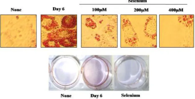
W e evaluated the effects of selenium on the inhibition of
adipocyte differentiation. Cultured 3T3-L1 adipocytes were exposed to selenate at different doses (at day 0), and cell differentiation was performed with a hormonal cocktail contained medium. At day 6, differentiations were ended and droplets of lipid were detected by oil staining. The treatment of adipocytes with selenate markedly suppressed adipocyte differentiation (Fig. 1). These results suggest that selenium might be an efficient blocker of adipocyte differ- entiation, and therefore, have a potential of anti-obesity agent.
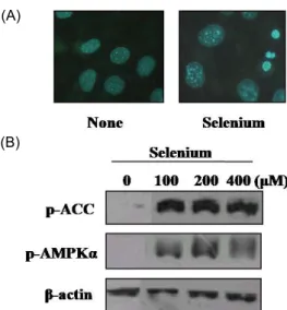
Selenium also induced mature adipocyte apoptosis The apoptotic ability of mature adipocytes were next evaluated, and 3T3-L1 adipocytes were differentiated with hormonal cocktail treatment, and mature cells were ex- posed to selenium. After treatment, apoptotic pattern was observed with hoechst 33342 dye, and the apoptotic bod- ies were found in selenium-exposed mature adipocytes (Fig. 2A).
Selenium was found to activate AMPK in the process of adipocyte differentiation blockage
We investigated the involvement of AMPKα in the proc- ess of selenium-induced inhibition of adipocyte differentia- tion. AMPK activation and its substrate acetyl-CoA carbox- ylase (ACC) phosphorylation were examined. The AMPK ac- tivation was observed directly by examining the increase of phosphorylated AMPK and indirectly by observing the phosphorylation level of acetyl-CoA carboxylase serine
79, the best-characterized phosphorylation site by the activated AMPK. The activation of AMPKα following selenium treat- ment (100-400 µM) was noticeable as shown in Fig. 2B.
Fig. 1. The effects of selenium on 3T3-L1 adipocyte differentia- tion. Postconfluent 3T3-L1 cells were differentiated with hormonal mixtures in the absence or presence of selenium (100-400 µM ) for 6 days. The morphological changes were photographed after Oil-Red O staining.
(A)
(B)
Fig. 2. Selenium activates AM PK and induces apoptosis in dif- ferentiated 3T3-L1 adipocytes. Postconfluent 3T3-L1 cells were differentiated with hormonal mixtures in the ab- sence or presence of selenium for 6 day. Then cells were incubated with 10 µM Hoechst 33342 and fixed with 3.7%
formaldehyde and fluorescence microscopic images were obtained (A). The levels of phospho-AM PKα and phos- pho-A cc were determined by W estern blot analysis.
Blockage of adipocyte differentiation by selenium was comparable to the adipocyte differentiation inhibition by either resveratrol or AICAR
The inhibition of adipocyte differentiation of AMPK by selenium was comparable to the treatment with grape phy- tochemical resveratrol or AICAR, a synthetic AMPK activa- tor in 3T3-L1 adipocytes (Fig. 3).
The involvement of AMPK activation in adipocyte differentiation process was evaluated with AICAR and Compound C
As shown in Fig. 4, the treatment of 3T3-L1 adipocytes with AICAR (1 mM and 2 mM) and Compound C, an AMPK inhibitor, revealed that AICAR activated AMPK, and the activation of AMPK by AICAR was inhibited by co-treatment with Compound C. AICAR, a synthetic form of AMPK activator, has shown to be an inhibitor of cell anabolism, especially in cancerous cells. This result in- dicates that AMPK is involved in the inhibition process of adipocyte differentiation.
Discussion
Correct assessment of the underlying mechanisms of an- ti-obesity effect of dietary agent is inevitable in considering
Fig. 3. The effects of resveratrol and AICAR on 3T3-L1 adipo- cyte differentiation. Postconfluent 3T3-L1 cells were dif- ferentiated with hormonal m ixtures in the absence or presence of resveratrol (50, 100 µM ) and AICAR (0.5, 1 mM) for 6 days. The morphological changes were photo- graphed after Oil-Red O staining.
Fig. 4. The role of AMPKα in differentiated adipocytes. Postcon- fluent 3T3-L1 cells were differentiated with hormonal m ixtures for 6day. A fter differentiation, cells were pre- treated with Compound C (10 µM) for 30 min and treated with AICAR (1, 2 mM ) for 6 hr. The levels of phos- pho-AM PKα and phospho-Acc were determ ined by W estern blot analysis.
the application to human intervention trials. In the present
study the molecular basis of selenium with emphasis on
their ability to control intracellular signaling cascades of
AMP activated kinase (AMPK) responsible for the inhibition
of adipogenesis was investigated. The evolutionarily con-
served serine/threonine kinase, AMPK, is known as a pri-
mary cellular homeostasis sensor and effecter modulating
energy balance and a possible target molecule of anti-obesity
[9]. Hypothalamic AMPK was found to integrate nutritional
and hormonal signals modulating feeding behavior and en-
ergy expenditure [10]. Moreover, adipocyte-derived hor-
mones such as leptin and adiponectin are shown to activate
AMPK [11,12]. The mechanism by which affects AMPK reg-
ulation with physiological stimuli or anti-obesity agents
might present a promising target for the development of strategies for the treatment of obesity. In present study, we investigated the effects of selenium on adipocyte differ- entiation in relation to AMPK activation. We have observed that adipogenesis was induced during adipocyte differ- entiation by hormonal cocktail, and selenium significantly inhibited the process of adipocyte differentiation and led to mature adipocyte apoptosis. The previous report from our laboratory has shown the possibility of ROS as an upstream signal of AMPK in the process of adipocyte control by the naturally occurring compounds such as genistein, EGCG (epigallocatechin-3-gallate) and capsaicin [13]. From the present study it was suggested that the activation of AMPK was necessary for the inhibition of adipogenesis in 3T3-L1 cells, and AMPK is a novel and critical component of both inhibition of adipocyte differentiation and apoptosis of ma- ture adipocyte by selenium implying AMPK as a prime tar- get of obesity control. Obesity is considered to be resulting from the imbalance between energy intake and energy ex- penditure that may be linked to a pathologic overgrowth of adipose cells [22]. The mass of adipose tissue is thought to be modulated by the blocking of adipogenesis from pre- cursors, and the control of the size of mature adipocytes.
Therefore, obesity is caused by the hypertrophy of adipo- cytes as well as the recruitment of new adipocytes from pre- cursor cells, and these two processes are critical for the adi- pocyte differentiation [22]. AMPK is known to play a major role in energy homeostasis by coordinating adaptive re- sponses in ATP-depleting metabolic states of exercise [8].
Furthermore, the persistent activation of AMPK showed to be connected to p53-dependent cellular senescence suggest- ing its role as an intrinsic regulator of the cell cycle in mam- malian cells [14]. Recently, AMPK cascades have emerged as novel targets for the treatment of obesity and type 2 diabetes. AMPK is known to be activated with 5-ami- no-imidazole-4-carboxamide riboside (AICAR), which is converted to a nucleotide that mimics the effect of AMP, and the long term treatment with AICAR prevented the de- velopment of diabetes in animal models [15]. Also, the proa- poptotic potential of the activated was observed in the AMPK over-expressed conditions of various cells [16,17].
Our results show that selenium activates AMPK, blocks adi- pocyte differentiation comparable to resveratrol or AICAR, and induces apoptosis of adipocytes. The anti-proliferatory and lipolytic effects of selenium have been attributed to their ability to modulate various signaling pathways, specially,
the control of cell proliferation and survival. However, the precise target of their anti-proliferatory effect remained unresolved. Here, we introduce AMPK as a possible main target of these compounds in their anti-obesity activity.
AMPK is activated by various stimuli including exercise, heat shock and ROS [15]. The activated for blocks the ana- bolic pathways and promotes catabolic pathway, and thus activation of AMPK is linked to inhibition of cell pro- liferation and apoptosis. AMPK is known to be involved in the oxidative stress-induced cellular apoptosis through the inhibition of specific protein trans-elongation factor 2 when it is phosphorylated [18,19]. The more recent studies suggest that AMPK plays a critical role in the inhibition of cellular protein synthesis as well as stress-induced apoptosis [20].
It has been reported that AMPK activation is necessary for the therapeutic effect of metformin and troglitazone [23]. The mechanism by which affects AMPK regulation with physio- logical stimuli or anti-obesity agents might present a promis- ing target for the development of strategies for the treatment of obesity. AMPK cascades have been postulated to respond to the intracellular level of AMP or AMP:ATP ratio, and to be highly sensitive to the oxidative stress [21]. The exact mechanism to stimulate preadipocyte mitosis and differ- entiation in vivo remains exclusive. However, it is proposed that hypertrophy of fat cells grown beyond a certain size might propagate to differentiate by sending specific signals.
Adipocyte inducers stimulate preadipocytes to undergo mi-
totic clonal expansion before transcriptional activation of
adipocyte genes before anchoring adipocyte phenotypes
[24]. The results indicated that adipogenesis was induced
during adipocyte differentiation by hormonal cocktail, and
selenium inhibited the adipocyte differentiation of lead to
apoptosis of mature adipocytes. Also we have tested wheth-
er AICAR has the similar effect on adipocyte differentiation
and AMPK activation in comparision with selenium, and we
have found that selenium and AICAR inhibited the differ-
entiation and early clonal expansion of pre-adipocytes. W e
have also found that AMPK inhibitor Compound C could
block the inhibitory effect of AMPK on adipocyte
differentiation. These results strongly suggested that the acti-
vation of AMPK was necessary for the inhibition of adipo-
genesis in 3T3-L1 cells, and AMPK is a novel and critical
component in adipogenesis, also AMPK is necessary for the
inhibition of both adipocyte differentiation and apoptosis of
mature adipocyte by selenium and resveratrol implying its
involvement in dietary agent-inhibited adipogenesis.
Acknowledgement
This work was supported by Hannam University Fund 2009.
References
1. Bays, H. E., J. M . Gonzalez-Campoy, G. A. Bray, A . E.
Kitabchi, D. A. Bergman, A. B. Schorr, H. W . Rodbard, and R. R. Henry. 2008. Pathogenic potential of adipose tissue and metabolic consequences of adipocyte hypertrophy and increased visceral adiposity. Expert. Rev. Cardiovasc. Ther. 6, 343-368.
2. Block, K. I., A. C. Koch, M . N. M ead, P. K. Tothy, R. A.
Newman, and C. Gyllenhaal. 2008. Impact of antioxidant supplementation on chemotherapeutic toxicity: a systematic review of the evidence from randomized controlled trials.
Int. J. Cancer. 123, 1227-1239.
3. Browne, G. J., S. G. Finn, and C. G. Proud. 2004. Stimulation of the AM P-activated protein kinase leads to activation of eukaryotic elongation factor 2 kinase and to its phosphor- ylation at a novel site, serine 398. J. Biol. Chem. 279, 12220-12231.
4. Cherukuri, D. P. and M . A. Nelson. 2008. Role of reactive oxygen species (ROS) and JNKs in selenite-induced apopto- sis in HepG2 cells. Cancer. Biol. Ther. 7, 689-696.
5. Dagon, Y., Y. Avraham, and E. M. Berry. 2005. AMPK acti- vation regulates apoptosis, adipogenesis, and lipolysis by eIF2alpha in adipocytes. Biochem. Biophys. Res. Commun. 340, 43-47.
6. Daval, M ., F. Foufelle, and P. Ferre. 2006. Functions of AM P-activated protein kinase in adipose tissue. J. Physiol.
574, 55-62.
7. Erbayraktar, Z., O. Yilmaz, A. T. Artmann, R. Cehreli, and C. Coker. 2007. Effects of selenium supplementation on anti- oxidant defense and glucose hom eostasis in experim ental diabetes m ellitus. Biol. Trace. Elem. Res. 118, 217-226.
8. Gao, S., K. P. Kinzig, S. Aja, K. A. Scott, W. Keung, S. Kelly, K. Chohnan, S. Strynadka, W . W . Smith, K. L. Tamashiro, E. E. Ladenheim, G. V. Tu, Y. Ronnett, M . J. Birnbaum, G.
D. Lopaschuk, and T. H. M oran. 2007. Leptin activates hy- pothalamic acetyl-CoA carboxylase to inhibit food intake.
Proc. Natl. Acad. Aci. USA 104, 17358-17363.
9. Gray, S. L. and A . J. Vidal-Puig 2007. A dipose tissue ex- pandability in the m aintenance of metabolic homeostasis.
Nutr. Rev. 65, 7-12.
10. Hardie, D. G., D. Carling, and M . Carlson 1998. The AM P- activated/SNF1 protein kinase subfamily: metabolic sensors of the eukaryotic cell? Annu. Rev. Biochem. 67, 821-855.
11. Horman, S., G. Browne, U. Krause, J. Patel, D. Vertommen, L. Bertrand, A. Lavoinne, L. Hue, C. Proud, and M . Rider.
2002. Activation of AM P-activated protein kinase leads to the phosphorylation of elongation factor 2 and an inhibition of protein synthesis. Curr. Biol. 12, 1419-1423.
12. Huypens, P., E. Quartier, D. Pipeleers, and M . Van de
Casteele. 2005. M etformin reduces adiponectin protein ex- pression and release in 3T3-L1 adipocytes involving activa- tion of AM P activated protein kinase. Eur. J. Pharmacol. 518, 90-95.
13. Hwang, J. T., I. J. Park, J. I. Shin, Y. K. Lee, S. K. Lee, H.
W . Baik, J. Ha, and O. J. Park. 2005. Genistein, EGCG, and capsaicin inhibit adipocyte differentiation process via acti- vating AM P-activated protein kinase. Biochem. Biophys. Res.
Commun. 338, 694-696.
14. Joens, R. G., D. R. Plas, S. Kubek, M . Buzzai, J. M u, Y. Xu, M . J. Birnbaum, and C. B. Thompson. 2005. AM P-activated protein kinase induces a p-53 dependent m etabolic checkpoint. Mol. Cell 18, 283-293.
15. Kemp, B. E., D. Stapleton, D. J. Campbell, Z. P. Chen, S.
M urthy, M . W alter, A. Gupta, J. J. Adams, F. Katsis, B. van Denderen, I. G. Jennings, T. Iseli, B. J. M ichell, and L. A . W itters. 2003. AMP-activated protein kinase, super metabol- ic regulator. Biochem. Soc. Trans. 31, 162-168.
16. Kola, B. 2008. Role of AM P-activated protein kinase in the control of appetite. J. Neuroendocrinol. 20, 942-951.
17. Kola, B., A. B. Grossman, and M . Korbonits. 2008. The role of AM P-activated protein kinase in obesity. Front. Horm.
Res. 36, 198-211.
18. Kubota, N., W . Yano, T. Kubota, T. Yamauchi, S. Itoh, H.
Kumagai, H. Kozono, I. Takamoto, S. Okamoto, T. Shiuchi, R. Suzuki, H. Satoh, A. Tsuchida, M . M oroi, K. Sugi, T.
Noda, H. Ebinuma, Y. Ueta, T. Kondo, E. Araki, O. Ezaki, R. Nagai, K. Tobe, Y. Terauchi, K. Ueki, Y. M inokoshi, and T. Kadowaki. 2007. A diponectin stimulates AM P-activated protein kinase in the hypothalamus and increases food intake. Cell Metab. 6, 55-68.
19. M acDougald, O. A . and S. M andrup. 2002. Adipogenesis:
forces that tip the scales. Trends. Endocrinol. Metab. 13, 5-11.
20. M eisse, D., M . Van de Casteele, C. Beauloye, I. Hainault, B. A. Kefas, M . H. Rider, F. Foufelle, and L. Hue. 2002.
Sustained activation of AM P-activated protein kinase in- duces c-Jun N-terminal kinase activation and apoptosis in liver cells. FEBS. Lett. 526, 38-42.
21. M ukherjee, P., T. J. M ulrooney, J. M arsh, D. Blair, T. C.
Chiles, and T. N. Seyfried. 2008. Differential effects of en- ergy stress on AM PK phosphorylation and apoptosis in ex- perim ental brain tumor and normal brain. Mol. Cancer 7, 37-51.
22. Shimomura, I., R. E. Hammer, J. A. Richardson, S. Ikemoto, Y. Bashmakor, J. L. Goldstein, and M . S. Brown. 1998.
Insulin resistance and diabetes m ellitus in transgenic m ice expressing nuclear SREBP-1c in adipose tissue: m odel for congenital generalized lipodystrophy. Genes Dev. 12, 3182-3194.
23. Song, X. M ., M . Fiedler, D. Galuska, J. W . Ryder, M . Fernstrom, A. V. Chibalin, H. W allberg-Henriksson, and J.
R. Zierath. 2002. 5-Amino-imidazole-4-carboxamide ribonu- cleoside treatment im proves glucose homeostasis in in- sulin-resistant diabetic (ob/ob) mice. Diabetologia. 45, 56-65.
24. Sorisky, A. 1999. From pre-adipocyte to adipocyte: differ- entiation-directed signals of insulin from the cell surface to the nucleus. Crit. Rev. Clin. Lab. Sci. 36, 1-34.