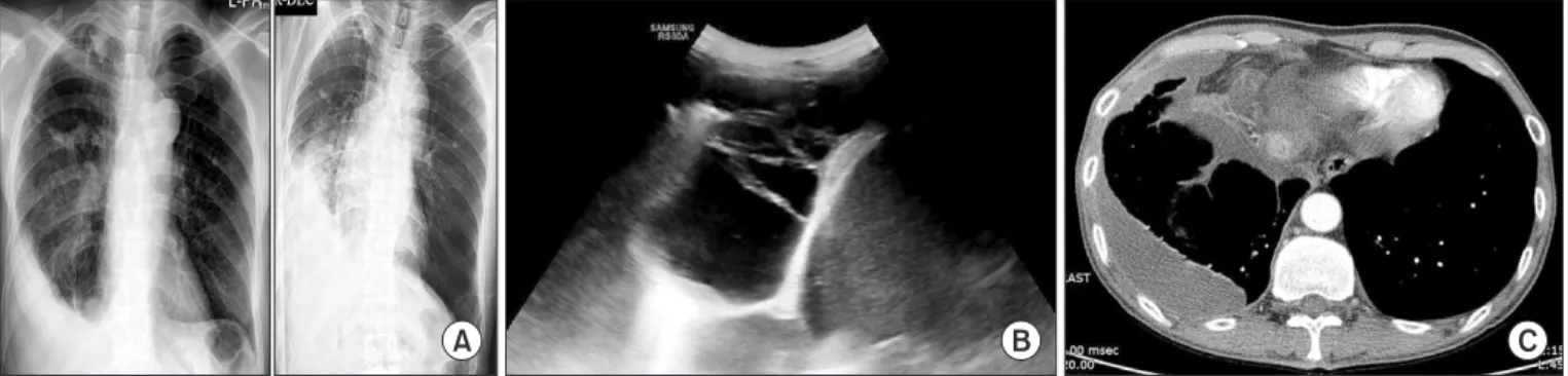
Loculated Tuberculous Pleural Effusion:
Easily Identifiable and Clinically Useful
Predictor of Positive Mycobacterial Culture from Pleural Fluid
Yousang Ko, M.D.
1,2,*, Changhwan Kim, M.D., Ph.D.
3,*, Boksoon Chang, M.D., Ph.D.
4, Suh-Young Lee, M.D.
1, 2, So Young Park, M.D.
1,2, Eun-Kyung Mo, M.D., Ph.D.
1,2, Su Jin Hong, M.D., Ph.D.
5, Myung Goo Lee, M.D., Ph.D.
2,6, In Gyu Hyun, M.D., Ph.D.
2,7and Yong Bum Park, M.D.
1,21
Division of Pulmonary, Allergy and Critical Care Medicine, Department of Internal Medicine, Hallym University Kangdong Sacred Heart Hospital, Hallym University College of Medicine, Seoul,
2Lung Research Institute, Hallym University College of Medicine, Chuncheon,
3Department of Internal Medicine, Jeju National University Hospital, Jeju,
4Department of Pulmonary and Critical Care Medicine, Kyung Hee University Hospital at Gangdong, Kyung Hee University School of Medicine, Seoul,
5
Department of Radiology, Hallym University Kangdong Sacred Heart Hospital, Hallym University College of Medicine, Seoul,
6
Division of Pulmonary, Allergy and Critical Care Medicine, Hallym University Chuncheon Sacred Heart Hospital, Chuncheon,
7
Division of Pulmonary, Allergy and Critical Care Medicine, Hallym University Dongtan Sacred Heart Hospital, Hwaseong, Korea
Background: Isolation of M. tuberculosis (MTB) is required in cases of Tuberculous pleural effusion (TBPE) for confirming diagnosis and successful therapy based on drug sensitivity test. Several studies have focused on predictors of MTB culture positivity in TBPE. However, the clinical role of loculated TBPE as a predictor of MTB cultivation from TBPE remains unclear. The aim of this study was to examine possible predictors including loculation of TBPE of MTB culture positivity in TBPE.
Methods: We retrospectively examined associations between clinical, radiological, microbiological, and laboratory characteristics and positive MTB culture from TBPE to determine a potent predictor of culture positivity.
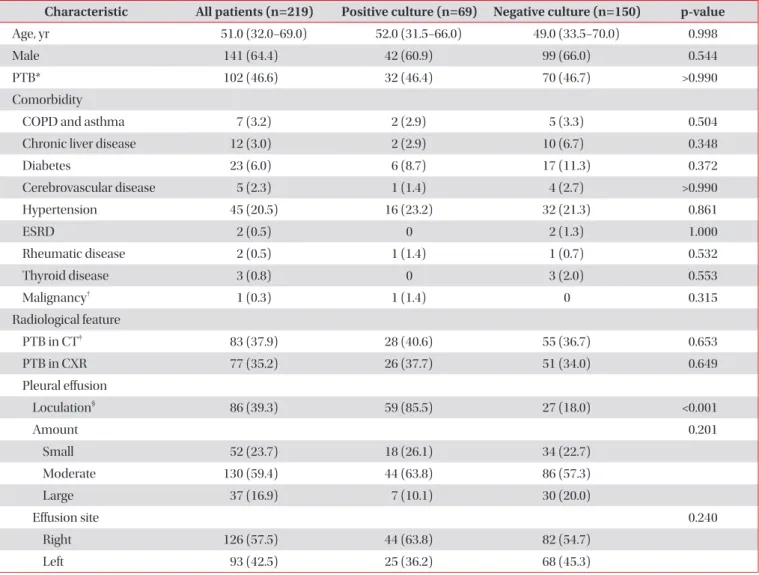
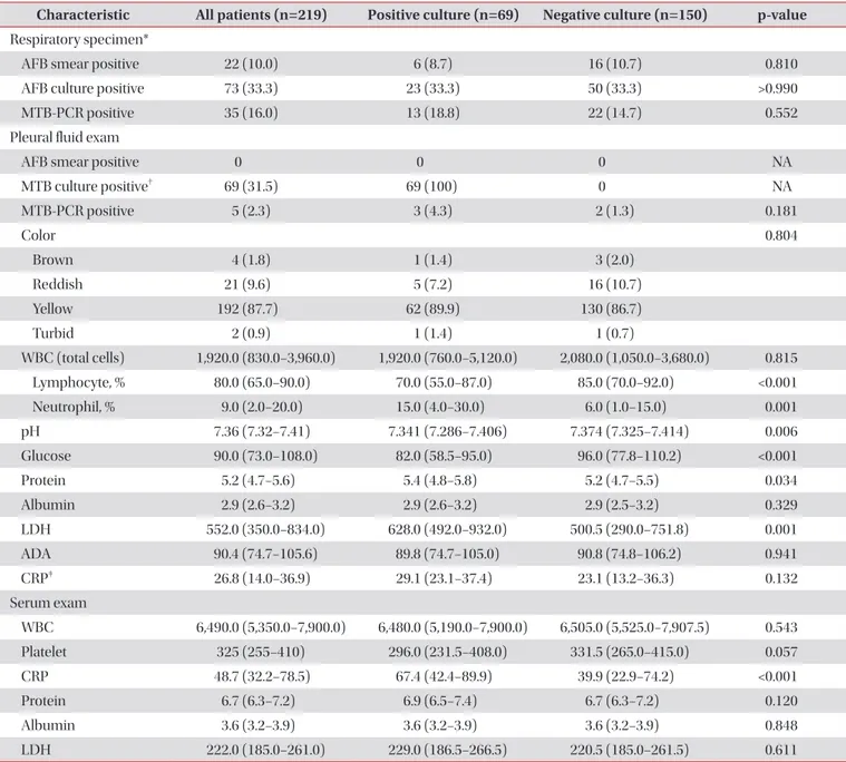
Results: From January 2011 to August 2015, 232 patients with TBPE were identified. Of these, 219 were finally analyzed.
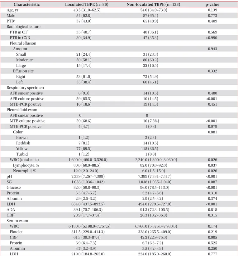
Among them, 69 (31.5%) were culture positive for MTB in TBPE and 86 (39.3%) had loculated TBPE. In multivariate logistic regression analysis, the loculation of TBPE was independently associated with culture positivity for MTB in TBPE (adjusted odds ratio [OR], 40.062; 95% confidence interval [CI], 9.355–171.556; p<0.001). In contrast, the lymphocyte percentage of TBPE (adjusted OR, 0.934; 95% CI, 0.899–0.971; p=0.001) was inversely associated with culture positivity for MTB in TBPE.
Conclusion: In clinical practice, identification of loculation in TBPE is easy, reliable to measure, not uncommon and may be helpful to predict the possibility of positive mycobacterial culture.
Keywords: Tuberculosis; Pleural Effusion; Pleurisy
Address for correspondence: Yong Bum Park, M.D.
Division of Pulmonary, Allergy and Critical Care Medicine, Department of Internal Medicine, Hallym University Kangdong Sacred Heart Hospital, 150 Seongan-ro, Gangdong-gu, Seoul 05355, Korea
Phone: 82-2-2224-2561, Fax: 82-2-488-6925, E-mail: bfspark2@gmail.com
*Yousang Ko and Changhwan Kim contributed equally to this work.
Received: Jul. 29, 2016, Revised: Aug. 30, 2016, Accepted: Sep. 1, 2016
cc It is identical to the Creative Commons Attribution Non-Commercial License (http://creativecommons.org/licenses/by-nc/4.0/).