Ⅰ. 서 론
1965년 Urist가 탈회된 골 기질에서 골유도 능력이 있는 단백
질을 추출하여 이를 골형성 단백질(Bone morphogenetic protein,BMP)이라 명명한 이래
1), 현재까지 최소한 18종류가 밝혀졌으
며, 재조합 유전자 기술의 발달로 골유도능을 갖고 있는 다양
한 recombinant human BMP를 생산할 수 있게 되었다. BMP-2 부 터 BMP-8까지는 TGF-β(Transforming growth factor-β)의 상과
(Superfamily)에 속하는 이량체 분자이며
2), BMP 중에서 BMP-2
와 3이 가장 활성이 뛰어난 것으로 여겨지고 있다3). 특히 구강
악안면 영역에서 BMP-2의 재생능력을 나타내는 상악동저의 거상, 치조골 유지, 국소적인 골 재생에 의한 치주 수복등에 관 한 임상 실험이 보고되었다4-6).
대다수의 연구에 의하면, 줄기 세포의 주화성, 증식과 분화와 같은 골 유도 조절이 BMP에 의 해 조절된다고 여겨지고 있다7). 그리고 BMP가 줄기세포가 골
형성 계통으로 분화하도록 자극함으로써 골치유를 촉진함에 중요한 역할을 한다고 보고된 바 있다8). 그러나 임상 실험에서
척추의 유합 유도에 많은 양의 BMP가 필요하다고 보고되었으 며, 일회성의 BMP 투여가 골결손이 크거나 숙주의 생물학적BMP-2에 의한 협부 지방 성체 줄기세포의 골형성
김창현∙박철헌*∙이일규**∙표성운 가톨릭대학교 의과대학 치과학교실 구강악안면외과
*가톨릭대학교 임상치과학 대학원 임프란트학과, **가톨릭대학교 대학원 통합의학과
Abstract (J. Kor. Oral Maxillofac. Surg. 2008;34:412-418)
표 성 운
420-717
경기도 부천시 원미구 소사동2
가톨릭대학교 성가병원 구강악안면외과 Sung-Woon PyoDept. of OMFS, Holy Family Hospital, The Catholic University of Korea 2 Sosa-dong, Wonmi-ku, Puchon, Kyunggi-do, 420-717, Korea Tel: +82-32-340-2130 Fax: +82-32-340-2255
E-mail: [email protected]
OSTEOGENESIS BY BMP-2 IN ADULT STEM CELL DERIVED FROM BUCCAL FAT PAD
Chang-Hyen Kim, Cheol-Hun Park
*, Il-Kyu Lee
**, Sung-Woon Pyo
Department of Oral and Maxillofacial Surgery, College of Medicine, The Catholic University of Korea
*Department of Implantolgoy, Graduate School of Clinical Dental Science, The Catholic University of Korea
**Department of Integrative Medicine, Graduate school, The Catholic University of Korea
Bone morphogenetic proteins (BMPs) in combination with stem cells gain more significance for their use in bone tissue engineering. The mes- enchymal stem cell can be differentiated into osteoblast by the treatment of BMP. The aim of this study is to characterize the osteogenic differentiation process of adult stem cells derived from buccal fat pad according to BMP-2 within culture media and decide the appropriate concentration of BMP-2 to facilitate osteogenesis.
The authors procured the stem cell from buccal fat pad and analyzed for presence of stem cell by flow cytomety against CD-34, CD-105 and STRO- 1. The buccal fat derived stem cells (BFDC) were treated by application of the different concentration with BMP-2 of 0, 10, 50, 100 and 200ng/ml, respectively. And their ability to differentiate into osteogenic pathway were checked by alkaline phosphatase(ALP) staining, Alizarin red staining and RT-PCR for osteocalcin(OC) gene expression at 7, 14 and 21day of culture.
Flow cytometric analysis and biochemical assays demonstrated that BFDC might be a distinguished stem cells, and mineralization was accompa- nied in proportion to BMP-2 concentration. However, with 100ng/ml concentration of BMP-2, the BFDC demonstrated most efficent staining pattern of ALP and Alizarin red. The feasibilty of the osteogenic differentiation in the group of both 50ng/ml and 200ng/ml of BMP-2 showed similar activity and relatively weaker than that of 100ng/ml.
These results suggest that the BMP-2 stimulate osteogenesis by BFDC effectively and that bone induction might be controlled through negative reg- ulatory feedback in higher concentration.
Key words: Stem cell, BMP-2, Osteogenesis, Differentiation
※본 연구는 보건복지부 보건의료기술 진흥사업 기능성 세포 치료제 개발(0405-DB01-0104-0006) 지원에 의하여 이루어졌음.
잠재성이 낮은 경우에 충분한 골유도 자극이 되지 못할 수도
있다9)
. 즉 골유도 과정에서 필요한 BMP의 양과 적용 시간에 대
하여는 충분히 논의된 바 없다.
줄기 세포를 이용한 생체 조직 공학을 위해서는 공여부의 결 손을 최소화하면서 충분한 양을 얻을 수 있어야 하며 쉽게 채 취할 수 있는 줄기 세포의 공급원이 요구된다. 지방 조직은 골 수와 같이 간엽 조직(mesenchymal tissue)으로부터 기원하며, 쉽 게 분리되는 지지 간질조직(supporting stroma)을 함유하고 있다.
Zuk 등은 지방 흡입술에서 얻어진 지방조직 내에서 줄기 세포
의 가능성이 있는 세포군 (Processed lipo-aspirated cell)을 분리 배 양하여 확인하였으며10), 이들은 다시 흡입 지방세포를 계통 특
이적 배지 (lineage specific media)를 이용하여 조골모 세포로 분 화시켰다11). 그리고 Halvorsen 등은 사람의 지방 조직에서 얻어
진 세포를 배양하여 지방 조직으로 분화할 수 있는 표지 (mark-er)와 골조직으로 분화할 때 나타나는 표지자와 경조직의 형성
을 확인한 바 있다12). 본 저자들은 전신 지방과는 다른 별개의
특성을 갖는 협부지방에서 줄기세포를 추출하여 이를 조골모 세포(osteoblast)로 분화시킨 바 있으며 골생성을 확인하였다13).
이번 연구에서는 협부지방 조직에서 추출된 세포가 성체 줄 기세포임을 확인한 후, BMP-2를 처리하여 조골모세포로의 분 화를 유도하고 골신생능을 평가하였다. 또한, BMP-2의 농도를 다르게 줄기세포 배양액에 투여하여 조골모세포로의 분화를 유도하는 가장 적절한 농도를 찾기 위하여 시행하였다.
Ⅱ. 방 법
본 연구에서 수행된 절차는, 가톨릭대학교 의과학연구원의
IRB 승인을 얻었고, 세포 제공자에게 연구 목적이 고지된 동의
서를 취득한 후 연구를 시행하였다.1. 세포의 분리 및 배양
수술실에서 분리된 협부 지방조직을 1% antibiotic & antimy-
cotic (Gibco BRL, Grand Island, USA)이 포함된 PBS (Gibco BRL)
에 담아 무균 상태를 유지하며 실험실로 운반하였다. 협부 지 방조직을 항진균, 항세균제가 포함된 PBS로 세척하고 잘게 자 르고 0.06% collagenase (Type I; Invitrogen Corporation, Carlsbad,USA)로 처리하여 37℃, 5% CO
2가 함유된 배양기 (FormaScientific Inc, Marietta, USA)에서 12시간동안 진탕한 후, 1000 RPM으로 10분간 원심분리하고 침전물을 10% FBS (Hyclone, Logan, USA)과 1% P/S (Penicillin/Streptomycin; Gibco BRL)가 함
유된 α-MEM (Minimum Essential Medium α ; Hyclone)에 부유시키
고, 70㎛ cell strainer (BD Bioscience, Bedford, USA)로 걸러 협부 지방세포가 아닌 조직을 걸러내고 필터를 빠져 나온 용액을 배양기에서 배양하였다. 이후 매일 형태학적 특성을 도립현미 경 하에서 관찰하였으며, 2일에 1회씩 배양액을 교환해 주었 다. 세포가 90% 포화상태에 이르면 Trypsin-EDTA (Gibco BRL)로 세포를 분리시킨 후, 3차례 계대배양하여 협부 지방세포군
(BFDC; Buccal fat-derived cell)을 얻었다.
2. Investigation of stem cell by flow cytometry
BFDC가 성체 줄기세포로서의 역할 가능성이 있는 세포인지
를 확인하기 위하여, 골수기원 다능성 줄기세포(multipotentialbone marrow stromal cell)를 선택하기 위해 사용되는 세포 표면
항원으로 알려져 있는 CD34, CD105와 STRO-1의 발현 여부를 유세포 분석기를 이용하여 수행하였다. 3회 계대 배양 된 협부 지방 세포군을 유세포 분석 72시간 전부터 일반 배양액에서 배 양하였다. 0.25% trypsin/EDTA (Gibco BRL)로 분리하여 1×10⁵ 개씩 tube (BD company, Franklin Larks, USA)에 분주하고 2%formaldehyde (Sigma, St. Louis, USA)로 30분간 고정하였다. 세척
완충액(0.2% BSA, 0.1% NaN3, 0.5 mM EDTA)으로 세척하고, FITC-conjugated monoclonal 1차 항체인 CD34 (BD Bioscience), CD105 (Serotec Ltd, Oxford, UK), STRO-1 (R&D system, Minneapolis, USA)와 배경 형광을 배제하기 위하여 PE-conjugat- ed nonspecific IgG 로 1시간동안 부란하고 세척완충액으로 세척
한 후, FACscan argon laser cytometer (Beckton Dickinson, SanDiego, USA)를 이용하여 관찰하였다. 대조군으로는 1차 항체
를 생략한 채 유세포 분석기를 통과시켰다.3. 골분화 배지와 골형성 단백질-2을 처리한 세포에서의 골분화 유도
3회 계대배양 된 협부지방세포를 12 well plate에 well당 2×10
5 개씩 분주하고 24시간 후 BMP-2 (에디포젠, 서울, 한국)를 0, 10,50, 100, 200ng/ml 의 농도로 처리하였다. BMP-2는 100㎍/㎖의
농도로 1 % BSA를 포함한 PBS에 수화시켜 분주한 후 사용할 때 까지 -80 ℃에 보관하여 사용하였다. 대조군으로 BFDC를 골분화 유도 배양액으로 교환하여 배양하였다. 골분화 유도 배양액의 조성은 DMEM (Dulbecco's Modified Eagle Medium),10% FBS, 0.1uM Dexamethasone (Sigma), 10mM glycerol phosphate (Sigma), 50uM L-ascorbic acid 2-phosphate (Sigma)이었다. 이 배양
액을 3일에 1회씩 교환해주며 90%의 세포 포화도를 보일 때까 지 배양하면서 도립현미경(Olympus, Tokyo, Japan) 아래에서 형 태학적 특성을 관찰하였다.4. 조직화학적 염색
A. Alkaline phosphatase assay
골분화 표지자인 세포내 alkaline phosphatase 활성을 분석하기 위하여 BMP-2 (에디포젠)를 0, 10, 50, 100, 200ng/ml 의 농도로 처리하고, 멸균 3차 증류수로 세척한 후, ALP staining kit (Sigma-
Aldrich, Steinheim, Germany)을 사용하여 염색하였다. Citrate-
Acetone-Formaldehyde 고정액에 30초간 담가 고정하고 탈이온
수로 세척하였다. 슬라이드를 alkaline dye mixture에서 실온에 서 15분간 부란하고 다시 세척한 후 Neutral Red solution으로 대 조 염색하였다.
B. Alizarin red 염색
BMP-2(에디포젠)를 0, 10, 50, 100, 200ng/ml의 농도로 처리하
고, 골이 형성되었는지 확인하기 위하여 세포외 기질의 석회 화를 나타내는 Alizarin red 염색을 시행하였다. 실온에서 10%formalin으로 30분간 고정하고, 멸균 3차 증류수로 세척한 후 2% alizarin red(Sigma-Aldrich)
로 실온에서 30분간 염색하고 다 시 멸균 3차 증류수로 세척하고 도립현미경으로 관찰하였다.5. Osteocalcin expression analysis by RT-PCR
BMP-2(에디포젠)를 0, 10, 50, 100, 200ng/ml 의 농도로 처리한
세포를 21일간 배양하고 PBS로 세척한 후 cell scraper(Sarsted,Inc, Newton, USA)를 이용하여 수확하고, QIAGEN RNA extrac- tion kit(QIAGEN, Hilden, Germany)을 이용하여 RNA를 분리한
후 RT-PCR premix(Bioneer, 대전, 한국)를 이용하여 RT-PCR을 수행하였다. 분리한 총 RNA 5㎍을 42℃에서 60분간 M-MuLV 역전사 효소를 이용하여 cDNA로 역전사한 후, primer쌍을 이 용하여 증폭하였다. 사용한 primer의 서열은 forward 5'-ctg cat
tct gcc tct ctg ac-3' 와 reverse 5'-cta ttc acc acc tta ctg ccc-3' 이며, 내
부 내조군은 GAPDH를 사용하였다. PCR의 반응 조건은 94℃에서 5분간 변성한 후, 94℃ 1분, 55℃ 1분 30초, 72℃ 1분간 30 주기 실시하고 마지막 주기에서 72℃ 5분간 연장하였다. 반응 산물은 2% agarose gel에서 전기영동 후 Ethidium bromide로 염 색하여 UV lamp에서 확인하였다.
Ⅲ. 결 과
1. Flow Cytometry를 이용한 줄기세포 확인
3계대 배양한 협부지방 세포군의 표면에 FITC conjugated anti-CD34, CD105, STRO-1 항체를 이용하여 염색한 후, 각각의
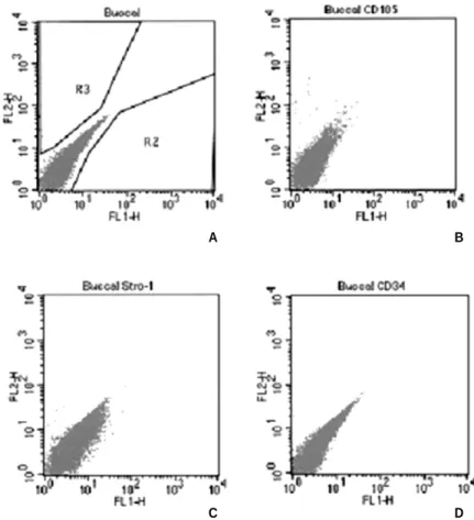
발현 유무를 확인한 결과, BMP-2 처리를 하지 않거나, 골분화Fig. 1. FACs result of marker expression in buccal fat pad derived cells(BFDC). Immune phenotype of BFDC were labeled with antibodies against the indicated antigens. (A) negative control, (B) CD105, (C) stro-1 and (D) CD34.
A B
C D
배지에서 배양하지 않은 대조군과 비교하였을 때, CD 34는 발 현 양상을 보이지 않았으나 CD105와 STRO-1의 경우 각각
20%, 19%로 발현 양상을 보였다(Fig. 1). 이는 협부 지방세포내
에 성체줄기 세포가 포함되어 있음을 시사하였다.2. Alkaline Phosphatase 염색
협부지방 세포군에 BMP-2를 0, 10, 50, 100, 200ng/ml의 농도로 각각 처리하고 7, 14, 21일 경과 후 Alkaline phosphatase를 염색한 결과, 50ng/ml 농도 이상에서 14일부터 염색되는 것을 확인하
Fig. 2. Alkaline phosphatase staining result of buccal fat pad derived cells after 7, 14, 21days of culture according to BMP-2 concentration. A, F and K: Treatment with 0ng/ml of BMP-2 after 7, 14, 21days, respectively ; B, G and L: Treatment with 10ng/ml of BMP-2 after 7, 14, 2days ; C, H and M: Treatment with 50ng/ml of BMP-2 after 7, 14, 21 days; D, I, N: Treatment with 100ng/ml of BMP- 2 after 7, 14, 21days, E, J and O: Treatment with 200ng/ml of BMP-2 after 7, 14, 21days.
Fig. 3. Alizarin Red staining result of buccal fat pad derived cells after 3, 7, 21days of culture according to BMP-2 concentration. A, F and K: Treatment of 0ng/ml of BMP-2 after 7, 14, 21days, respectively ; B, G and L: Treatment with 10ng/ml of BMP-2 after 7, 14, 21days ; C, H and M:
Treatment with 50ng/ml of BMP-2 after 7, 14, 21days; D, I, N: Treatment with 100ng/ml of BMP-2
였으며 21일후 염색된 영역이 확대되어 있었고 염색 농도가 증 진되어 있음을 확인하였다. 또한 처리한 BMP-2의 농도에 따라 염색 강도가 높아졌으며, 100ng/ml 농도에서 가장 진한 발현을 보였다(Fig. 2).
3. Alizarin red S 염색
BMP-2농도가 50ng/ml 이상에서 14일째부터 발현 양상을 보
이기 시작하였으며 21일째에서 확연한 발현 양상을 보였고100ng/ml 농도에서 가장 높은 발현 양상을 보였다(Fig. 3).
4. Osteocalcin 발현 유무 확인
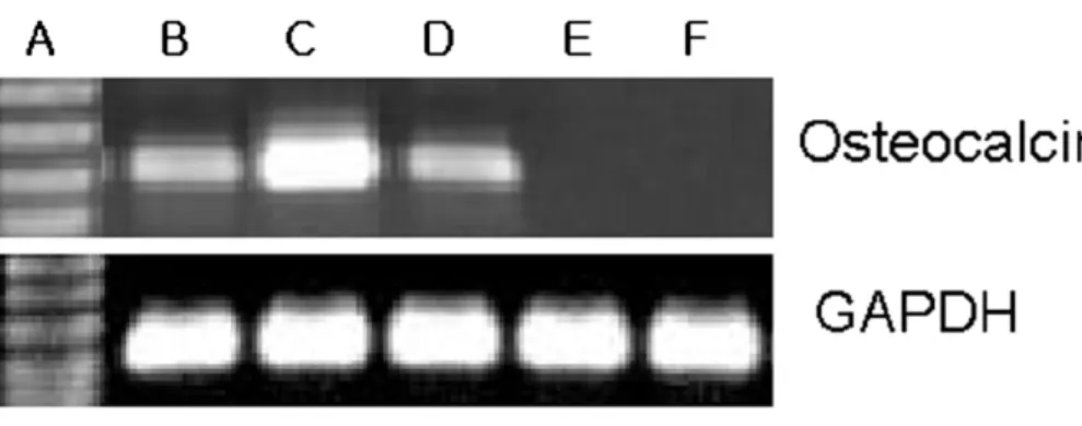
BMP-2 처리후 21일째에서 대부분의 실험군에서 osteoclacin
의 발현 양상을 보였으며, Osteocalcin의 농도는 배지내에 투여 한 BMP-2의 농도에 의존적으로 증가하였으나, 100ng/ml의 농 도 이후에는 감소하는 것을 관찰하였다(Fig. 4).Ⅳ. 고 찰
골형성 단백질(bone morphogenetic protein, BMP)는 다능성 중 배엽세포(multipotential mesenchymal cell)이 골-연골성(osteochon-
drogenic)과 전구 골모세포(osteoblast precusor cell)로 분화를 유도
한다14). 여러 종류의 BMP가 규명되었고, 이들은 서열의 유사 성(sequence homology)에 따라 하위 그룹(subgroup)으로 구분된 다. BMP-2 와 BMP-4는 BMP-2/-4 하위 그룹에 속하며, BMP-5,-6, -7, and -8는 osteogenic protein-1 그룹에 속하게 되며, 이 두 군
이 주로 생체내에서 골과 연골의 형성을 유도한다. BMP는 세 포막의 제 1형 또는 2형 serine/threonine kinase 수용체와 결합하 여, cytoplasmatic proteins mother against decapentaplegic(smad)를 인 산화 한다15).
BMP는 성체줄기세포의 침윤, 연골 형성, 혈관 형성, 골 형성
과 신생골의 개조를 야기할 수 있다16)
. BMP-2는 골표지자(bone marker) 유전자의 발현을 상향 조절하고 alkaline phospha- tase(ALP)의 활성을 증가시키므로써, 조골모세포 표현형(phe- notype)으로의 향상된 계통적 발전(lineage progression)을 야기
한다17).
이 실험에서는 협부지방조직에서 얻어진 세포를 조골모세 포로 분화시키기 위하여 BMP-2를 처리하였다. 기존의 연구에 따르면, BMP의 농도를 달리하여 설치류의 골막하에 이식하였 을 때, BMP의 농도에 비례하여 골이 형성되었고, 높은 농도의
BMP에서 골의 형성이 더 많았고, 더 빠른 골유도가 발현되었
다18).
다른 연구에서 주장된 바에 의하면, 골형성 단백질을 이 용한 골유도의 과정에서 BMP-3, Smad-6 그리고 Smad-7과 같은 골유도를 방해하는 단백질들이 발견되며, 특히 골형성 전구세 포들이 골형성 단백질-2에 노출되면서 이러한 단백질들의 발 현 증가되었다고 보고되었다19,20). 이것은 국소적인 골 유도에
서 BMP 자체가 BMP의 작용을 막는 negative regulatory feedback 에 의해 자가 조절된다는 것을 의미한다. 하지만, 정상 세포 수 준보다 수백만 배 높은 농도의 BMP를 처리하면 제어할 수 없 는 과도한 골유도가 나타난다21).
이번 실험에서 BMP-2에 의한 골모세포로의 분화와 골기질 의 생산이 초기 보다는 후기로 갈수록 확실해 지는 것으로 관 찰되었다. 그러나 Zachos 등22)에 따르면 BMP-2의 초기 발현이 줄기세포의 분화 과정에서 골전구 세포(osteo progenitor cell)로 의 유도에 필요 충분조건이 된다고 하였으며, 후기에서 나타 나는 BMP는 골수기원 간엽줄기세포의 autocrine 또는 paracrine 효과로 여겨진다고 주장한 바 있어, 본 연구의 결과와 일치하 지 않아 이에 따른 후속 연구가 필요할 것으로 생각된다.
골의 생성 과정은 기질이 분비되는 증식, 기질의 성숙, 기질 의 석회화의 단계로 이루어진다. 일반적으로 줄기세포가 골모 세포로 기능적인 발달 촉진은 ALP의 발현으로 나타나며23)
, BMP에 의한 전형적인 골분화 증거는 광물체(mineralized body)
의 형성으로 나타난다. ALP는 세포막에 붙어있는 효소로서 골Fig. 4. Osteocalcine expression of buccal fat pad derived cells after treatment with BMP-2 for 21days.
A: 1Kb Size marker; B: treatment with 200ng/ml BMP-2; C: treatment with 100ng/ml BMP-2, D:
treatment with 50ng/ml BMP-2, E: treatment with 10ng/ml of BMP-2; F: No treatment with BMP-2.
형성의 초기에 풍부하게 나타난다. 저자들은 초기 골형성의 증거로 ALP staining을 사용했고, 50ng/ml의 BMP 처리 배지에서 배양 14일차에서 염색이 나타나기 시작했으며, 21일차에는 염 색된 영역이 확대되었으며 시간이 지날수록 짙은 염색이 나타 났다. 투여한 BMP의 농도에 따라 ALP의 발현이 농도 의존적 인 경향이 나타났으나, 200ng/ml에서의 발현 정도가 오히려
100ng/ml보다 낮음이 관찰되었다. 또한 이 실험에서 Alizarin red
로 칼슘을 염색하여 세포외 기질의 석회화를 파악했으며, 이 때에도 14일차부터 발현이 시작되어 21일에 현저하게 발현되 었다. 농도에 따른 비교에서 ALP의 경우와 유사하게 농도와 비례하여 광물체의 형성이 시작되었으나, 100ng/ml의 농도에 서 가장 염색 강도가 높았다.Osteocalcin(OC)
은 골모 세포에서 분비되는 표지자이며, 최종 골모 세포분화의 신호이다24). RNA
를 분리하여 RT-PCR을 시행 하여 OC유전자의 발현을 확인한 결과, OC를 encoding하는 많 은 양의 유전자가 발현됨을 확인하여 골형성이 유도되고 있음 을 확인할 수 있었다. 100ng/ml농도의 BMP-2를 협부지방조직 에서 얻어진 세포에 전사했을 때 상대적으로 많은 발현이 관 찰되었다. 이번 연구에서는 BMP-2의 농도에 비례하여 BFDC 의 골화정도가 증가되었으나, 200ng/ml에서는 오히려 골화의 정도가 감소되었다. 이는 전술한 바와 같이 BMP-2의 농도가100ng/ml보다 높으면 BMP 자체가 BMP의 작용을 막는 세포외
억제물을 유도하여 negative regulatory feedback되는 것으로 사료 된다21).
이번 연구에서는 dexamethasone과 ascorbic acid가 포함되어 줄 기 세포가 골모세포로 분화를 유도하는 골분화 유도배지
(osteogenic media)에 BMP를 첨가한 상태에서 세포를 배양하지
않았다. 향후, 협부지방에서 얻어진 줄기 세포를 조골모세포 로 분화를 유도하는 과정에서 이번 연구에서 유효한 것으로 밝혀진 100ng/ml 농도의 BMP-2와 골분화 유도배지(osteogenicmedia)를 함께 처리하여 기존의 연구와 비교하여 볼 필요가 있
을 것으로 사료된다.임상적으로 골결손 부위를 수복하기 위하여 BMP와 줄기세 포를 이용한 조직공학적 기법을 사용하려면, BMP의 계속적인 발현 여부와 이를 위한 전달 방법에 대한 포괄적 연구가 필요 할 것으로 사료된다. BMP-2 유전자를 함유하는 아데노바이러 스(adenovirus)에 의한 줄기 세포의 transduction은 BMP의 계속적 인 발현에 유리할 것으로 사료되어 연구의 대상이 되고 있다25)
.
그러나 바이러스 transfection에 의한 BMP의 발현은 안정성 문 제가 지적되고 있어 아직 제한적으로 실험실내에서만 사용되 고 있는 실정이다. Knippenberg의 연구에 따르면 rhBMP를10ng/ml의 농도로 15분간 지방줄기세포에 적용하였을 때, 골생
성이 나타남을 밝힌 바 있다26). 이번 연구에서는 BMP의 적정
농도의 결정을 위하여 BMP의 단일 투여방법을 이용하였으나, 향후 연구를 통해서 BMP를 계속적으로 유리할 수 있는 담체(scaffold)의 개발과 적정 투여 기간의 결정에 대한 연구가 필요
할 것으로 사료된다.Ⅴ. 결 론
협부지방조직에서 성체줄기세포를 추출하고 골형성단백 질-2를 이용하여 조골모세포로의 분화를 유도한 후, 골형성단 백질-2의 농도에 따른 골화 정도를 비교하였다. 골형성단백질-
2는 50ng/ml 농도 이상에서 줄기 세포의 분화를 촉진하였으며,
투여 14일부터 골신생 소견이 관찰되었다. 이상의 결과로 줄기 세포와 BMP를 이용한 시도는 구강악안면영역의 조직공학적 골재생에 중요한 도구가 될 것으로 사료된다.참고문헌

![director [deg.]](data:image/gif;base64,R0lGODlhAQABAIAAAP///wAAACH5BAEAAAAALAAAAAABAAEAAAICRAEAOw==)