† 주 저자 (e-mail: [email protected]) call: 043-299-8495
Vol. 45, No. 4, December 2019, 373-380 http://dx.doi.org/10.15230/SCSK.2019.45.4.373
AFM을 이용한 얼굴과 하박내측 각질세포 표면 특성 비교연구
장 민 열*,**,***,†
*서원대학교 바이오코스메틱학과
**글로벌피부임상센터
***충남대학교 의과대학
(2019년 11월 18일 접수, 2019년 12월 12일 수정, 2019년 12월 20일 채택)
5)
Study on the Surface Properties of Corneocyte between Face and Forearm Using Atomic Force microscopy (AFM)
Minyoul Chang*,**,***,†
*Department of Bio-Cosmetic Science, Seowon University, 377-3 Musimseoro, Seowon-gu, Cheongju, Chungbuk, 28674, Korea
**Global Skin Research Center
***Department of Dermatology, School of Medicine, Chungnam National University (Received November 18, 2019; Revised December 12, 2019; Accepted December 20, 2019)
요 약: 얼굴과 하박내측의 피부는 경피수분손실량(TEWL), 피부 수분량, 탄력 등에서 많은 차이를 보이고 있다. 특히, 이전 연구 결과에서 얼굴피부와 하박내측 피부는 수화(hydrating) 과정에 따른 탄력특성의 차이를 보여 주었다. 이에 본 연구에서는 신체부위에 따른 피부특성 차이는 각 신체부위를 구성하고 있는 각질세포 특성과 연관성이 있을 것이라는 가정하에 atomic force microscopy (AFM)을 이용하여 각질세포 표면 특성을 비교 연구하였다. 각질세포 표면의 거칠기(roughness)와 villus-like projections (VPs)을 이용하여 비교 평가 하였다. 더 나아가 얼굴피부, 하박내측, 입술 피부의 각질세포 표면을 정성적으로 비교해 보았다. 각질세포는 8명의 피험자의 얼굴과 하박내측 피부에서 tape-stripping을 이용하여 채취하여, AFM을 이용하여 40 μm x 40 μm 크기로 각질세포의 아랫면 표면 특성(bottom surface of corneocyte)을 측정하였다. 그 결과, 얼굴 각질세포 아랫면 표면 거칠기는 388.34 ± 86.189 nm이었고, 하박내측 각질세포 아랫면 표면 거칠기는 662.27
± 224.257 nm로 하박내측 각질세포가 얼굴 각질세포보다 더 거친 표면 특성임을 확인하였다(p < 0.001).
관찰된 VPs의 양을 비교해보면, 입술 각질세포가 가장 많았고, 그 다음이 얼굴 각질세포이며, 하박내측 각질세 포는 낮은 수준이었다. 이러한 결과를 통해 볼때, 각질세포 표면 특성이 얼굴과 하박내측 피부 사이에 보이는 특성 차이에 어느 정도 관여하고 있음을 확인할 수 있었으며, VPs는 부위별 피부 특성 연구에 유용한 parameter가 될 수 있는 가능성도 확인할 수 있었다. 그리고, AFM은 각질세포 표면의 미세한 구조적 차이를 비교 연구하는데 매우 유용한 기기임을 알 수 있었다. 향후 조금 더 많은 연구가 진행된다면 각질세포에 대한 새로운 화장품 효능 평가법이 개발될 것으로 사료된다.
Abstract: There are many differences in tran-epidermal water loss (TEWL), skin water contents, and skin elasticity, etc between face and forearm skin. In particular, our previous studies showed that elasticity of face skin was significantly differed from forearm depending on full hydration. So, we have studied the surface properties of corneocyte using atomic force microscopy (AFM), assuming that the differences between face and forearm skin would be associated with the
surface properties of corneocyte. The surface roughness of corneocyte and villus-like projections (VPs) were measured.
Furthermore, qualitative comparison among the surface of face, forearm, and lip corneocyte was performed. Corneocytes were collected by tape-stripping on both face and forearm of 8 volunteers, and the bottom surface of corneocytes were measured at 40 μm × 40 μm using AFM. Results showed that the lower surface roughness of face corneocytes was 388.34 ± 86.189 nm, and that of forearm corneocytes was 662.27 ± 224.257 nm, which confirmed that the lower surface of forearm corneocytes was more rough than that of face corneocytes (p < 0.001). Compared with the amount of VPs, lip corneocytes were the highest followed by face corneocytes, and forearm corneocytes were the lowest. From these results, it is conclued that the surface properties of corneocytes are somewhat involved in the property differences between the face and the forearm skin and VPs can be a useful parameter for the study of corneocyte by site. In addition, AFM is a very useful device for the comparative study of nano-structural differences on the surface of corneocytes.
More studies can lead to develop a new evaluation method of corneocytes.
Keywords: corneocyte, surface roughness, atomic force microscopy (AFM), villus-like projections (VPs), face skin, and forearm skin
1. 서 론
얼굴피부의 각질층 두께와 세포층수는 각각 12.8 ± 1.5 μm, 10 ± 3 layers인 반면에 하박내측 피부는 각각 19.5 ± 2.8 μm, 16 ± 4 layers라고 연구 되었고[1], 한국인 107명을 대상으로 한 대한화장품산업연구원의 ‘2017 국가별 피부특 성은행 구축사업 보고서 ’의 결과를 보면, 피부 수분량, 경 피수분손실량 (TEWL), 피부 pH 등에서 얼굴피부의 측정값 이 하박내측 피부 측정값보다 높다 . 보고서에 따르면 얼굴 피부 (뺨)의 피부 수분량과 TEWL은 각각 59.57 ± 10.04와 13.86 ± 2.45 g/m
2h이고, 하박내측(전박)의 피부 수분량와 TEWL은 각각 35.34 ± 7.77, 6.73 ± 1.15 g/m
2h)이다. 또한, 한국인 여성 96명을 대상으로 한 연구결과에서도 하박내 측 피부의 탄력 특성 값이 얼굴 피부의 탄성 특성값보다 높았다 [2]. 그리고 얼굴피부를 구성하는 각질세포의 크기 도 하박내측 피부 각질세포보다 작으며 , 각질박리 속도도 하박내측 피부보다 빠르다 [3]. 이렇게 부위에 따라 피부 특 성이 다른 이유로는 피부의 두께 , 각질세포의 분화 정도, 그리고 각질층에서 각질세포들의 구조적인 치밀함 정도 (structural integrity) 등 여러 환경에 따른 결과라고 할 수 있다 [4]. 또한, 동일한 각질층 수화(hydration) 환경에 대하 여 얼굴 피부의 탄성 특성은 하박내측 피부의 탄성 특성보 다 더 큰 폭으로 변화되었다는 연구결과를 통해 [5], 동일한 자극에 대해 얼굴 피부와 하박내측 피부의 반응이 다르게 나타남은 얼굴 피부의 각질층 구조와 하박내측 피부의 각 질층 구조에 차이가 있을 것이라 생각할 수 있다 .
최근 피부연구에 새로운 기기들이 이용되고 있다 . 대 표적인 예로 in vivo confocal scanning microscopy와
confocal Raman microscopy등이 있다. 하지만 atomic force microscopy (AFM)을 이용한 피부 연구는 아직까지 광범위 하게 이루어지고 있지는 않다 . AFM은 probe (cantilever)와 샘플 표면사이에 작용하는 힘 (force)을 기본으로 샘플 표면 을 scanning하는 기기로서[6,7], 높은 해상력과 3차원 분석 [8,9], 그리고 샘플의 변형 없이 분석이 가능하다는 것이 장점이다 [10]. 그래서 샘플 표면의 거칠기 등을 고해상도 로 분석이 가능하다 [11]. 이에 AFM을 이용한다면 각 부위 의 각질세포 표면의 거칠기 분석이 가능하리라 생각할 수 있다 .
최근의 scanning electron microscopy (SEM)기기를 이용한 연구에서 아토피성 피부의 각질세포 표면에서 비정상적인 구조물이 보고되었는데 , 이를 villus-like projections (VPs)라 고 하였다 [12]. VPs와 비슷한 구조를 protrusions[13], rough corneocytes[14], bead- or nipple-like elevations[15], nano-scale protrusions[16] 이라고도 불리기도 한다. VPs는 주로 아토 피성 피부 , ichthyosis vulgaris, psoriasis 등의 질환에서 주로 관찰되고 , 건강한 피부의 하박내측 피부에서는 거의 관찰 되지 않는다고 [13] 한다. 또한 최근의 논문에서는 VPs와 피부장벽 (skin barrier) 기능 사이의 연관성이 연구되고 있 다 [17]. 하지만, 정상피부에서 부위별 VPs에 대한 연구는 많지 않다 .
본 연구는 각질층을 구성하는 각질세포의 표면 특성이
부위별 피부 특성에 영향을 미칠 수 있을 것이라는 가정
하에 , AFM을 이용하여 얼굴 피부 각질세포와 하박내측 각
질세포의 표면 거칠기를 비교 연구하였다 . 그리고 얼굴,
하박내측 , 그리고 입술 각질세포에서 VPs를 비교 연구하
였다 .
2. 재료 및 방법
2.1. 피험자
총 8 명의 건강한 성인을 대상으로, 얼굴(cheek)과 하박 내측 (volar forearm), 그리고 입술 부위를 선정하여 시험을 진행하였다 . 이 연구에 대하여 참여의사 확인서를 받은 후, 서원대학교 내 임상윤리심의위원회 (IRB, institutional review board)의 허가를 득하여 수행하였다(승인번호 SWHIRB-20 190908).
2.2. 각질세포의 채취
얼굴부위와 하박내측 피부 각질세포는 D-squame
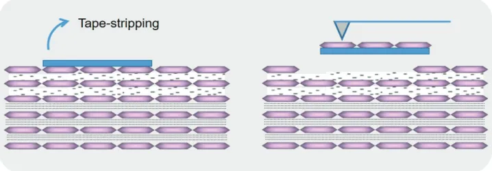
Ⓡtape (CuDerm corporation, USA)를 이용하여 채취하였다. 동일한 조건으로 각질세포를 채취하기 위해 원통모양의 100 g 무 게의 도구를 이용하였다 . Tape를 피부에 붙인 후 3 s 동안 100g 무게의 도구로 힘을 가한 후 tape stripping을 진행하 였다 . 각질세포가 있는 tape를 slide glass위에 고정한 후 AFM을 이용하여 각질세포 표면을 측정하였다(Figure 1).
입술 각질세포는 100 g 무게의 도구를 사용하지 않고 동 일한 시험자가 동일한 조건으로 tape stripping을 실시하 였다 .
각질세포 채취 과정은 항온항습 조건이 유지되는 실험 실 (22 ± 2 ℃, 50 ± 5% relative humidity)에서 진행되었다.
2.3. 각질세포 표면 측정
각질세포 표면은 AFM (Park systems, Korea)을 이용하여 측정하였다 (Figure 2). 40 μm x 40 μm 면적을 non-contact
cantilever를 이용하여 측정하였고, 분석은 AFM 보유 자체 분석 프로그램 중 거칠기를 나타내는 Rq (nm) parameter를 이용하였다 .
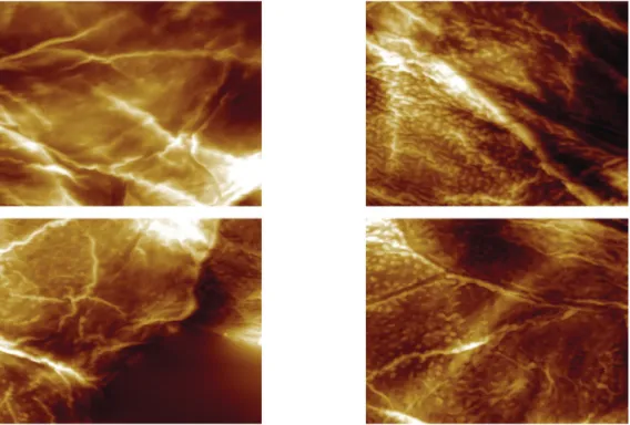
측정 부위는 각질세포의 아랫면 표면 (bottom surface of corneocyte)이며(Figure 1), sample당 임의로 3곳을 선정하여 측정하였고 , 그 평균값을 이용하여 거칠기를 비교분석하였 다 . 또한 AFM을 통해 얻어진 영상을 이용하여 villus-like projections (VPs)을 비교하였다(Figure 3).
2.4. 통계 분석
통계 분석 결과는 95% 신뢰구간에서 유의성 여부를 확 인하였으며 , 결과값에 대한 정규성 검증은 Kolmogorov- Smirnov, Shapiro-Wilk 방법으로 실시하였다. 본 데이터는 정규성을 만족하여 independent t-test를 사용하여 군간 비교 분석을 실시하였다 . 통계분석 프로그램은 SPSS 23.0 (IBM SPSS Statistics, USA)을 사용하였다.
3. 결과 및 고찰
3.1. 각질세포 표면 거칠기 결과
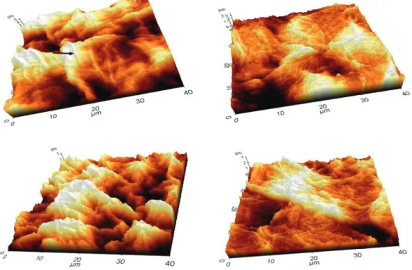
AFM을 이용하여 측정한 얼굴 각질세포와 하박내측 각 질세포 표면 거칠기 결과는 Figure 4, Figure 5와 같다. 하 박내측 각질세포의 표면은 얼굴 각질세포의 표면에 비해 거친 특성을 보여 주고 있다 . 화살표가 가리키는 구조물은 아직 명확하게 밝혀진 것은 아니지만 filamentous structures [18]라 할 수 있으며, 이 구조물이 하박내측 각질세포에서 좀 더 명확한 형태를 갖추고 있음을 알 수 있다 .
Figure 1. Example of AFM scanning. Superficial corneocytes are obtained by tape stripping. The bottom surface of corneocytes adhering to the tape is imaged by AFM.
Figure 2. Atomic Force Microscopy (Park systems, Korea).
Figure 3. The representative AFM images of corneocytes. (left) General bottom surface images of corneocytes of forearm and face. (right) VPs images of corneocytes of face and lip.
Figure 4. The 3-dimentional analysis of surface roughness on corneocytes of forearm and face. (left) Forearm corneocytes (right) face corneocytes (black arrow) filamentous structures[18].
Figure 5. The graph for bottom surface roughness comparison between forearm and face corneocytes. Each values represents mean ± SD. (***: forearm vs face, independent t-test, p <
0.001).
AFM 분석 프로그램을 이용해 각질세포 아랫면 표면 거 칠기 측정값을 정량화하여 비교한 결과 , 얼굴 각질세포는 388.34 ± 86.189 nm이었고, 하박내측 각질세포 는 662.27 ± 224.257 nm이었다(p < 0.001). AFM 사진에서 확인되었듯이 표면 거칠기 측정값은 통계적으로 유의한 차이를 보여주 고 있다 (Figure 5).
M. Gorcea등의 연구에 의하면 얼굴피부는 하박내측 피 부에 비해 각질층수가 얇고 , TEWL이 높으며, 각질세포 표 면 면적이 적고 , 크기가 작다 [18]. 얼굴 피부 각질세포 표 면적은 754.68 ± 126 μm
2이고, 하박내측 각질세포 표면적 은 1000.18 ± 164 μm
2이다 . 연속적으로 하박내측 피부를 tape-stripping 20회 했을 때의 각질세포 표면적(650 ± 20 μm
2)과 얼굴 피부 tape-stripping 6회 했을 때의 각질세포 표면적 (623 ± 20 μm
2)은 비슷한 수준이었다[18]. 각질세포 표면적을 기준으로 판단해 때 , 하박내측 피부의 분화 정도 는 얼굴 피부에 비해 더 오래도록 진행되었음을 알 수 있 다 . 이러한 연구 결과를 통해 볼 때, 얼굴 피부 특성과 하 박내측 피부 특성 간의 차이에 분화 정도가 영향을 미칠 수 있음을 시사해 준다.
따라서 위의 결과에서 알 수 있듯이 얼굴 각질세포 표 면 거칠기가 하박내측 각질세포에 비해 거칠지 않음은 이 러한 분화정도의 차이와 관련이 있을 것이라 판단된다.
3.2. 각질세포 표면의 Villus-like Projections (VPs) 결과
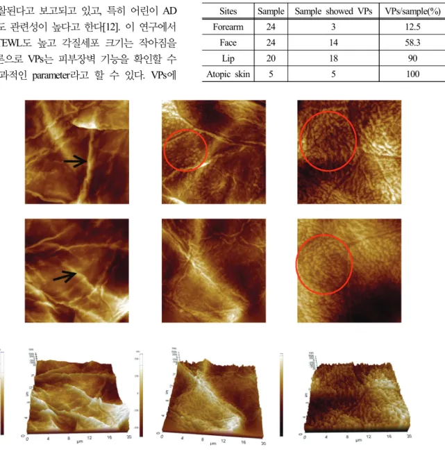
Figure 6은 하박내측, 얼굴, 그리고 입술 각질세포 표면
에서 관찰된 VPs의 대표사진을 보여주고 있다. 하박내측
각질세포에서는 24 샘플 중에 3 샘플에서 VPs가 관찰되었
고 (12.5 %), 얼굴 각질세포는 24 샘플 중에 VPs가 14 샘플 에서 관찰되었으며 (58.3 %), 입술 각질세포는 20 샘플 중 에 18 샘플에서 VPs가 관찰되었다 (90 %)(Table 1). 하지만, 입술 각질세포에서 VPs라고 판단하지 않은 2 샘플의 경우 는 정상적인 각질세포 표면 형태가 아니었으며 , 비정상적 인 형태로 보아 변형된 형태의 VPs가 아닌가 조심스럽게 판단해 본다 (Figure 7). 또한 아토피성 피부에서 5 샘플을 채취해서 VPs를 관찰해 본 결과, 5샘플 모두에서 VPs가 관찰되었다 (100%).
VPs는 주로 건선, 아토피 피부염, 지루성 피부염, 민감 성 피부에서 관찰된다고 보고되고 있고 , 특히 어린이 AD 의 증상 정도와도 관련성이 높다고 한다 [12]. 이 연구에서 VPs가 높으면 TEWL도 높고 각질세포 크기는 작아짐을 확인하였고 , 결론으로 VPs는 피부장벽 기능을 확인할 수 있는 빠르고 효과적인 parameter라고 할 수 있다. VPs에
대해서는 아직까지 그 생성원인과 기능에 대해 밝혀진 것 은 없지만, immature fragile cornified envelopes와 관련되거 나 [19,20], cytoskeleton이 분열된 desmosome과 결합된 것이 거나 desomosome이 붙는 자리 등[20]의 가정이 제안되고 있다 .
Sites Sample Sample showed VPs VPs/sample(%)
Forearm 24 3 12.5
Face 24 14 58.3
Lip 20 18 90
Atopic skin 5 5 100
Table 1. The number of samples showing VPs are calculated.
There are unidentified images of 2 samples in lip corneocytes If these 2 images are calculated as VPs, all lip corneocytes will be showed VPs
Figure 6. The representative images of VPs on forearm and face and lip corneocytes. (left) Forearm corneocyte. no VPs. Filamentous structures (black arrow) are shown. (middle) face corneocyte. VPs (circle) are shown. (right) lip corneocyte. VPs (circle) are shown. Most bottom surface of lip corneocytes are covered with VPs.
Figure 7. Unidentified images are detected on lip corneocytes.
These images are supposed as another form of VPs.