INTRODUCTION
Marfan syndrome (MFS) is an autosomal dominant connec-
tive tissue disorder usually caused by a mutation in the FBN1 gene that encodes fibrillin-1. Fibrillin-1 regulates TGF-β sig- naling, in addition to being a major structural component of extracellular matrix microfibrils.1-4 Fibrillin-1 is important in multiple organ systems, including the musculoskeletal, ocu- lar, pulmonary, cardiovascular, and central nervous systems.
Defects in the cardiovascular system cause death in MFS pa- tients. Early detection of MFS-related cardiovascular disease and prophylactic surgery have been shown to increase life ex- pectancy up to 60 years old.5-7 The progressive aortic dilatation process continues even after aortic surgery, necessitating long- term medical therapy.8,9 β-blockers are known to slow the rate of aortic dilatation by reducing aortic wall stress through their negative chronotropic effect. This is the only medication cur-
The Beneficial Effect of Renin-Angiotensin-
Aldosterone System Blockade in Marfan Syndrome Patients after Aortic Root Replacement
Seung-Jun Lee
1, Jaewon Oh
1, Young-Guk Ko
1, Sak Lee
2, Byung-Chul Chang
2, Do Yun Lee
3, Young-Ran Kwak
4, and Donghoon Choi
11Cardiology Division, Department of Internal Medicine, Severance Cardiovascular Hospital, Yonsei University Health System, Seoul;
2Department of Cardiovascular Surgery, Severance Cardiovascular Hospital, Yonsei University Health System, Seoul;
3Department of Radiology, Research Institute of Radiological Science, Yonsei University Health System, Seoul;
4Department of Anesthesiology and Pain Medicine, Severance Cardiovascular Hospital, Yonsei University Health System, Seoul, Korea.
Purpose: In this study, we evaluated the long term beneficial effect of Renin-Angiotensin-Aldosterone System (RAAS) blockade therapy in treatment of Marfan aortopathy.
Materials and Methods: We reviewed Marfan syndrome (MFS) patients who underwent aortic root replacement (ARR) between January 1996 and January 2011. All patients were prescribed β-blockers indefinitely. We compared major aortic events including mortality, aortic dissection, and reoperation in patients without RAAS blockade (group 1, n=27) to those with (group 2, n=63). The aortic growth rate was calculated by dividing the diameter change on CT scans taken immediately post-operatively and the latest scan available.
Results: There were no differences in clinical parameters except for age which was higher in patients with RAAS blockade. In group 1, 2 (7%) deaths, 5 (19%) aortic dissections, and 7 (26%) reoperations occurred. In group 2, 3 (5%) deaths, 2 (3%) aortic dis- sections, and 3 (5%) reoperations occurred. A Kaplan-Meier plot demonstrated improved survival free from major aortic events in group 2. On multivariate Cox, RAAS blockade was an independent negative predictor of major aortic events (hazard ratio 0.38, 95% confidence interval 0.30–0.43, p=0.002). Mean diameter change in descending thoracic and supra-renal abdominal aorta was significantly higher in patients without RAAS blockade (p<0.05).
Conclusion: In MFS patients who underwent ARR, the addition of RAAS blockade to β-blocker was associated with reduction of aortic dilatation and clinical events.
Key Words: Marfan syndrome, angiotensin receptor blocker, ACE inhibitor, RAAS blockade Yonsei Med J 2016 Jan;57(1):81-87
http://dx.doi.org/10.3349/ymj.2016.57.1.81 pISSN: 0513-5796 · eISSN: 1976-2437
Received: April 7, 2015 Revised: May 28, 2015 Accepted: June 5, 2015
Corresponding author: Dr. Donghoon Choi, Cardiology Division, Department of Internal Medicine, Severance Cardiovascular Hospital, Yonsei University Health System, 50-1 Yonsei-ro, Seodaemun-gu, Seoul 03722, Korea.
Tel: 82-2-2228-8530, Fax: 82-2-2227-7732, E-mail: [email protected]
•The authors have no financial conflicts of work.
© Copyright: Yonsei University College of Medicine 2016
This is an Open Access article distributed under the terms of the Creative Com- mons Attribution Non-Commercial License (http://creativecommons.org/ licenses/
by-nc/3.0) which permits unrestricted non-commercial use, distribution, and repro- duction in any medium, provided the original work is properly cited.
rently proven in prospective randomized settings to reduce aortic dilatation and clinical events, and to improve survival.10 However, about one out of ten MFS patients who underwent aortic surgery experiences re-operation in spite of continuous β-blocker therapy.11-13
Previous researches have shown that propranolol has no beneficial effects on elastic fiber fragmentation or aortic wall structure, whereas losartan-treated mice show definite im- provement in both parameters.14 In a non-randomized, retro- spective study involving 18 pediatric MFS patients who had prominent aortic dilatation despite β-blocker therapy, the ad- dition of an angiotensin receptor blocker (ARB) clearly re- duced the rate of aortic root dilatation.15 Moreover, angioten- sin converting enzyme inhibitors (ACEIs) also reduced aortic stiffness and dilatation in MFS patients.16,17 However, long-term data supporting the efficacy of Renin-Angiotensin-Aldosterone System (RAAS) blockade in combination with β-blockers in MFS patients who have undergone aortic root replacement is lacking. Therefore, we evaluated the long term beneficial effect of RAAS blockade therapy in treatment of Marfan aortopathy.
MATERIALS AND METHODS
Study design and patients
This was a retrospective study of patients seen at a 2000-bed tertiary teaching hospital. The study protocol was approved by the Institutional Review Board of Severance Cardiovascu- lar Hospital (# 4-2013-0524) and was in compliance with the Declaration of Helsinki. Between January 1996 and January 2011, using ICD-9 codes, we identified 143 consecutive pa- tients diagnosed with MFS (code 759.82) who had undergone aortic root replacement (ARR) operation with continuous β-blocker therapy after the operation. All patients met the re- vised Ghent criteria7 and had pathology consistent with me- dial degeneration. Patients with the diagnosis of Loeys-Dietz syndrome or Ehlers-Danlos syndrome were not included in this study. Of the 143 patients, we excluded 22 patients with prior aortic dissection, 4 patients who underwent ARR under the age of 18 years, 11 patients who had a change in β-blocker or RAAS blockade prescription status, 2 patients with mental retardation (due to the concern of uncertain drug compliance), 4 patients who were pregnant during follow up, 4 patients with more than 1 graft, 3 patients who had overlapping of Dacron graft with pulmonary artery (PA) bifurcation, and 3 patients with insufficient clinical data. As a result, we enrolled 90 MFS patients who underwent ARR in our study. Of these patients, 62 had Bentall composite graft and 28 had underwent valve- sparing ARR.
Clinical data collection
Patient medical records were reviewed for information on pa- tient age at the time of operation, gender, weight, height, blood
pressure, heart rate, drug therapy, echocardiographic results and computed tomography scan results. Patient databases were searched to identify known or putative risk factors for hy- pertension, dyslipidemia, atrial fibrillation (AF), congestive heart failure, congenital heart disease (CHD) other than MFS, and smoking status. AF was documented with 12-lead elec- trocardiography or 24 hr Holter recording. Heart failure was defined as satisfying the previously published Framingham criteria18 (presence of 2 major or 1 major with 2 minor criteria).
CHD included repaired or non-cyanotic atrial septal defect, ventricular septal defect, tetralogy of Fallot, or coarctation of the aorta. We analyzed heart rate and blood pressure, which were averaged with more than ten measurements at post-op- erative outpatient visit.
Clinical endpoint definition
We evaluated major aortic events including all-cause mortali- ty, newly developed aortic dissection, and reoperation for re- current valvular regurgitation or progression distal aorta pa- thology. Indications for the reoperation consisted of recurrent aortic regurgitation of G3 (central jet width/LVOT width=0.50, n=1), progressive aneurysmal enlargement of the downstream aorta greater than 55 mm in 2 patients, rapidly expanding an- eurysm (>5 mm in 6 months) in 6 patients, and impending aortic rupture in 2 patients. Follow-up was censored after the first aortic event. Patients who were alive and aortic event free at last follow-up were censored.
Imaging data collection and analysis
The maximum aortic diameters were measured at five levels of the aorta preoperatively; 1) ascending and 2) descending thoracic aorta (DTA) at the level of PA bifurcation, 3) aortic arch, 4) the mid-section between the diaphragm neck and right renal artery ostium, and 5) the inferior mesenteric artery (IMA) ostium level. Pulmonary artery bifurcation level corre- sponded to a level above Dacron graft in all patients. Imaging studies included CT scan, trans-esophageal echocardiography, magnetic resonance imaging or angiography performed with- in 3 months before ARR. In case of patients who were evaluat- ed their aortic dimension by two or more modalities, we se- lected the maximal diameter.
Postoperative aortic growth rate of each group was com- pared in patients who have undergone repeated CT scanning after the operation [n=19 (70%) for group 1 vs. n=43 (68%) for group 2]. All of these patients took the first scan one week after the operation and another follow-up scan at least 1 year after operation. We compared the change in the maximal aortic di- ameter perpendicular to the aortic centerline at 3 levels of the aorta. The measurements were obtained from cranial to cau- dal at the 1) PA bifurcation level, 2) the mid-section between the diaphragm neck and right renal artery ostium, and 3) the IMA ostium level. The rate of aortic diameter change was cal- culated by dividing the increment of the aortic maximal diam-
eter by the time interval on the CT scans. All collected imaging data were read in consensus by 2 radiologists who were blind- ed to the patients’ clinical data.
Statistical analysis
Continuous variables that were normally distributed were re- ported as mean±SD, and were compared using Student’s t- tests for parametric data and Mann-Whitney tests for non- parametric data. Categorical variables were reported as counts (percentages) and compared using chi-squared or Fisher’s exact tests. Kaplan-Meier survival curves were plotted for β-blocker- only patients (group 1) or RAAS blockade-combination pa- tients (group 2) and compared by means of the log-rank test.
To identify independent predictor variables, we performed multivariate Cox proportional-hazards analysis using the fac- tors predictive in univariate analysis (p<0.05). The Wilcoxon signed rank test was used to compare the rates of aortic diam- eter change in patients with or without RAAS blockade. A p- value<0.05 was considered statistically significant. SPSS statis- tical package (SSPS Inc., Chicago, IL, USA) version 18.0 was used to perform all statistical analysis.
RESULTS
Study population and baseline characteristics
Clinical characteristics and echocardiographic parameters are presented in Table 1. Of the 90 patients, group 1 patients (n=27) had no history of RAAS blockade prescription and group 2 patients (n=63) were prescribed β-blocker and RAAS blockade concomitantly after ARR. The rationale for prescrip- tion of RAAS blockade in group 2 patients according to chart review are as follows; control of blood pressure (n=22), re- duced left ventricular systolic function (n=15), attendant’s clinical decision (n=14), and not clearly documented on the chart (n=12). Of the 90 patients with MFS, 60 patients (67 per- cent) were male. There were no differences in the clinical characteristics of the two groups, except for age at the time of ARR. On average, group 2 patients were 4.4 years older than group 1 patients (34.8 years vs. 39.2 years; p=0.001). In family history, there was no difference in the presence of aortic dis- section, rupture, sudden death or FBN1 mutation. There were no differences in echocardiographic parameters including aortic annulus diameter, sinus of Valsalva diameter, or ejection fraction. Mean systolic blood pressure and heart rate were not different between the two groups. The frequency of other pre- scription medications, including statins, digoxin, calcium channel blockers, and diuretics, were not significantly differ- ent between the two groups. More detailed demographic data that subdivided group 2 into patients with ACEI and ARB are shown in Supplementary Table 1 (only online). There was no difference in the preoperative aortic dimension between the two groups, except for the supra-renal abdominal aorta, which
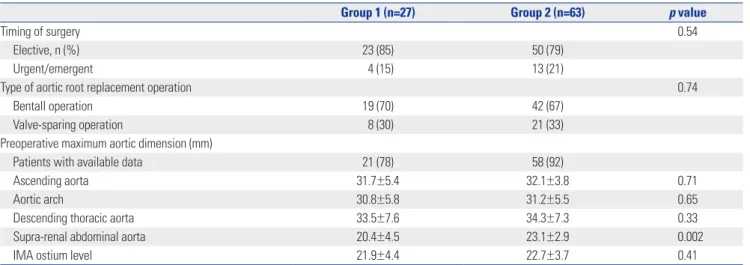
was higher in group 2 (Table 2). We evaluated the preopera- tive aortic dimension within 3 months before ARR in 24 pa- tients (89%) of group 1 and 58 patients (92%) of group 2. There was no difference in the baseline aortic dimensions between the groups, except for the supra-renal abdominal aorta level, which was higher in patients with RAAS blockade (p=0.002).
Clinical outcomes
The incidence rates of major aortic events are presented in Ta- ble 3. During the mean 82±60 months of follow-up, 2 (7%) and 3 (5%) patients died of cardiac or non-cardiac causes in group 1 and 2, respectively (p=0.32). The development of aortic dis- section was significantly higher in group 1 (19% vs. 3%, p=0.02).
Preoperative maximal diameters of distal aorta of the patients who developed new aortic dissection were 45.9 mm; 43.6 mm;
42.1 mm; 38.6 mm; 36.3 mm in group 1 and 44.2 mm; 37.0 mm in group 2, respectively. Of those 7 patients who developed new aortic dissection, 2 patients (40%) of group 1 and 1 patient of group 2 (50%) had positive family history of aortic dissection.
The risk of re-operation for recurrent aortic regurgitation or distal aortic pathology was also higher in group 1 (p=0.003), especially at the aortic arch or DTA level (p=0.01). Conse- quently, there was a significant difference in the incidence of major aortic events between the two groups (p=0.001). In Ka- plan-Meier analysis, group 2 patients had higher cumulative event-free survival than group 1 (p=0.008) (Fig. 1). Clinical outcome of patients treated with ACEI or ARB were not differ- ent, which was significantly better than that of group 1 pa- tients (Supplementary Fig. 1, only online). We used univariate and multivariate Cox regression modeling to test the ability of potential baseline risk factors to predict major aortic events.
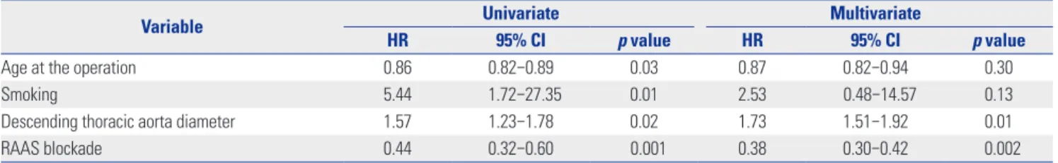
Table 4 shows several variables that were associated with a higher risk of major aortic events. RAAS blockade therapy, in addition to β-blocker was an independent negative predictor of major aortic events after adjustment for age, smoking and descending thoracic aorta diameter (hazard ratio=0.38, 95%
confidence interval 0.30–0.43, p=0.002).
Aortic dilatation rate
A total of 62 patients (69%) had a CT scan performed more than twice after ARR: 19 (70%) in group 1 and 43 (68%) in group 2. Rates of aortic diameter change measured at 3 posi- tions are depicted in Fig. 2. At the pulmonary artery bifurcation level, the growth rate was 2.7±1.4 mm/yr in group 1 and 1.1±0.7 mm/yr in group 2 (p<0.001). At the mid-section between the diaphragm and the right renal artery ostium level, the growth rate was 2.5±1.0 mm/yr in group 1 and 0.9±0.7 mm/yr in group 2 (p<0.001). There was no significant difference in the rate of aortic growth at the IMA ostium level between the groups (0.9±0.6 mm/yr in group 1 and 0.8±0.6 mm/yr in group 2, p=0.73).
DISCUSSION
Although β-blockers have been proven to decrease the rate of
aortic root dilatation and reduce the risk of clinical events by reducing tensile stress on the aorta, the risk of aortic events or re-operation is still high. This is particularly true in MFS pa- Table 1. Baseline Clinical Characteristics and Prescription Status
Variable Group 1 (n=27) Group 2 (n=63) p value
Female, n (%) 10 (37) 20 (32) 0.53
Age (at aortic root replacement) 34.8±9.3 39.2±12.2 0.001
Risk factors
Hypertension, n (%) 10 (37) 25 (40) 0.92
Dyslipidemia 6 (22) 11 (17) 0.52
Atrial fibrillation 4 (15) 14 (22) 0.47
Congestive heart failure 7 (27) 17 (27) 0.99
Congenital heart disease 1 (4) 6 (10) 0.38
Smoking 19 (71) 37 (59) 0.29
Calendar year of operation 0.83
1996–2000 7 (26) 18 (29)
2001–2005 9 (33) 20 (32)
2006–2011 11 (41) 25 (40)
Family history
Family history of Marfan syndrome 12 (45) 31 (49) 0.68
Family history of aortic dissection or rupture 11 (39) 28 (44) 0.75
Family history of sudden death 6 (21) 15 (24) 0.88
Presence of casual FBN1 mutation 0.70
Present 9 (32) 18 (28)
Absent 2 (9) 5 (8)
Unknown 16 (59) 40 (64)
Echocardiographic parameters
Mean initial aortic annulus diameter (mm) 33.8±13.0 31.8±10.4 0.51
Mean initial sinus of valsalva diameter (mm) 61.2±13.5 61.4±13.1 0.99
Ejection fraction (%) 56.7±14.0 56.3±11.5 0.89
Aortic insufficiency, n (%) 0.43
Grade 1 5 (18) 6 (10)
Grade 2 4 (15) 6 (10)
Grade 3 10 (37) 23 (36)
Grade 4 8 (30) 28 (44)
Mean blood pressure and heart rate
Systolic blood pressure (mm Hg) 118±11 117±10 0.69
Diastolic blood pressure (mm Hg) 71±10 71±8 0.81
Heart rate (beats/min) 59±10 63±11 0.14
Baseline renal function
Serum creatinine 1.37±0.50 1.26±0.43 0.71
Estimated GFR – mL/min/1.73 m2 68.3±22.7 70.6±21.4 0.87
Estimated GFR rate <60 mL/min/1.73 m2 9 (34) 16 (26) 0.23
Serum potassium – mmol/L 4.5±0.9 4.3±0.7 0.26
β-blocker dose standardized to carvedilol equivalent (mg) 20.8±12.8 13.5±10.1 0.01
Concomitant drug use
Statin 5 (19) 14 (22) 0.68
Digoxin 5 (19) 6 (10) 0.24
Calcium channel blocker 4 (15) 11 (17) 0.63
Diuretic 3 (11) 13 (21) 0.52
GFR, glomerular filtration rate.
tients who have already undergone aortic operation.19,20 Cur- rent guidelines on medical treatment after aortic surgery are rare and not based on randomized clinical trials.21,22 RAAS blockade are attracting much attention in MFS due to their ability to modulate excessive TGF-β signaling. There are sev- eral experimental evidences that supports the superiority of ARB, selective angiotensin II receptor type (AT) I blocker, com- pared with non-selective angiotensin receptor blocking ACEI, as the downstream signaling of AT2 ameliorated aneurysm progression in rodent Marfan model. However, some contro- versy exists about the beneficial effect of AT2, as it can also in- duce vascular smooth muscle cell apoptosis which is prone to progression of aneurysm.23 Until now, there is no solid evi- dence that supports the superior efficacy of ARB compared with ACEI, especially in MFS patients who have replaced their aortic root. In this study, both ACEIs and ARBs were found to be associated with reducing aortic dilatation and major aortic events in Marfan aortopathy patients who were previously on β-blockers. This phenomenon was independent of the blood pressure reduction that can be explained by these molecular mechanisms, which are supported by recent evidence in hu- man and animal studies.24-26
In addition to its molecular mechanism, RAAS blockade is Table 2. Operative Information
Group 1 (n=27) Group 2 (n=63) p value
Timing of surgery 0.54
Elective, n (%) 23 (85) 50 (79)
Urgent/emergent 4 (15) 13 (21)
Type of aortic root replacement operation 0.74
Bentall operation 19 (70) 42 (67)
Valve-sparing operation 8 (30) 21 (33)
Preoperative maximum aortic dimension (mm)
Patients with available data 21 (78) 58 (92)
Ascending aorta 31.7±5.4 32.1±3.8 0.71
Aortic arch 30.8±5.8 31.2±5.5 0.65
Descending thoracic aorta 33.5±7.6 34.3±7.3 0.33
Supra-renal abdominal aorta 20.4±4.5 23.1±2.9 0.002
IMA ostium level 21.9±4.4 22.7±3.7 0.41
IMA, inferior mesenteric artery.
Table 3. Major Aortic Events during Follow-Up
Major aortic event, n (%) Group 1 (n=27) Group 2 (n=63) p value
All-cause mortality 2 (7) 3 (5) 0.32
New aortic dissection 5 (19) 2 (3) 0.02
Re-operation 7 (26) 4 (6) 0.003
Aortic valve 0 1 0.52
Ascending aorta 2 1 0.16
Aortic arch or DTA 4 1 0.01
Abdominal aorta 1 1 0.54
Total 14 (52) 8 (13) 0.001
DTA, descending thoracic aorta.
The numbers in parentheses represent percentages.
100
80
60
40
20
00
β-blocker (group 1)
β-blocker+RAAS blockade (group 2) p=0.008
No. at risk
Group 1 27 23 18 15 11 7 5 4 Group 2 63 49 39 27 22 17 10 6
2 4 6
Follow up (years)
8 10 12 14
Survival free from major aortic events
Fig. 1. Kaplan-Meier curves for cumulative survival free from major aortic events. Patients without RAAS blockade (group 1) had lower cumulative survival free of major aortic events (p=0.008). RAAS, Renin-Angiotensin- Aldosterone System.
expected to have beneficial effects on Marfan aortopathy by reducing hemodynamic stress. In MFS patients, aortic stiffness increases and distensibility decreases over time.27,28 In a previ- ous study, decreased aortic distensibility was an independent predictor of progressive aortic dilatation in MFS patients.29 Early reflection of the pressure wave from the stiffened periph- eral arteries increases load on the central arteries and possibly increases the risk of aortic rupture.30,31 ARBs and ACEIs have a greater effect on aortic stiffness than β-blockers despite their similar effects on peripheral blood pressure.32-34 Yetman, et al.16 compared the efficacy of enalapril to that of β-blockers in a non-randomized, open label study and reported that enala- pril improved the aortic stiffness and distensibility in MFS pa- tients and decreased aortic root dilatation and clinical events.
In our study, the concomitant prescription of RAAS block- ade with β-blocker was negatively correlated with the risk of aortic dissection development and re-operation, especially at the aortic arch and descending thoracic aorta. The reduction in aortic growth rate by RAAS blockade was definite in the DTA and abdominal aorta at the supra-renal level, as opposed to at the IMA ostium level where there was no difference be- tween groups. The additive effect of RAAS blockade in MFS patients who have undergone ARR might be explained by a re- duction in shear stress on the central arteries through an im- provement in aortic stiffness and distensibility, in addition to a molecular mechanism.
Study limitations
There are several limitations to this study. First of all, this was a retrospective study conducted at a single center, carrying all the limitations of this study design. Dose titration of RAAS blockade was almost entirely dependent on clinician’s decision and blood pressure measurement. However, the blood pressure measure- ment did not strictly meet the principle to measure 3 times over 3-min interval with constant time gap since last medica- tion, which could have influenced the outcome. Second, the mean age of group 1 patients at the time of ARR was 4.4 years younger than group 2 patients, suggesting that the natural progression of disease is more rapid in group 1 than in group 2. On univariate analysis, operation at older ages was nega- tively associated with major aortic events or re-operation. Al- though we adjusted for age in the multivariate Cox regression analysis, there is still the possibility of selection bias. Third, this study covers a relatively long time period, which neces- sarily includes changes in operation technique and graft pros- thesis material, both of which could influence clinical results.
Finally, a histologic study of re-operated patients that might show the structural or molecular effects of RAAS blockade on the aortas of MFS patients was not performed. A measure- ment of central pressure, augmentation index, or pulsed wave velocity that could explain this result from a hemodynamic perspective was not performed in most of patients due to it’s retrospective nature.
To conclude, in MFS patients who underwent ARR, the ad- dition of RAAS blockade to β-blocker was associated with re- duction of aortic dilatation and clinical events. This result might be explained by improvement in aortic stiffness or dis- tensibility, as well as inhibition of TGF-β signaling. Further evaluation in a prospective setting is needed to confirm the benefit.
ACKNOWLEDGEMENTS
This study was supported by a grant of the Korea Health 21 R&D Project, Ministry of Health &Welfare, Republic of Korea (A085136).
REFERENCES
1. Cañadas V, Vilacosta I, Bruna I, Fuster V. Marfan syndrome. Part 1:
pathophysiology and diagnosis. Nat Rev Cardiol 2010;7:256-65.
Table 4. Univariate and Multivariate Predictors of Major Aortic Events
Variable Univariate Multivariate
HR 95% CI p value HR 95% CI p value
Age at the operation 0.86 0.82–0.89 0.03 0.87 0.82–0.94 0.30
Smoking 5.44 1.72–27.35 0.01 2.53 0.48–14.57 0.13
Descending thoracic aorta diameter 1.57 1.23–1.78 0.02 1.73 1.51–1.92 0.01
RAAS blockade 0.44 0.32–0.60 0.001 0.38 0.30–0.42 0.002
RAAS, Renin-Angiotensin-Aldosterone System; HR, hazard ratio; CI, confidence interval.
Fig. 2. Mean annual rate of change in aortic diameter after aortic root re- placement. Significant reduction of aortic dilatation rate by the addition of RAAS blockade was observed in descending thoracic aorta and supra- renal abdominal aorta. RAAS, Renin-Angiotensin-Aldosterone System.
Descending thoracic aorta
Supra-renal abdominal aorta
Inferior mesenteric artery ostium
Change in aortic diameter (mm/yr)
0 1 2 3 4
p=0.73 p<0.001 p<0.001
5 β-blocker (n=19)
β-blocker+RAAS blockade (n=43)
2. Shin MS, Park HY, Lim Y, Shin GJ, Jang Y, Jang BC, et al. Identifica- tion of Molecular Defects in Korean Patients with Marfan Syn- drome. Korean Circ J 2003;33:1018-27.
3. Jondeau G, Michel JB, Boileau C. The translational science of Marfan syndrome. Heart 2011;97:1206-14.
4. Sawaki D, Suzuki T. Targeting transforming growth factor-β sig- naling in aortopathies in Marfan syndrome. Circ J 2013;77:898-9.
5. Finkbohner R, Johnston D, Crawford ES, Coselli J, Milewicz DM.
Marfan syndrome. Long-term survival and complications after aortic aneurysm repair. Circulation 1995;91:728-33.
6. Judge DP, Dietz HC. Marfan’s syndrome. Lancet 2005;366:1965- 76.
7. Loeys BL, Dietz HC, Braverman AC, Callewaert BL, De Backer J, Devereux RB, et al. The revised Ghent nosology for the Marfan syndrome. J Med Genet 2010;47:476-85.
8. Pyeritz RE. Marfan syndrome: current and future clinical and ge- netic management of cardiovascular manifestations. Semin Tho- rac Cardiovasc Surg 1993;5:11-6.
9. Gott VL, Greene PS, Alejo DE, Cameron DE, Naftel DC, Miller DC, et al. Replacement of the aortic root in patients with Marfan’s syn- drome. N Engl J Med 1999;340:1307-13.
10. Shores J, Berger KR, Murphy EA, Pyeritz RE. Progression of aortic dilatation and the benefit of long-term beta-adrenergic blockade in Marfan’s syndrome. N Engl J Med 1994;330:1335-41.
11. Volguina IV, Miller DC, LeMaire SA, Palmero LC, Wang XL, Con- nolly HM, et al. Valve-sparing and valve-replacing techniques for aortic root replacement in patients with Marfan syndrome: analy- sis of early outcome. J Thorac Cardiovasc Surg 2009;137:1124-32.
12. Karck M, Kallenbach K, Hagl C, Rhein C, Leyh R, Haverich A.
Aortic root surgery in Marfan syndrome: Comparison of aortic valve-sparing reimplantation versus composite grafting. J Thorac Cardiovasc Surg 2004;127:391-8.
13. de Oliveira NC, David TE, Ivanov J, Armstrong S, Eriksson MJ, Ra- kowski H, et al. Results of surgery for aortic root aneurysm in pa- tients with Marfan syndrome. J Thorac Cardiovasc Surg 2003;125:
789-96.
14. Habashi JP, Judge DP, Holm TM, Cohn RD, Loeys BL, Cooper TK, et al. Losartan, an AT1 antagonist, prevents aortic aneurysm in a mouse model of Marfan syndrome. Science 2006;312:117-21.
15. Brooke BS, Habashi JP, Judge DP, Patel N, Loeys B, Dietz HC 3rd.
Angiotensin II blockade and aortic-root dilation in Marfan’s syn- drome. N Engl J Med 2008;358:2787-95.
16. Yetman AT, Bornemeier RA, McCrindle BW. Usefulness of enala- pril versus propranolol or atenolol for prevention of aortic dila- tion in patients with the Marfan syndrome. Am J Cardiol 2005;95:
1125-7.
17. Groenink M, den Hartog AW, Franken R, Radonic T, de Waard V, Timmermans J, et al. Losartan reduces aortic dilatation rate in adults with Marfan syndrome: a randomized controlled trial. Eur Heart J 2013;34:3491-500.
18. McKee PA, Castelli WP, McNamara PM, Kannel WB. The natural history of congestive heart failure: the Framingham study. N Engl J Med 1971;285:1441-6.
19. Geisbuesch S, Schray D, Bischoff MS, Lin HM, Di Luozzo G, Gri- epp RB. Frequency of reoperations in patients with Marfan syn- drome. Ann Thorac Surg 2012;93:1496-501.
20. Gott VL, Cameron DE, Alejo DE, Greene PS, Shake JG, Caparrelli
DJ, et al. Aortic root replacement in 271 Marfan patients: a 24- year experience. Ann Thorac Surg 2002;73:438-43.
21. Nienaber CA, Von Kodolitsch Y. Therapeutic management of pa- tients with Marfan syndrome: focus on cardiovascular involve- ment. Cardiol Rev 1999;7:332-41.
22. Ades L, CSANZ Cardiovascular Genetics Working Group. Guide- lines for the diagnosis and management of Marfan syndrome.
Heart Lung Circ 2007;16:28-30.
23. Habashi JP, Doyle JJ, Holm TM, Aziz H, Schoenhoff F, Bedja D, et al. Angiotensin II type 2 receptor signaling attenuates aortic an- eurysm in mice through ERK antagonism. Science 2011;332:361- 5.
24. Nagashima H, Sakomura Y, Aoka Y, Uto K, Kameyama Ki, Ogawa M, et al. Angiotensin II type 2 receptor mediates vascular smooth muscle cell apoptosis in cystic medial degeneration associated with Marfan’s syndrome. Circulation 2001;104:I282-7.
25. Moltzer E, te Riet L, Swagemakers SM, van Heijningen PM, Ver- meij M, van Veghel R, et al. Impaired vascular contractility and aortic wall degeneration in fibulin-4 deficient mice: effect of an- giotensin II type 1 (AT1) receptor blockade. PLoS One 2011;6:
e23411.
26. Iida Y, Xu B, Schultz GM, Chow V, White JJ, Sulaimon S, et al. Effi- cacy and mechanism of angiotensin II receptor blocker treatment in experimental abdominal aortic aneurysms. PLoS One 2012;7:
e49642.
27. Adams JN, Brooks M, Redpath TW, Smith FW, Dean J, Gray J, et al. Aortic distensibility and stiffness index measured by magnetic resonance imaging in patients with Marfan’s syndrome. Br Heart J 1995;73:265-9.
28. Segers P, De Backer J, Devos D, Rabben SI, Gillebert TC, Van Bor- tel LM, et al. Aortic reflection coefficients and their association with global indexes of wave reflection in healthy controls and pa- tients with Marfan’s syndrome. Am J Physiol Heart Circ Physiol 2006;290:H2385-92.
29. Nollen GJ, Groenink M, Tijssen JG, Van Der Wall EE, Mulder BJ.
Aortic stiffness and diameter predict progressive aortic dilatation in patients with Marfan syndrome. Eur Heart J 2004;25:1146-52.
30. Groenink M, de Roos A, Mulder BJ, Verbeeten B Jr, Timmermans J, Zwinderman AH, et al. Biophysical properties of the normal- sized aorta in patients with Marfan syndrome: evaluation with MR flow mapping. Radiology 2001;219:535-40.
31. Groenink M, Langerak SE, Vanbavel E, van der Wall EE, Mulder BJ, van der Wal AC, et al. The influence of aging and aortic stiff- ness on permanent dilation and breaking stress of the thoracic de- scending aorta. Cardiovasc Res 1999;43:471-80.
32. Dhakam Z, McEniery CM, Yasmin, Cockcroft JR, Brown MJ, Wilkinson IB. Atenolol and eprosartan: differential effects on cen- tral blood pressure and aortic pulse wave velocity. Am J Hyper- tens 2006;19:214-9.
33. Mackenzie IS, McEniery CM, Dhakam Z, Brown MJ, Cockcroft JR, Wilkinson IB. Comparison of the effects of antihypertensive agents on central blood pressure and arterial stiffness in isolated systolic hypertension. Hypertension 2009;54:409-13.
34. Takami T, Shigemasa M. Efficacy of various antihypertensive agents as evaluated by indices of vascular stiffness in elderly hy- pertensive patients. Hypertens Res 2003;26:609-14.