Korean Circulation Journal
Introduction
Familial Mediterranean fever (FMF) is an autosomal recessive auto-inflammatory disorder characterized by recurrent, episodic fever and acute episodes of serosal membrane inflammation (sterile pleuritis, peritonitis, arthritis, etc.). The clinical disease most commonly occurs in Jews, Turks, Armenians, and Arabs.
1)The incidence of FMF is reportedly between 5/1000 in Jews and 1/1000 in Turks.
2)The gene causing FMF, also designated as the Mediterranean fever (MEFV) gene, is located on the short arm of chromosome 16. This gene encodes a protein called pyrin that plays an important role in the inhibition of pro-inflammatory
Print ISSN 1738-5520 • On-line ISSN 1738-5555
Aortic Flow Propagation Velocity in Patients with Familial Mediterranean Fever: an Observational Study
Kayihan Karaman, MD
1, Arif Arisoy, MD
1, Aysegul Altunkas, MD
2, Ertugrul Erken, MD
3, Ahmet Demirtas, MD
3, Mustafa Ozturk, MD
4, Metin Karayakali, MD
1, Safak Sahin, MD
3, and Atac Celik, MD
11
Department of Cardiology, Gaziosmanpasa University School of Medicine, Tokat,
2Department of Radiology, Gaziosmanpasa University School of Medicine, Tokat,
3
Department of Internal Medicine, Gaziosmanpasa University School of Medicine, Tokat,
4Department of Cardiology, Erzurum Territorial Training and Research Hospital, Erzurum, Turkey
Background and Objectives: Systemic inflammation has an important role in the initiation of atherosclerosis, which is associated with arterial stiffness (AS). Aortic flow propagation velocity (APV) is a new echocardiographic parameter of aortic stiffness. The relationship between systemic inflammation and AS has not yet been described in patients with familial Mediterranean fever (FMF). We aimed to investigate the early markers of AS in patients with FMF by measuring APV and carotid intima-media thickness (CIMT).
Subjects and Methods: Sixty-one FMF patients (43 women; mean age 27.3±6.7 years) in an attack-free period and 57 healthy individuals (36 women; mean age 28.8±7.1 years) were included in this study. The individuals with atherosclerotic risk factors were excluded from the study. The flow propagation velocity of the descending aorta and CIMT were measured to assess AS.
Results: APV was significantly lower (60.2±16.5 vs. 89.5±11.6 cm/sec, p<0.001) and CIMT was significantly higher (0.49±0.09 vs. 0.40±0.10 mm, p<0.001) in the FMF group compared to the control group. There were significant correlations between APV and mean CIMT (r=-0.424, p<0.001), erythrocyte sedimentation rate (ESR) (r=-0.198, p=0.032), and left ventricle ejection fraction (r=0.201, p=0.029). APV and the ESR were independent predictors of FMF in logistic regression analysis (OR=-0.900, 95% CI=0.865-0.936, p<0.001 and OR=-1.078, 95% CI=1.024-1.135, p=0.004, respectively). Mean CIMT and LVEF were independent factors associated with APV in linear regression analysis (β=-0.423, p<0.001 and β=0.199, p=0.017, respectively)
Conclusion: We demonstrated that APV was lower in FMF patients and is related to CIMT. According to our results, APV may be an independent predictor of FMF. (Korean Circ J 2017;47(4):483-489)
KEY WORDS: Familial Mediterranean fever; Aortic flow propagation velocity; Arterial stiffness; Carotid intima-media thickness.
Received: November 11, 2016 Revision Received: February 3, 2017 Accepted: March 11, 2017
Correspondence: Kayihan Karaman, MD, Department of Cardiology, Gaziosmanpasa University School of Medicine, Training and Research Hospital, Tokat 60100, Turkey
Tel: 90-546-802-3542, Fax: 90-356-212-2142 E-mail: [email protected]
• The authors have no financial conflicts of interest.
This is an Open Access article distributed under the terms of the Creative Commons Attribution Non-Commercial License (http://creativecommons.org/
licenses/by-nc/3.0) which permits unrestricted non-commercial use,
distribution, and reproduction in any medium, provided the original work is
properly cited.
cytokines, and increases the discharge of anti-inflammatory mediators by inflammatory cells such as neutrophils, eosinophils, and monocytes.
3)Mutations in MEFV are responsible for regional and systemic inflammation.
Cardiovascular disease (CVD) is a common cause of death in patients with auto-inflammatory disorders.
4)Recent data have demonstrated that atherosclerosis and arterial stiffness (AS) play a major role in the pathogenesis of CVD. AS, which reflects arterial compliance, is progressively being accepted as an independent predictor of cardiovascular death.
5)Chronic systemic inflammation in FMF may increase AS by accelerating the natural process of atherosclerosis. Aortic flow propagation velocity (APV), which has been used as a new echocardiographic index of AS, is a noninvasive technique for measuring the distensibility of the aorta. Several studies have investigated the relationship between APV and endothelial dysfunction, and carotid intima-media thickness (CIMT) has been found to be associated with APV.
6)7)In this study, we aimed to investigate the early markers of AS in patients with FMF by measuring APV and CIMT.
Subjects and Methods
Patient population
This cross-sectional and case-control study included 61 patients without cardiovascular involvement in an attack-free period (18 men, 43 women; mean age, 27.3±6.7 years) diagnosed with FMF in our center between August 2014 and August 2015. The diagnosis of FMF was made according to the Tel-Hashomer criteria.
8)Fifty-seven age- and sex-matched control healthy subjects (21 men, 36 women;
mean age, 28.8±7.1 years) were included from our cardiology outpatient clinics. All of the patients with FMF were under regular colchicine treatment. We excluded patient and control subjects with a history of coronary artery disease, arterial hypertension, diabetes mellitus, smoking, hypercholesterolemia, left ventricle (LV) ejection fraction (LVEF) <50%, hypertrophic or dilated cardiomyopathies, valvular heart disease, prosthetic heart valve, bundle branch block, atrial fibrillation and other electrocardiographic abnormalities, renal failure, chronic obstructive lung disease, thyroid dysfunction, other known inflammatory disease, malignancy, amyloidosis, or echocardiographic imaging that was technically insufficient.
Hemoglobin, white blood cell (WBC) count, neutrophil count, lymphocyte count, neutrophil-lymphocyte ratio (NLR), erythrocyte sedimentation rate (ESR; reference range: 0-20 mm/hr), and C-reactive protein (CRP; reference range: 0-5 mg/L) levels were obtained from the laboratory records. Ethics committee approval and informed consent were obtained for all patients.
Echocardiographic evaluation
Conventional echocardiographic parameters of the patient and control subjects were performed in the left lateral decubitus position.
Standard two-dimensional images were acquired by two experienced echocardiographers who were blinded to the clinical data, using a 2.5-3.5-MHz transducer (General Electric Vivid S5, Milwaukee, WI, USA). LV diameter and thickness were measured from M-mode traces from the parasternal long axis view. LVEF was assessed using the biplane-modified Simpson method. LV end-systolic and end-diastolic volumes (LVESV and LVEDV) were calculated using the biplane- modified Simpson’s rule in the apical 4- and 2-chamber views. For pulse wave Doppler measurements, the sample volume was placed in the middle of the LV inflow tract 1 cm below the plane of mitral annulus in the apical four-chamber view. Mitral early diastolic velocity (E), late diastolic velocity (A), and E/A ratio were determined.
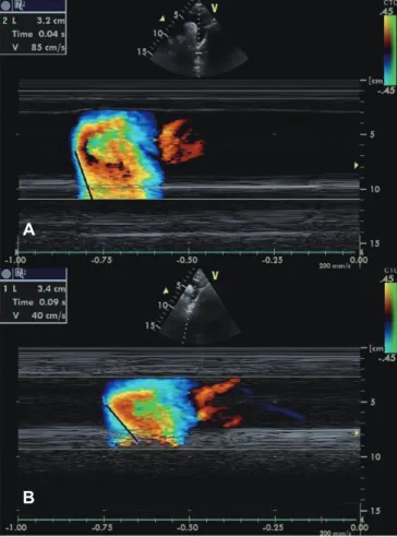
Color M-mode Doppler measurements were obtained on the suprasternal view and recordings were obtained with the cursor parallel to the main flow of direction in the descending thoracic aorta. The color Doppler Nyquist limit is adapted to 30-50 cm/s, and switching to M mode with a recorder sweep rate of 200 mm/s, an M-mode spatio-temporal velocity map with the shape of a flame is displayed (Fig. 1). If the slope of the flame was unclear, the baseline shifting was used to change the aliasing velocity until a clear delineation of isovelocity slope was obtained. APV was then calculated by dividing the distance between the points corresponding to the beginning and end of the propagation slope by the duration between the corresponding time points. Thus, APV corresponds to the velocity at which the flow is propagating down the artery. The mean of at least three measurements was recorded as the APV value.
Carotid intima-media thickness measurement
Carotid intima-media thickness (CIMT) measurements of the participants were performed by physicians who were blinded to the patients and obtained APV values. Both common carotid arteries were analyzed with a Toshiba Aplio 500 (Toshiba Medical System, Tokyo, Japan) ultrasound device with a 7.5 MHz linear probe, and the CIMT was automatically measured with software. CIMT was measured at the posterior wall when they were clearly visible in the longitudinal plane at 1–1.5 cm below bifurcation. The CIMT measurement was obtained from three contiguous sites at 1-mm intervals. The average of the three measurements and the mean of the left and right common carotid arteries were used for analyses.
Pearson correlation analysis was performed to assess intra-
and interobserver variability. Intraobserver variability for APV and
CIMT were 3.1% and 1.8%, respectively (p>0.05). In addition, the
interobserver variability for APV and CIMT were 3.4% and 2.0%,
respectively (p>0.05).
Statistical analysis
All tests were performed using PASW Statistics v.18.0. Variables with a normal distribution were analyzed using the Kolmogorov–
Smirnov test. Continuous variables are expressed as mean±standard deviation, while categorical variables are presented as numbers and percentages. Differences between independent groups were assessed by Student’s t-test for normally distributed continuous variables and Mann-Whitney’s U-test for variables without normal distribution. Categorical variables were assessed by the chi-square test. Correlations between parameters with normal and non-normal distribution were computed through Pearson’s and Spearman’s correlation analysis, respectively. Logistic regression analysis was used to analyze independent predictors of FMF, and linear regression analysis was used to analyze independent predictors of APV. The sensitivity and specificity of APV to predict FMF was evaluated with receiver operative characteristic (ROC) analysis. The area under curve (AUC) was obtained. All results were considered statistically significant at the level of p<0.05.
Results
Clinical characteristics and laboratory findings from patients with FMF and control subjects are shown in Table 1. The distributions of age, sex, body mass index (BMI), systolic and diastolic blood pressure, lipid profile, CRP, WBC count and NLR were similar between the two groups. However, ESR was significantly higher in the FMF patients compared with the control group. The two- dimensional echocardiography and the Doppler study variables are shown in Table 2. LV diameters, LV volumes, LVEF, interventricular septum (IVS) thickness, mitral E and A velocity ratio were similar in both groups. However, the mean CIMT was significantly higher, and APV was significantly lower in FMF patients. Mean disease and treatment durations in patients with FMF were 95.9±59.9 and 52.9±52.7 months, respectively.
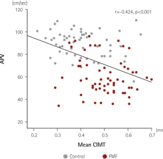
There were significant correlations between APV and mean CIMT (r=-0.424, p<0.001) (Fig. 2), ESR (r=-0.198, p=0.032), and LVEF (r=0.201, p=0.029). However, there was no correlation among APV, age, CRP, NLR, and WBC count (Table 3). Moreover, mean CIMT was significantly correlated with age (r=0.360, p<0.001), BMI (r=0.208, p=0.024), total cholesterol (r=0.207, p=0.024), LDL cholesterol (r=0.252, p=0.006), and ESR (r=0.351, p<0.001).
APV (OR=-0.900, 95% CI=0.865-0.936, p<0.001) and the ESR (OR=-1.078, 95% CI=1.024-1.135, p=0.004) were independent predictors associated with FMF in logistic regression analysis (Table 1, 2). Receiver–operating characteristic (ROC) curve analysis suggested that the optimum APV cutoff point for patients with FMF was 69.6 cm/s, predicted FMF with 77.1% sensitivity and 96.5% specificity. The area under curve was 0.912 (95% CI = 0.846- 0.956, p<0.001) (Fig. 3). Mean CIMT and LVEF were the independent factors associated with APV (β=-0.423, p<0.001 and β=0.199, p=0.017, respectively) (Table 3). On the other hand, APV and mean CIMT (r=0.245, p=0.057 and r=0.074, p=0.573, respectively) values showed no significant correlation with mean disease and treatment durations in patients with FMF.
Discussion
In this study, the presence of subclinical atherosclerosis was first demonstrated in patients with FMF using the APV method. The flow propagation velocity parameter in the arterial lumen derived from the APV method was decreased in FMF patients with no clinical symptoms of heart disease compared to the control group.
Moreover, we showed that APV was correlated with mean CIMT, ESR, and LVEF and it was an independent predictor associated with FMF.
Fig. 1. Measurement of APV in a subject in the control group (A) and in a patient with FMF (B). APV: aortic flow propagation velocity, FMF: familial Mediterranean fever.
A
B
FMF is characterized by localized and systemic inflammation that affects serous cavities such as the peritoneum, pleura, pericardium, and joints.
1)In addition, an increased risk of ventricular arrhythmia,
9)conduction defects,
10)and cardiac autonomic dysfunction
11)has been described in these patients. Subclinical inflammation continues during the attack-free period in patients with FMF. However, the levels of some inflammatory markers, such as CRP and fibrinogen, are lower during attack-free periods. The levels of these markers increase during attack periods; however, they are usually normal in between attacks. Impaired cardiovascular function in FMF patients may be associated with continuing subclinical inflammation.
12)Therefore, because it is related to atherosclerosis, early diagnosis
of subclinical inflammation has become increasingly important.
Subclinical inflammation plays a major role in the pathogenesis of atherosclerosis in different auto-inflammatory diseases, and increases the incidence of cardiovascular events and mortality.
13)Atherosclerosis is a systemic inflammatory disease that not only affects medium-sized vessels, but also wide vessels, such as the thoracic aorta.
14)Endothelial dysfunction is an early indicator of prior vascular damage and development of the subclinical atherosclerotic process.
15)Endothelial damage increases vascular fibrosis of wide arteries, resulting in a decrease in arterial elasticity.
For these reasons, it ultimately leads to increased AS.
16)There have been a few studies examining the relationship between AS and auto- Table 1. Demographic and biochemical characteristics in patients with FMF and controls
FMF Patients (n=61) Control group (n=57) p* OR (95% CI) p**
Age (year) 27.3±6.7 28.8±7.1 0.251
Female 43 (70.5) 36 (63.2) 0.397
BMI (kg/m
2) 23.6±4.7 23.9±3.3 0.667
Systolic BP (mmHg) 109.6±11.2 111.5±9.4 0.330
Diastolic BP (mmHg) 70 (70-80) 70 (65-80) 0.839
Heart rate (beats/min) 77.4±12.8 79.6±12.4 0.339
Total cholesterol (mg/dL) 158 (145-178) 164 (142-180) 0.499
Triglyceride (mg/dL) 89 (73-110) 105 (62-134) 0.279
HDL-C (mg/dL) 43.3±16.4 46.4±12.6 0.248
LDL-C (mg/dL) 101.9±26.6 102.8±35.6 0.877
CRP (mg/dL) 3.75 (3.40-9.65) 3.45 (3.30-4.43) 0.110
ESR (mm/h) 9 (4-21) 5 (2-8) <0.001 1.078 (1.024-1.135) 0.004
NLR 1.78 (1.41-2.53) 1.82 (1.39-2.05) 0.190
WBC count (×10
9/L) 7.21±1.92 7.54±1.82 0.346
Values are presented as mean±standard deviation or number (%). *Independent samples t-test or Mann-Whitney U test, **logistic regression analysis. FMF:
familial Mediterranean fever, OR: odds ratio, CI: confidence interval, BP: blood pressure, BMI: body mass index, HDL-C: high-density lipoprotein-cholesterol, LDL-C: low-density lipoprotein-cholesterol, CRP: C-reactive protein, ESR: erythrocyte sedimentation rate, NLR: neutrophil-lymphocyte ratio, WBC: white blood cell
Table 2. Echocardiographic findings in patients with familial Mediterranean fever and controls
FMF patients (n=61) Control group (n=57) p* OR (95% CI) p**
LVEDD (mm) 45.1±2.9 45.2±3.6 0.830
LVESD (mm) 30 (29-32) 29 (27-32) 0.166
LVEDV (mL) 85.4±16.3 88.8±13.8 0.232
LVESV (mL) 35.7±9.0 35.5±7.5 0.918
LVEF 58.5±5.1 60.3±4.9 0.053
IVS (mm) 8 (8-9) 9 (8-10) 0.111
E/A 1.64±0.5 1.53±0.44 0.237
Mean CIMT (mm) 0.49±0.09 0.40±0.10 <0.001 15.11 (0.035-6471.62) 0.38
APV (cm/sec) 60.2±16.5 89.5±11.6 <0.001 0.900 (0.865-0.936) <0.001
Values are presented as mean±standard deviation or number (%). *Independent samples t-test or Mann-Whitney U test, **logistic regression analysis.
LVEDD: left ventricular end-diastolic diameter, LVESD: left ventricular end-systolic diameter, LVEDV: left ventricular end-diastolic volume, LVESV: left ven-
tricular end-systolic volume, LVEF: left ventricular ejection fraction, IVS: interventricular septum thickness, E/A: mitral E and A velocity ratio, CIMT: carotid
intima–media thickness, APV: aortic propagation velocity
inflammatory diseases. For example, Demiralp et al.
17)investigated changes in aortic elasticity in ankylosing spondylitis patients. They reported that aortic elasticity decreased independently of disease duration, and that the mean aortic stiffness index was increased, as compared to the control group. Similar to the above study, Bicer et al.
18)found that the aortic stiffness index in psoriasis vulgaris patients was higher than in the control group. These modifications in the arterial wall are mediated by the development of atherosclerosis and can be explained by inflammation. However, the exact mechanism underlying the association between FMF and the cardiovascular involvement is not fully understood.
Prior studies have shown that increased AS is an important marker of increased cardiovascular mortality and morbidity.
19)20)Therefore, several noninvasive methods, such as CIMT, ankle brachial index, and carotid-femoral pulse wave velocity (PWV), were developed to detect AS in daily practice. Color M-mode propagation velocity provides the spatiotemporal map of blood flow velocities along the arterial lumen. Increased AS may decrease the flow propagation velocity with increased downstream resistance within the arterial lumen. Based on this, we investigated the association between APV with CIMT and inflammatory markers in patients with FMF.
Previous studies have shown a strong association between Table 3. Relationship between APV and clinical and echocardiographic variables
Pearson correlation
coefficient p Spearman correlation
coefficient p Beta regression
coefficient p
Mean CIMT -0.424 <0.001 -0.423 <0.001
LVEF 0.201 0.029 0.199 0.017
Age -0.018 0.849
BMI -0.035 0.708
IVS -0.030 0.743
E/A 0.023 0.803
WBC -0.023 0.803
CRP -0.059 0.529
ESR -0.198 0.032 0.154 0.187
NLR 0.004 0.968
Pearson and spearman correlation analyses, and linear regression analysis. APV: aortic propagation velocity, CIMT: carotid intima-media thickness, LVEF:
left ventricular ejection fraction, BMI: body mass index, IVS: interventricular septum thickness, E/A: mitral E and A velocity ratio, WBC: white blood cell, CRP: C-reactive protein, ESR: erythrocyte sedimentation rate, NLR: neutrophil-lymphocyte ratio
Fig. 3. Receiver–operating characteristic curve analysis showing the sensitivity and specificity of APV ≤69.6 for predicting the CIMT. APV: aortic flow propagation velocity, AUC: area under curve, CI: confidence interval.
120
100
80
60
40
20
0.2 0.3 0.4 0.5 0.6
Control FMF
0.7 r=-0.424, p<0.001
Sensitivity: 77.1%
Specificity: 96.5%
AUC: 0.912 p<0.001 CI 95%: 0.846-0.956
APV
1.0
0.8
0.6
0.4
0.2
0.0
Sensitivity
APV
0.0 0.2 0.4 0.6 0.8 1.0
Mean CIMT 1-Specificity
(mm) (cm/sec)