Veterinary Science
http://dx.doi.org/10.4142/jvs.2011.12.3.273
Received: 31 Mar. 2010, Accepted: 19 Oct. 2010
Original Article
*Corresponding author
Tel: +88-01714-073953; Fax: +88-091-61510
E-mail: [email protected]
Biomarkers for identifying the early phases of osteoarthritis secondary to medial patellar luxation in dogs
Md. Rafiqul Alam
1,*, Joong Ryong Ji
2, Min Su Kim
3, Nam Soo Kim
3
1
Department of Surgery and Obstetrics, Faculty of Veterinary Science, Bangladesh Agricultural University, Mymensingh-2202, Bangladesh
2
Paek Kwang C&S, Sungnam 463-824, Korea
3
Department of Surgery, College of Veterinary Medicine, Chonbuk National University, Jeonju 561-756, Korea
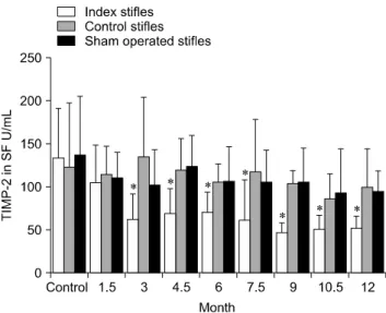
The levels of tartrate resistant acid phosphatase (TRAP), matrix metalloproteinase-2 (MMP-2), and tissue inhibitor of matrix metalloproteinase-2 (TIMP-2) in synovial fluid (SF) and serum in cases of canine osteoarthritis (OA) were measured. OA was induced by a surgically-created medial patellar luxation in the left stifle of 24 dogs. SF and blood samples were collected at 1.5- and 3-month intervals, respectively. Every 3 months, one dog was euthanatized to collect tissue samples from both stifles. TRAP levels in SF and serum were measured using a spectrophotometer, and TRAP-positive cells in joint tissues were identified by enzyme histochemistry. MMP-2 and TIMP-2 in SF and serum were detected by Western blotting and ELISA, respectively. TRAP in SF from the stifles and serum was significantly increased (p < 0.05) after 3 months. TIMP-2 in SF and serum was significantly decreased (p < 0.05), whereas MMP-2 in SF was significantly increased (p < 0.05) during the progression of OA. Histochemistry revealed an increased number of TRAP-positive cells in tissues from OA-affected joints. Assays measuring TRAP, MMP-2, and TIMP-2 in SF and serum, and methods that detect in- creased numbers of TRAP-positive cells in the joint tissues can play an important role in identifying the early phases of degenerative changes in canine joint components.
Keywords: MMP-2, osteoarthritis, synovial fluid, TIMP-2, TRAP
Introduction
Patellar luxation (PL) is one of the most common orthopedic disorders in dogs [18]. PL can result in the development of degenerative joint disease, pain, and
lameness. It has been reported that medial patellar luxation (MPL) increases the stress on the cranial cruciate ligament (CCL), predisposing the structure to degeneration and rupture. Secondary osteoarthritis (OA) is a common result of MPL [20].
OA is a slowly progressive disease with different etiologies that finally converge on the same pathogenic pathway that is hallmarked by characteristic changes in cartilage, subchondral bone, and synovial membrane. The insidious onset and “silent” progression of OA not only prevent early diagnosis of this disease, but also delay treatment that may help prevent further cartilage destruction and joint failure [16]. Hence, determinants or markers that may be used to detect and monitor molecular events early in the pathogenesis of OA are of considerable interest as to detect preclinical disease, determine prognosis, and monitor the response to drugs and therapy [10].
Although the cause of the synovial inflammation associated with OA is unknown, recent work has shown that the synovial intima contains large numbers of dendritic cells and macrophage-like cells, which express tartrate-resistant acid phosphatase (TRAP), together with increased immunoglobulin deposition [17]. TRAP- positive mononuclear cells within the synovium typically express many degradative collagenolytic enzymes including cathepsin K, cathepsin S, and matrix metalloproteinases (MMPs). The presence of TRAP- positive cells within the synovium is a characteristic feature of arthritis, and is considered to be a key factor for promoting progressive and irreversible articular cartilage and joint destruction [23]. Production of MMPs, cathepsins, and TRAP at sites of inflammation potentially contributes to articular cartilage matrix degradation [8,23].
It is well documented that osteoblasts express and secrete a large amount of TRAP. This factor has been shown to be a useful histochemical marker for identifying osteoclasts and a biochemical index for osteoclastic resorption [9,11].
Cartilage destruction is a common pathological feature in