Surface characteristics and bioactivity of minocycline-treated Ti-6Al-4V alloy
11
0
0
전체 글
(2) MC improved Ti surface characteristics. tetracycline, saline, chlorhexidine, hydrogen peroxide and doxycycline were commonly employed [6].. treatment to titanium surface. In this study, MC treatment was performed to remove. The tetracyclines are antibiotics that act by inhibition of. bacterial bioflms and to improve biocompatibility of. protein synthesis. Besides for their antibiotic properties,. titanium surface. The purpose of this study was to evaluate. tetracyclines possess several unique characteristics like. surface characteristics and the biocompatibility of MC. collagenase inhibition, inhibition of neutrophil chemotaxis,. treated smooth titanium alloy surface.. anti-inflammatory effects and root surface conditioning. Those characteristics could be account for their efficacy in periodontal treatment [7]. Data from clinical researches on the treatment of peri-. MATERIALS AND METHODS Titanium disks. implantitis suggest different methods by using chemical agents. In the clinical study by Mellonig et al. [8], the. Titanium alloy (Ti-6Al-4V) samples were manufactured. titanium surface was detoxified with a solution of 50. as disks in two different diameters (15 mm diameter and 4. mg/mL tetracycline-HCl, in contact with the titanium. mm thickness, 4 mm diameter and 4mm thickness) from. surface remaining for 2 to 3 minutes. Renvert et al. [9]. Titanium Center (Gwangju, Korea). In this study, Ti-6Al-. observed that both probing pocket depth and bleeding. 4V alloy was used rather than pure titanium for assumption. on probing scores were reduced by using local delivery of. of clinical situation. Dental implant abutment, the most. minocycline (MC) microspheres. Zhou et al. [10] reported. portion of contacting chemical agent, was generally made. that local delivery of MC-HCl (Periocline; Dongkook Co.,. of Ti-6Al-4V [13]. Titanium alloy disks were wet ground. Seoul, Korea) could be effectively used in treating peri-. by using progressively finer diamond paper (Deerfos Co.,. implantitis in cases without peri-implant fistula. A method. Incheon, Korea) up from 800 to 2,000 grit size. Disks were. of local drug delivery can attain higher concentrations in. ultrasonically degreased in 99.8% acetone and followed by. subgingival sites which was 100-fold higher than that of a. distilled water for 10 minutes respectively. The washing. systemic drug delivery method. For example, local delivery. procedures were repeated three times.. of MC-HCl can show drug concentrations in excess of 1500 μm/mL in gingival cervicular fluid after 24 hour of drug administration. MC concentrations in the periodontal pockets decreased gradually for 7 days after administration. MC treatment Each sample was soaked with MC-HCl (Dongkook Co.) solution in different conditions. Specimens were divided. [11]. In the study reported by Mouhyi et al. [12], treatment of. into 6 groups according to their concentration and treating. CA to titanium surface derived a result of cleaner titanium. time (Table 1). Group I was non-treated control group.. surface as observed by scanning electron microscopy. Group II, III, IV, V, and VI were MC or CA-treated groups.. (SEM) than the surface of untreated control [12]. Overall,. The immersion time of each group was varied with the. the use of antibiotics like MC and chemical agents resulted. concentration, which set up conditions reference to clinical. in improvement of soft tissue inflammation around peri-. situations associated with implant surface treatment, such. implant lesion.. as MC-HCl ointment local delivery or diluted irrigation of. Although the majority of studies on implant. MC-HCl solution [8-10].. detoxification are focused on the removal of bacterial biofilms, there has been little emphasis placed on the. SEM. changes in morphological surface properties that can occur when an implant surface is subjected to chemical. The surface morphology of MC-treated Ti-6Al-4V alloy. detoxifacation. In addition to, there has been a lack of. disks was observed by SEM (S-4700; Hitachi, Tokyo, Japan).. comprehensive understanding on the effectiveness of MC. 188. www.chosunobr.org.
(3) Jung-Hyuk Lee, et al. Table 1. Treatment condition of each group Group. Inc., Seoul, Korea) and using software (Surften.4.5; OEG,. Chemical concentration. Immersion time. Non treated Smooth titanium surface. -. Group I. Berlin, Germany).. Cell culture. Group II. MC 1.5 mg/mL. 1h. Group III. MC 1.5 mg/mL. 24 h. Group IV. MC 15 mg/mL. 10 min. Group V. MC 100 mg/mL. 5 min. Group VI. Citric acid (pH 1). 3 min. MC, minocycline.. Mouse MC3T3-E1 cells (ATCC; Rockville, MD, USA) were an immortalized cell-line as pre-osteoblast. The cells were cultured in T-75 flasks in alpha minimum essential medium (α-MEM; Invitrogen, Carlsbad, CA, USA) supplemented with 10% heat-inactivated fetal bovine serum (FBS; Invitron Co., St. Louis, MO, USA), 100 mg/mL penicillin, and 100 mg/ mL streptomycin (Invitrogen, Carlsbad, CA, USA) at 37° C in. Roughness test Roughness test was performed using an electronic. a humidified atmosphere of 5% CO2-95% air.. Evaluation of cell attachment. portable surface roughness tester (Diavite dh-7; ASMETO AG, Basel, Switzerland) for MC-treated titanium alloy surface. Triplicate specimens of all groups were included.. The titanium alloy disks (15 mm diameter and 4 mm thickness) were placed, under aseptic conditions, in the bottom of 12-well culture dishes, and then rinsed three. X-ray diffractometer (XRD) test. times in 70% ethanol, exposed to ultraviolet light for 1418 hours, and air dried in the cell culture hood. Cells were. To analyze the variation of crystal form of each group,. seeded in a 12-well plate at a density of 1×10⁴ cells/mL in. the XRD test was performed. The surface of the MC-. α-MEM medium containing 10% FBS. After incubation for 1 day, the dishes were washed three times with phosphate. treated Ti-6Al-4V alloy disks were analyzed with an XRD (DMAX/1200; Rigaku, Tokyo, Japan) using CuKα1 incident radiation, a tube voltage of 40 kV and a current of 40 mA.. buffered saline and fixed with 2.5% glutaraldehyde in 100. Scanning speed was 2° /min and the scanning angle was. increasing concentrations of ethanol (30%, 60%, 95%, and. ranged from 20°to 90°2θ.. 100%), immersed in hexamethyldisilazane (Sigma-Aldrich. mM cacodylate buffer. The samples were dehydrated in. Co., St. Louis, MO, USA) for 15 minutes, air-dried, and. X-ray photoelectron spectroscopy (XPS) test. immediately mounted on aluminum stubs and coated with carbon. Mouse MC3T3-E1 cells were evaluated for cell. XPS test was executed to analyze the surface composition. attachment and growth patterns by analyzing SEM images.. of oxide and OH groups. The surface composition was examined by XPS (Multilab 2000; Thermo Electron,. Cell viability test. Waltham, MA, USA). MTT assay (CellTiter 96 Aqueous one; Promega, Madison,. Contact angle test. Wi, USA) was used to evaluate cell viability after incubation for 3 days. Titanium disks (4 mm diameter and 4mm. The contact angles were determined after dropping. thickness) were placed in a 96-well plate. The cells (1×10⁴/. distilled water at room temperature. The images of the. mL) were seeded and expected to attach on titanium disks. distilled water droplet were taken at 30 seconds after. for 3 days in alpha-MEM solution. The number of viable. delivery. The contact angle of each sample was measured. cells was estimated by the amount of Formazan reduction.. using an image analyzing microscope (Camscope, Sometech. Formazan accumulation was quantified by the absorbance. 189.
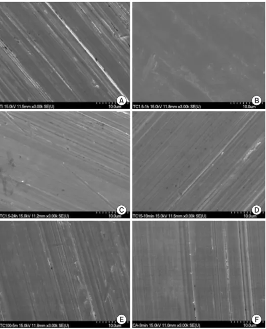
(4) MC improved Ti surface characteristics. at 490 nm using an spectrophotometeric microplate reader (VERSAmax; Molecular Device, Sunnyvale, CA, USA) and analyzed. The experiments were carried out in triplicate.. RESULTS Surface and microstructural characteristics (SEM analysis). Statistical analysis The surface morphology of each group was shown in One-way analysis of variance and Tukey's post hoc test. Fig. 1. Non-treated control Ti-6Al-4V surface showed. for repeated measurements were performed to examine. a uniform texture with straight coarse line. MC-treated. the data for surface roughness, contact angle measurement,. specimens showed narrow gap between lines. CA-treated. MTT assay with SPSS ver. 12.0 (SPSS Inc., Chicago, IL, USA).. specimen showed more dense structure than MC-treated. The results were considered to be significant at p <0.05.. titanium.. A. B. C. D. E. 190. www.chosunobr.org. F. Fig. 1. The scanning electron microscopy images of titanium surface with or without minocycline (MC) treatment. (A) Group I: control, (B) group II: MC 1.5 mg/mL 1 hour, (C) group III: MC 1.5 mg/mL 24 hour, (D) group IV: MC 15 mg/mL 10 minutes, (E) group V: MC 100 mg/mL 5 minutes, (F) group VI: citric acid 3 minutes..
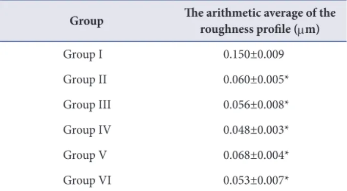
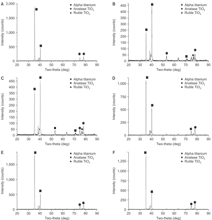
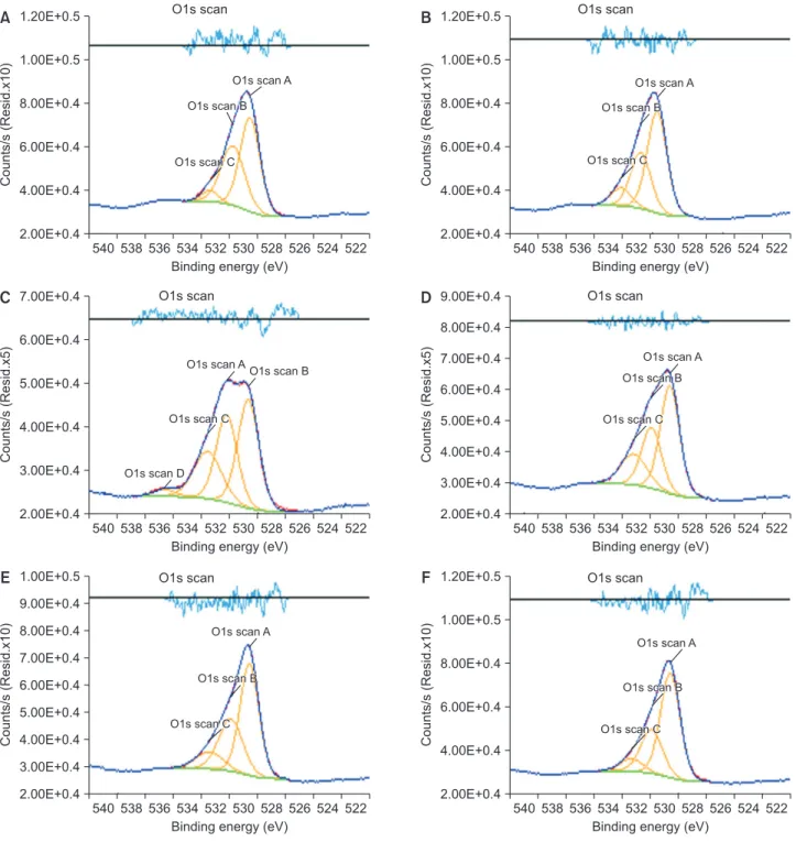
(5) Jung-Hyuk Lee, et al.. Roughness test. than that of the other groups.. The result of roughness test in each group was shown. Contact angle analysis. in Table 2. Roughness of group I, II, III, IV, V, and VI was 0.150, 0.060, 0.056, 0.048, 0.068, and 0.053 µ m, respectively. MC-treated groups and CA-treated group. Group II, III, IV, and V have significantly lower contact. have significantly lower roughness value compared to the. angle than control group and group VI (p <0.05).. Contact angle of each group was shown in Table 4.. control group (p <0.05).. Cell attachment XRD test For each sample, the cell morphology and cell attachment Fig. 2 showed XRD patterns of samples. The pattern of. were evaluated by SEM images (Fig. 4). All specimens have. the peaks demonstrated the portion of Ti-6Al-4V crystal. shown favorable cell adhesion and connection of dendritic. form. The peaks were classified with their two-θ degrees. For all the specimens, similar pattern of the peaks was. process. Cell spreading was increased in MC-treated. observed. Even though the most of peaks were composed. cell attachment was observed than the other groups and. of the alpha titanium peaks, group II and III showed the. the number of cells with polygonal shape increased.. titanium alloy surfaces. In the group II and group III, more. stronger intensity of the peaks belonging to rutile TiO2 (202) than the other groups. Furthermore, the intensity of the. MTT assay. peaks belonging to anatase TiO2 (105, 206) was increased in group II and III.. Cell viability was estimated by MTT assay (Table 5). On day 3, the MC-treated titanium surfaces were observed to. XPS analysis. have significantly increased cell proliferation compared with the control group and CA-treated group (p <0.05).. Fig. 3 and Table 3 showed XPS survey on titanium surface treated with MC. The variation in portion of hydroxide groups was detected at each group. Generally,. DISCUSSION. the portion of O1s Scan C was increased in MC- and CA-. Even though titanium has excellent biological properties,. treated groups. The portion of O1s Scan C which means. titanium implants may fail to osseointegrate commonly due. the portion of Ti-OH belonging group II and V was higher. to inflammation of peri-implant surface. Peri-implantitis was attributed to dental plaque formation. This oral. Table 2. Roughness value of each group (n=3) Group. The arithmetic average of the roughness profile (μm). biofilm was caused by oral bacterial colonization around the implant surface [14]. Removal of by-product from oral bacterial and reduction the level of micro-organisms is the crucial method in the treatment of peri-implant. Group I. 0.150±0.009. Group II. 0.060±0.005*. Group III. 0.056±0.008*. in removing bacterial biofilm, which may lead to re-. Group IV. 0.048±0.003*. osseointegration [16]. Many of treatments used to detoxify. Group V. 0.068±0.004*. titanium implants are either acidic or contain high. Group VI. 0.053±0.007*. concentrations of fluoride [17].. Values are presented as mean±standard deviation. *Significantaly different from the control group, p <0.05.. diseases [15]. Surface detoxification and debridement of contaminated implant surfaces can be effective way. In this study, MC was treated to Ti-6Al-4V alloy surface under different concentrations and immersion time to. 191.
(6) MC improved Ti surface characteristics 2,000. Alpha titanium Anatase TiO2 Rutile TiO2. 1,500. B. 1,000. Alpha titanium Anatase TiO2 Rutile TiO2. 450 400. Intensity (counts). Intensity (counts). A. 500. 350 300 250 200 150 100 50. 0. 0 20. 30. 40. 50 60 70 Two-theta (deg). C. 80. 90. Alpha titanium Anatase TiO2 Rutile TiO2. 450 400. 300 250 200 150. 30. 40. D. 50 60 70 Two-theta (deg). 80. 90. Alpha titanium Anatase TiO2 Rutile TiO2. 1,000. Intensity (counts). Intensity (counts). 350. 20. 750. 500. 250. 100 50 0. 0 20. 30. 40. 50 60 70 Two-theta (deg). E. 90. Alpha titanium Anatase TiO2 Rutile TiO2. 20. 1,000. 500. 30. 40. F. 50 60 70 Two-theta (deg). 80. 90. Alpha titanium Anatase TiO2 Rutile TiO2. 1,250. Intensity (counts). 1,500. Intensity (counts). 80. 1,000 750 500 250. 0. 0 20. 30. 40. 50 60 70 Two-theta (deg). 80. 90. 20. 30. 40. 50 60 70 Two-theta (deg). 80. 90. Fig. 2. X-ray diffractometer images of the specimens. (A) Group I: control, (B) group II: minocycline (MC) 1.5 mg/mL 1 hour, (C) group III: MC 1.5 mg/mL 24 hour, (D) group IV: MC 15 mg/mL 10 minutes, (E) group V: MC 100 mg/mL 5 minutes, (F) group VI: citric acid 3 minutes.. improve biocompatibility and increase wettability. It was. employed medications for treatment of the periodontal. found that the change of surface roughness and increased. diseases. As a group, the tetracyclines were varied with. wettability and favorable cell response with osteoblastic. their spectrum of antibacterial activity for gram-negative. cell on titanium surfaces treated with MC.. obligate anaerobes.. The MC is antibiotic agent that acts by inhibition of. MC appears to be more effective in inhibition of gram-. protein synthesis. MC was one of the mostly frequently. negative facultative anaerobes than tetracycline [18]. From. 192. www.chosunobr.org.
(7) Jung-Hyuk Lee, et al.. 1.20E+0.5. O1s scan. B. 1.00E+0.5 O1s scan A. 8.00E+0.4. O1s scan B. 6.00E+0.4 O1s scan C. 4.00E+0.4. Counts/s (Resid.x10). Counts/s (Resid.x10). A. 2.00E+0.4. 1.20E+0.5 1.00E+0.5. O1s scan A. 8.00E+0.4. 7.00E+0.4. O1s scan B. 6.00E+0.4 O1s scan C. 4.00E+0.4 2.00E+0.4. 540 538 536 534 532 530 528 526 524 522 Binding energy (eV). C. O1s scan. O1s scan. 540 538 536 534 532 530 528 526 524 522 Binding energy (eV). D. 9.00E+0.4. O1s scan. O1s scan A. O1s scan B. 5.00E+0.4 4.00E+0.4 3.00E+0.4. O1s scan C. Counts/s (Resid.x5). Counts/s (Resid.x5). 8.00E+0.4 6.00E+0.4. O1s scan D. 7.00E+0.4 6.00E+0.4 5.00E+0.4 4.00E+0.4. 2.00E+0.4 540 538 536 534 532 530 528 526 524 522 Binding energy (eV). 1.00E+0.5. O1s scan C. 3.00E+0.4. 2.00E+0.4. E. O1s scan A O1s scan B. O1s scan. 540 538 536 534 532 530 528 526 524 522 Binding energy (eV). F. 1.20E+0.5. O1s scan. 8.00E+0.4. O1s scan A. 7.00E+0.4 6.00E+0.4 5.00E+0.4. O1s scan B. O1s scan C. 4.00E+0.4. Counts/s (Resid.x10). Counts/s (Resid.x10). 9.00E+0.4 1.00E+0.5 O1s scan A. 8.00E+0.4 O1s scan B. 6.00E+0.4 O1s scan C. 4.00E+0.4. 3.00E+0.4 2.00E+0.4. 2.00E+0.4 540 538 536 534 532 530 528 526 524 522 Binding energy (eV). 540 538 536 534 532 530 528 526 524 522 Binding energy (eV). Fig. 3. X-ray photoelectron spectroscopy images of the specimens. (A) Group I: control, (B) group II: minocycline (MC) 1.5 mg/mL 1 hour, (C) group III: MC 1.5 mg/mL 24 hour, (D) group IV: MC 15 mg/mL 10 minutes, (E) group V: MC 100 mg/mL 5 minutes, (F) group VI: citric acid 3 minutes.. all these considerations, the MC was selected for agent to. treatments including tetracycline and CA on the oxide layer. treat the smooth titanium surface.. morphology of titanium surface. Discoloration, increase in. Previous studies discussed the influence of acidic. the depth of peaks and valleys and pits on the surface of. treatment in the corrosion behavior of titanium surface.. the samples were observed when acidic treatment coupled. Wheelis et al. [19] investigated the impact of acidic. with rubbing method. Suito et al. [20] immersed specimens. 193.
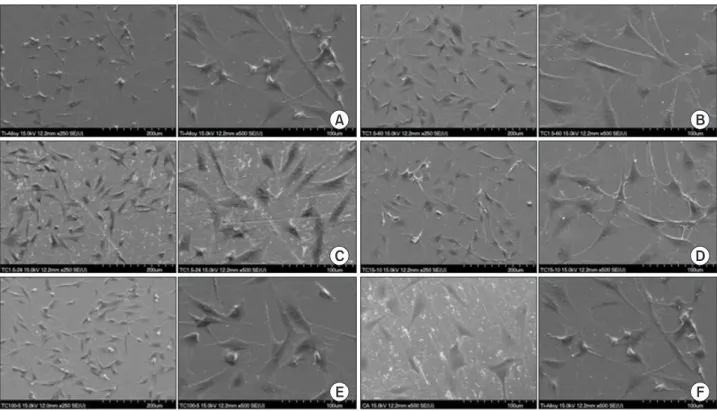
(8) MC improved Ti surface characteristics. of commercially pure titanium in an acidic solution whose. It could be assumed that relatively lower concentrations. pH value was controlled using either hydrochloric or lactic. of MC would be associated with the different tendency of. acid. Eluted titanium ions were observed after 24 hours (pH. current study from previous data. In addition, the higher. 2.0 and 3.0) and after 48 hours (pH 9.0). This means that. concentration groups (group IV and V) in this study have. delamination of the etched oxide layer could be occurred. shorter immersion time than in previous studies. Similar. by acidic treatment.. patterns were showed in SEM images of test groups. More. In this study, however, surface roughness was decreased and morphology of titanium surface was more smoothened. smoothened surfaces could be observed in MC- and CAtreated groups.. after MC treatment. MC- and CA-treated groups were shown to be significantly decreased in roughness. Table 4. Contact angle of groups (n=6) Group. Table 3. X-ray photoelectron spectroscopy analysis Group. TiO2 (%). TiOH (%). Ti-OH (%). Group I. 31.92. 23.86. 3.12. Group II. 13.55. 16.89. 10.67. Group III. 27.74. 16.44. 6.07. Group IV. 33.11. 18.91. 6.08. Group V. 23.21. 13.54. 8.97. Group VI. 32.82. 15.22. 4.61. Contact angle (°). Group I. 69.08±3.27. Group II. 58.87±4.29*. Group III. 56.90±3.85*. Group IV. 59.25±8.02*. Group V. 54.67±3.33*. Group VI. 62.40±5.62. Values are presented as mean±standard deviation. *Significantly different from the control group, p <0.05.. A. B. C. D. E. F. Fig. 4. The scanning electron microscopy images of cell attachment on Ti-6Al-4V alloy of each group (1: ×250, 2: ×500). (A) Group I: control, (B) group II: minocycline (MC) 1.5 mg/mL 1 hour, (C) group III: MC 1.5 mg/mL 24 hour, (D) group IV: MC 15 mg/mL 10 minutes, (E) group V: MC 100 mg/mL 5 minutes, (F) group VI: citric acid 3 minutes.. 194. www.chosunobr.org.
(9) Jung-Hyuk Lee, et al. Table 5. Cell viabillity assay of samples after 3 days of culture (n=3). seeded to titanium surfaces. Although all samples show. Group. Cell viability. favorable cell attachment and webbing of dendritic. Group I. 0.30±0.03. process, more osteoblastic cells were spreading on MC-. Group II. 0.94*±0.10. Group III. 0.78*±0.10. Group IV. 0.84*±0.10. Group V. 0.84*±0.11. Group VI. 0.32±0.05. Values are presented as mean±standard deviation. *Significantly different from the control group, p <0.05.. treated titanium alloy surfaces. In MC-treated groups, more cell adhesion was observed than the control group and the number of cells showing polygonal shape increased. This growth pattern could be understood by improved surface characteristics of MC treated titanium surface affect the cell attachment. MTT assay was performed to analyze the effect of MC treatment on cell viability of osteoblastic cells on the titanium surface. At 3 days of culture, all MC-treated group. To observe crystal structural change of titanium alloy surface, XRD analysis was performed. Results of analysis. showed significantly higher cell viability compared to the control group.. indicated that MC treatment could affect the anatase/. Similar tendency was observed on contact angle test.. rutile transition of TiO2. It has been reported that rutile. Significantly lower contact angle was obtained in MC-. has better dielectric and optical properties than anatase. treated groups. This may explain the improved cyto-. [21]. Rutile plane has the highest atomic density and more. compatibility of MC-treated titanium alloy surface.. thicken oxide layer [22]. The new peaks belonging to rutile. Although it was well conducted, the present study has. TiO2 could be seen in group II and III. Which suggested. some limitations. This study evaluated the biocompatibility. that oxide layer thickness was increased in group II and III. of the MC-treated Ti surface with osteoblast-like cells. With. compared to control group.. smooth titanium surface for material of implant abutment,. XPS analysis was also performed to analyze the surface. microbial attachment as well as fibroblasts or epithelial. oxide layer and the amount of OH group of TiO2. Titanium. cells attachment should be considered [3]. Various materials. dioxide surfaces have two hydroxide groups: an acidic. were used for fabricating implant fixtures and abutments.. hydroxide group (531.1 ev, TiOH) and a basic hydroxide. Zirconia has been used for implant fixtures or abutments.. group (532.4 eV, Ti-OH). Ti-OH group has higher reactivity. Therefore, future studies for surface with another material. with other anions than TiOH group [23]. MC- treated. are necessary.. groups showed more portion of Ti-OH than that of control. In conclusion, chemical agents including MC and CA. group. Group II and V have more basic group than the. were suggested to treat contaminated implant surface. In. other groups. This indicates that the reactivity of Ti-OH. this study, MC-HCl treatment was performed to improve. to other anions was increased in MC-treated groups. As a. biocompatibility of titanium surface. The purpose of. result, those groups might have higher hydrophilic surface.. this study was to evaluate the characteristics and the. Some studies reported the relationship between surface. biocompatibility of the Ti-6Al-4V surface treated by the. wettability and cell viability. Cell attachment or spreading and protein adsorption were influenced by surface. MC. The results as follows;. wettability [24]. Hydrophilic surface showed improved. Titanium surfaces treated with MC showed more. osteoblast differentiation and alkaline phosphatase activity.. smoothened surface microstructure and decreased. Liao et al. [25] reported hydrophilic surfaces are more easily. roughness. In XRD and XPS analysis, MC treated titanium. stimulate the differentiation of osteoblast in comparison. surface was shown improved osteogenic favorable surface. with hydrophobic surfaces.. modifications. Cell attachments and proliferation also. To analyze the relationship of surface modification by MC treatment and cell spreading, MC3T3-E1 cells were. showed significantly higher cell viability compared to control group at 3 days of culture.. 195.
(10) MC improved Ti surface characteristics. The surface roughness, crystal structure, surface hydrophilicity, cell viability of smooth titanium surface was improved by MC treatment. Compared with control group and CA treated group, smooth titanium surface immersed in MC-HCl showed improved surface characteristics and cell biocompatibility.. CONFLICT OF INTEREST The authors declare that they have no competing interests.. ORCID Jung-Hyuk Lee https://orcid.org/0000-0002-0413-6613 Young-Gon Sun https://orcid.org/0000-0002-9169-7522 Eui-Ri Na https://orcid.org/0000-0002-1584-8966 Jong-Wook Moon https://orcid.org/0000-0002-6688-0210 Young-Joon Kim https://orcid.org/0000-0003-1397-8166. REFERENCES 1. Gilbert JL. Mechanically assisted corrosion of metallic biomaterials. In: Narayan R, editor. ASM Handbook: Materials for Medical Devices. Ohio: ASM International; 2014. p. 79-89. 2. Yamazoe J, Nakagawa M, Matono Y, Takeuchi A, Ishikawa K. The development of Ti alloys for dental implant with high corrosion resistance and mechanical strength. Dent Mater J 2007;26:260-267. doi: 10.4012/dmj.26.260. 3. Kim YS, Shin SY, Moon SK, Yang SM. Surface properties correlated with the human gingival fibroblasts attachment on various materials for implant abutments: a multiple regression analysis. Acta Odontol Scand 2015;73:38-47. doi: 10.3109/00016357.2014.949845. 4. Pye AD, Lockhart DE, Dawson MP, Murray CA, Smith AJ. A review of dental implants and infection. J Hosp Infect 2009;72:104-110. doi: 10.1016/j.jhin.2009.02.010. 5. Berglundh T. Treatment of peri-implant mucositis and peri-implantitis. In: Lindhe J, Lang NP, eds. Clinical periodontology and implant dentistry. West Sussex: John. 196. www.chosunobr.org. Wiley & Sons; 2015. p. 861-869. 6. Valderrama P, Blansett JA, Gonzalez MG, Cantu MG, Wilson TG. Detoxification of implant surfaces affected by peri-implant disease: an overview of nonsurgical methods. Open Dent J 2014;8:77-84. doi: 10.2174/1874210601408010077. 7. Benet LZ, Oie S, Schwartz JB. Design and optimization of dosage regimens; pharmacokinetic data. In: Gilman AG, Goodman LS, Gilman AZ, Hardman JG, Molinoff PB, Limbird LE, editors. Goodman & Gilman's the pharmacological basis of therapeutics. New York: McGraw-Hill; 1996. p. 1707-1792. 8. Mellonig JT, Griffiths G, Mathys E, Spitznagel J Jr. Treatment of the failing implant: case reports. Int J Periodontics Restorative Dent 1995;15:384-395. doi: 10.11607/prd.00.0133. 9. Renvert S, Lessem J, Lindahl C, Svensson M. Treatment of incipient peri-implant infections using topical minocycline microspheres versus topical chlorhexidine gel as an adjunct to mechanical debridement. J Int Acad Periodontol 2004;6(4 Suppl):154-159. doi: 10.1111/j.1600051X.2006.00919.x. 10. Zhou L, Lin Y, Qiu LX, Chen B, Hu XL, Wang X. Clinical study of periocline on peri-implantitis treatment. Zhonghua Kou Qiang Yi Xue Za Zhi 2006;41:299-303. 11. Park YJ, Yeom HR, Lee SC, Lee SJ, Chung CP. Drug release kinetics, biodegradability, antimicrobial activity and cytotoxicity of 2% minocycline-loaded polysaccharide microcapsules. J Korean Soc Clin Pharmacol Ther 1995;3: 160-168. 12. Mouhyi J, Sennerby L, Pireaux JJ, Dourov N, Nammour S, Van Reck J. An XPS and SEM evaluation of six chemical and physical techniques for cleaning of contaminated titanium implants. Clin Oral Implants Res 1998;9:185-194. doi: 10.1034/j.1600-0501.1998.090306.x. 13. Elias CN, Lima JHC, Valiev R, Meyers MA. Biomedical applications of titanium and its alloys. Jom 2008;60:46-49. doi: 10.1007/s11837-008-0031-1. 14. Klinge B, Hultin M, Berglundh T. Peri-implantitis. Dent Clin North Am 2005;49:661-676. doi: 10.1016/ j.cden.2005.03.007. 15. Lindhe J, Meyle J. Peri-implant diseases: consensus report of the sixth european workshop on periodontology. J Clin Periodontol 2008;35(8 Suppl):282-285. doi: 10.1111/ j.1600-051X.2008.01283.x. 16. Schou S, Holmstrup P, Jørgensen T, Skovgaard LT, Stoltze K, Hjørting-Hansen E, Wenzel A. Implant surface preparation in the surgical treatment of experimental peri-implantitis with autogenous bone graft and ePTFE membrane in cynomolgus monkeys. Clin Oral Implants Res 2003;14:412-422. doi: 10.1034/j.1600-0501.2003.00912.x. 17. Gosau M, Hahnel S, Schwarz F, Gerlach T, Reichert TE, Bürgers R. Effect of six different peri-implantitis.
(11) Jung-Hyuk Lee, et al. disinfection methods on in vivo human oral biofilm. Clin Oral Implants Res 2010;21:866-872. doi: 10.1111/j.16000501.2009.01908.x. 18. Goodson JM. Antimicrobial strategies for treatment of periodontal diseases. Periodontol 2000 1994;5:142-168. doi: 10.1111/j.1600-0757.1994.tb00022.x. 19. Wheelis SE, Gindri IM, Valderrama P, Wilson TG Jr, Huang J, Rodrigues DC. Effects of decontamination solutions on the surface of titanium: investigation of surface morphology, composition, and roughness. Clin Oral Implants Res 2016; 27:329-340. doi: 10.1111/clr.12545. 20. Suito H, Iwawaki Y, Goto T, Tomotake Y, Ichikawa T. Oral factors affecting titanium elution and corrosion: an in vitro study using simulated body fluid. PLoS One 2013;8:e66052. doi: 10.1371/journal.pone.0066052. 21. Byun C, Jang JW, Kim IT, Hong KS, Lee BW. Anatase-torutile transition of titania thin films prepared by MOCVD. Mater Res Bull 1997;32:431-440. doi: 10.1016/S00255408(96)00203-6.. 22. Jones P, Hockey JA. Infra-red studies of rutile surfaces. Part 2.—hydroxylation, hydration and structure of rutile surfaces. Trans Faraday Soc 1971;67:2679-2685. doi: 10.1039/TF9716702679. 23. Cui DZ, Park KD, Lee KK, Jung YS, Lee BA, Lee YJ, Kim OS, Chung HJ, Kim YJ. Surface characteristics and osteoblastic cell response to titanium-8tantalum-3neobium alloy. Appl Surf Sci 2012;262:107-109. doi: 10.1016/j.apsusc. 2012.02.112. 24. Wei J, Igarashi T, Okumori N, Igarashi T, Maetani T, Liu B, Yoshinari M. Influence of surface wettability on competitive protein adsorption and initial attachment of osteoblasts. Biomed Mater 2009;4:045002. doi: 10.10 88/1748-6041/4/4/045002. 25. Liao H, Andersson AS, Sutherland D, Petronis S, Kasemo B, Thomsen P. Response of rat osteoblast-like cells to microstructured model surfaces in vitro. Biomaterials 2003; 24:649-654. doi: 10.1016/S0142-9612(02)00379-4.. 197.
(12)
수치
+3

관련 문서
These results suggest that dentin-derived HA coating using the RF magnetron sputtering method has good surface characteristics and biocompatibility.. Key
