Random Amplified Polymorphic DNA Analysis for Typing Extended-Spectrum-β-Lactamase of Klebsiella pneumoniae
Byoung-Seon Yang
Department of Clinical Pathology, Jinju Health College, Jinju 660-757, Korea
Fifty-one extended-spectrum-β-lactamase(ESBL) producing Klebsiella pneumoniae strains were isolated from national university hospitals. All K. pneumoniae strains showed resistance to broad-spectrum antibiotic and most of them presented resistance to amikacin, gentamicin and ciprofloxacin. The results of amplified polymorphic DNA (RAPD) pattern for randomly isolated fifty-one strains were as follows; both twenty-one strains from Chungnam National University hospital and ten strains from Chungbuk National University hospital showed RAPD type Ia and Ib. However, twenty strains isolated from Gyeongsang National University hospital belonged to RAPD type IIa and IIb. All isolates were divided into four molecular types and showed high level of genetic diversity. These results suggested that RAPD analysis provided a rapid and simple method for analysing genotypes of ESBL.
Key Words : Klebsiella pneumoniae, Extended-spectrum-β-lactamase, Randomly amplified polymorphic DNA
대한임상검사학회지 : 37권 제3호, 149-154, 20051) I. INTRODUCTION
Klebsiella pneumoniae has been increasingly recognized as a cause of hospital-acquired infections internationally.
These organisms are resistant to a number of antibiotics, including extended-spectrum cephalosporins and amino- glycosides, because of the acquisition of plasmids which code for the production of extended-spectrum-β-lactamases (ESBL) and aminoglycoside-modifying enzymes (Jarlier et al, 1988; Hogg et al, 1993; Meyer et al, 1993). Traditional epidemiologic tools, including biotyping and serotyping, are not useful in distinguishing between strains of K.
pneumoniae. Molecular techniques, including plasmid analysis and ribotyping, have suggested very useful techniques in distinguishing between strains of K.
pneumoniae(Brousseau et al, 1933). Distinctive polymor- phisms generated by the random amplified polymorphic DNA analysis(RAPD) are now being utilized for differen-
Corresponding author: Byoung-Seon Yang,
Department of Clinical Pathology, Jinju Health College.
Tel: 055-740-1851, Fax: 055-740-1846 E-mail: [email protected]
tiating strains. The aim of this study is to suggests the use of RAPD analysis to a cluster of ESBL-producing K.
pneumoniae infections detected by routine infection control surveillance at the hospitals (Ellsworth et al, 1993).
Ⅱ. MATERIALS AND METHODS
1. Bacterial strains
Between February 2005 and May 2005, 51 K.
pneumoniae strains were isolated from the university hospitals(Table 1). They consisted of five consecutive clinical isolates(24 from sputum, 10 from urine, 3 from blood, 2 from mucosal swabs, and 12 from others).
2. Phenotyping
Routine identification and antibiotic susceptibility tests
of the isolates were performed with the automated Vitek
system. Susceptibility to antimicrobial agents was tested
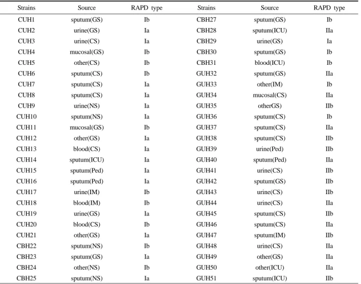
Table 1. RAPD genotyping of ESBL K. pneumoniae from various isolates
Strains Source RAPD type Strains Source RAPD type
CUH1 sputum(GS) Ib CBH27 sputum(GS) Ib
CUH2 urine(GS) Ia CBH28 sputum(ICU) IIa
CUH3 urine(CS) Ia CBH29 urine(GS) Ia
CUH4 mucosal(GS) Ib CBH30 sputum(GS) Ib
CUH5 other(CS) Ib CBH31 blood(ICU) Ib
CUH6 sputum(CS) Ib GUH32 sputum(GS) IIa
CUH7 sputum(CS) Ia GUH33 other(IM) Ib
CUH8 sputum(CS) Ia GUH34 mucosal(CS) IIa
CUH9 urine(NS) Ia GUH35 otherGS) IIb
CUH10 sputum(NS) Ia GUH36 sputum(CS) Ib
CUH11 mucosal(GS) Ib GUH37 sputum(CS) IIa
CUH12 other(GS) Ia GUH38 sputum(CS) IIb
CUH13 blood(CS) Ia GUH39 urine(Ped) IIb
CUH14 sputum(ICU) Ia GUH40 sputum(Ped) IIa
CUH15 sputum(Ped) Ia GUH41 urine(CS) IIb
CUH16 sputum(Ped) Ia GUH42 sputum(GS) IIb
CUH17 urine(IM) Ib GUH43 urine(CS) IIb
CUH18 blood(IM) Ib GUH44 urine(CS) IIa
CUH19 urine(GS) Ia GUH45 sputum(CS) IIb
CUH20 blood(CS) Ib GUH46 sputum(CS) IIa
CUH21 other(GS) Ia GUH47 sputum(IM) IIb
CBH22 sputum(NS) Ib GUH48 urine(CS) IIa
CBH23 sputum(GS) Ia GUH49 other(GS) IIa
CBH24 other(NS) Ib GUH50 other(ICU) IIa
CBH25 sputum(NS) Ia GUH51 sputum(ICU) IIb
CUH, Chungnam University Hospital; CBH, Chungbuk University Hospital; GUH, Gyoungsang University Hospital; GS, general surgery; ICU, intensive cure unit; CS, chest surgery; Ped, pediatrist; NS, neurosurgery; IM, internal medicine.
by the disk diffusion method on muller-hinton agar. The production of clavulanic acid-susceptible ESBL was detected by using the double-disk synergy test. Antibiotic susceptibility disks containing amoxicillin + clavulanate (AMC) were placed on the center of Petri dishes. Cefo- taxime (CTX) and ceftazidine (CAZ) disks were placed 25~30 mm apart circularly around the co-amoxiclav disk.
The agar plates were incubated for 24 hours at 35℃.
When the disk containing co-amoxiclav extended to any of the other antibiotic disk inhibition zones, ESBL production was inferred. Additionally, inhibition zone diameters of various β-lactams, including aztreonam and imipenem, aminoglycosides, chloramphenicol, and other
3. RAPD analysis
Bacteria were grown overnight on trypticase soy broth.
The broth was suspended of lysis buffer (50 mM Tris-Cl [pH 8], 50 mM EDTA, 1% sodium dodecyl sulfate, 30 ㎍ RNase per mL). The bacteria were then lysed, and RNA was digested by incubation of the lysate at 37℃ for 1h.
After incubation, the lysate was cleared by brief centrifugation and 0.5 mL was removed to a fresh tube.
One third volume of saturated ammonium acetate was added, to the content of the mix. DNA pellet was collected from the cleared lysate by ethanol precipitation.
The DNA pellet was dissolved in 100 μL on TE buffer
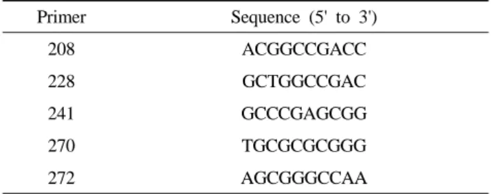
PCR : 208, 228, 241, 270 and 272 (Table 2). The reaction mixture contained Tris-HCl, MgCl
2, each primer, dNTP, Taq polymerase, and 4 μL of DNA extract in a final volume of 20 μL. Amplification was performed in a GeneAmp PCR 9600 thermal cycler with 45 consecutive cycles of 15s at 94℃, 15s at 36℃, and 70s at 72℃, with a single final extension step of 5 min at 72℃. PCR products were separated by electrophoresis in a 1.5%
agarose gel with 1⨉TBE running buffer at 90V for 1hr and stained with ethidium bromide and photographed under UV light.
Table 2. Oligonucleotide primers for RAPD for ESBL K.
penumoniae
Primer Sequence (5' to 3')
208 ACGGCCGACC
228 GCTGGCCGAC
241 GCCCGAGCGG
270 TGCGCGCGGG
272 AGCGGGCCAA
4. Statistical analysis
Polaroid photographs of the gels were scanned and saved. The images were normalized, a similarity matrix was produced by using the multivariate statistical package (MVSP), and dendrogram was constructed from the resulting data by the unweighted pair group method.
Ⅲ. RESULTS
1. Antimicrobial susceptibility test
Fifty-one strains of K. pneumoniae were isolated from three university hospitals. All strains exhibited a β-lactam susceptibility profile consistent with the production of ESBL: 1) decreased susceptibility or resistance to amoxi- cillin, piperacillin, cephalothin, cefamandole, extended- spectrum cephalosporins and aztreonam 2) full suscepti- bility to imipenem, temocillin and cephamycins 3) a positive disk synergy test with extended-spectrum cephalo- sporins(Fig. 1).
Fig. 1. Detection of ESBL production of CUH1 strain in double disc synergy tests.
(A) discs : left, ceftazidine; centre, amoxicillin+ clavulanate;
right, cefotaxime.
(B) discs : left, ceftazidine; centre, ticarcillin+ clavulanate; right, cefotaxime.
2. RAPD analysis
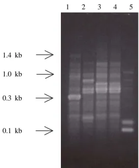
For each of the primers used, RAPD analysis yielded four groups of closely related fingerprints (major types Ia through IIb), each exhibited by CUH1, CBH22, GUH32, GUH33 and GUH50 strains respectively. For group I, two subtypes obtained with primer 208 and three subtypes obtained with primer 228 were distinguished by single-band variations of the core pattern. Primer 241 produced for clearly different groups of fingerprints based on bands with high intensity. Besides several common bands, one subtype (Ia) showed an additional fragment of high intensity (approximate molecular size of 0.7 kb) and the disappearance of a low-intensity fragment with a size<0.7 kb. For another subtype (Ib), the modification consisted of an additional fragment of low intensity with a molecular size of approximately 0.6 kb. The resulted of cluster analysis of the RAPD results with MVSP software.
21 strains from Chungnam National University Hospital
CUH2, CUH3 CUH7, CUH8 CUH9, CUH10 CUH12, CUH13 CUH14, CUH15 CUH16, CUH19 CUH21, CBH23 CBH25, CBH26 CBH29
Ia
CUH1, CUH4 CUH5, CUH6 CUH11, CUH17 CUH18, CUH20 CBH22, CBH24 CBH27, CBH30 CBH31, GUH33 GUH36
Ⅰb
CBH28, CUH32 CUH34, CUH37 CUH40, CUH44 CUH46, CUH48 CUH49, CUH50
Ⅱa
CUH35, CUH38 CUH39, CUH41 CUH42, CUH43 CUH45, CUH47 CUH51
Ⅱb
1 2 3 4 5
1.4 kb
1.0 kb
0.3 kb
0.1 kb
Fig. 2. RAPD patterns of K. pneumoniae isolates produced by using primer 241.
Lanes: 1, CUH1; 2, CBH22; 3, GUH32 4, GUH33; 5, GUH50.
and 10 strains from Chungbuk National University Hospital including the type strain were represented to Ia and Ib pattern but 20 strains from Gyeongsang National University Hospital belonged to IIa, IIb pattern (Fig. 2, 3).
Ⅳ. DISCUSSION
Nosocomial outbreaks caused by ESBL-producing K.
pneumoniae have been reported in Europe and in the United States. Infection caused by these strains, particularly in adult patients admitted to ICUs as observed in this study, have been described elsewhere (Baraniak et al, 2002; Bingen et al, 1993). In the past two decades, a significant number of nosocomial outbreaks of infection by K. pneumoniae have been reported, causing increasing concern in hospitals (Asensio et al, 2000). In this study, the RAPD patterns classified two major groups Ia, Ib and IIa, IIb. The first pattern limited in Chungnam, Chungbuk National University Hospital and type strain, but the second pattern only presented in Gyeongsang national university hospital. It appears from this study, the RAPD patterns were more identical in particular area not specimens or patients from isolated strains. As shown in Fig. 3 the test primers (208, 228, 270 and 272) were not clearly different groups of fingerprints (data not shown).
But, in the case of primer 241, the RAPD pattern is very good to fingerprints (Fig. 3). So, I think that this primer is very useful in distinguish among ESBL-producing K.
pneumoniae in our country for ribotyping.
RAPD is a new tool that is being used in such studies.
The simplicity and wide applicability of the method are
dependent on the use of short nucleotide primers which
are not related to known DNA sequences of the target
organism. Genetic mapping and determination of the
degree of relatedness between strains have been
performed, with validation by ribotyping (Welsh et al,
1990; Williams et al, 1990). The banding pattern derived
from this process allows the identification of similar strains by a method significantly less complicated and time-consuming than ribotyping. Analysis of an accurate antibiogram did not always reliably differentiate between strains.
In this study, it is suggested that RAPD analysis provides rapid and simple typing method of K. pneu- moniae strains for epidemiological studies and genotyping.
ACKNOWLEDGMENTS
This study was supported by a grant from Jinju Health College.
REFERENCES
1. Asensio A, Oliver C, Gonzalez-Diego P, Baquero F, Perez-Diaz C, Ros P, Cobo J, Palacios M, Lasheras D, Canton R. Outbreak of a multiresistant Klebsiella pneumoniae strain in an intensive care unit: antibiotic use as risk factor for colonization and infection. Clin Infect Dis 30:55-60, 2000.
2. Baraniak A, Sadowy E, Hryniewicz W, Gniadkowski M. Two different extended-spectrum beta-lactamases (ESBLs) in one of the first ESBL-producing salmonella isolates in Poland. J Clin Microbiol 40:1095-1097, 2002.
3. Bingen EH, Desjardins P, Arlet G, Bourgeois F, Mariani- Kurkdjian P, Lambert- Zechovsky Y, Denamur E, Philippon A, Elion J. Molecular epidemiology of plasmid spread among extended
broad-spectrum beta-lactamase-producing Klebsiella pneumoniae isolates in a pediatric hospital. J Clin Microbiol 31:179-184, 1993.
4. Brousseau R, Saint-Onge A, Prefontaine G, Masson L, Cabana J. Arbitrarily primed polymerase chain reaction, a powerful method to identify Bacillus thuringiensis serovars and strains. Appl Environ Microbiol 59:114-119, 1993.
5. Ellsworth DL, Rittenhouse KD, Honeycutt RL.
Artefactual variation in randomly amplified polymor- phic DNA banding patterns. BioTechniques 14:214-217, 1993.
6. Hogg GG, Forsyth JRL, Hibberd J, McBride J.
Importance of resistant Klebsiella species in Victoria.
Med J Aust 158:722 (Letter), 1993.
7. Jarlier V, Nicolas MH, Fournier G and Philippon A.
Extended broad-spectrum beta-lactamases conferring transferable resistance to newer beta-lactam agents in Enterobacteriacae: hospital prevalence and suscepti- bility patterns. Rev Infect Dis 10:867-878, 1988.
8. Meyer KS, Urban C, Eagen JA, Berger BJ, Rahal JJ.
Nosocomial outbreak of Klebsiella infection resistant to late-generation cephalosporins. Ann Intern Med 119:353-358, 1993.
9. Welsh JW, McClelland M. Fingerprinting genomes using PCR with arbitrary primers. Nucleic Acids Res 18:7213-7218, 1990.
10. Williams JGK, Kubelik AR, Livak KJ, Rafalski JA, Tingey SV. DNA polymorphisms amplified by arbitrary primers are useful as genetic markers.
Nucleic Acids Res 18:6531-6515, 1990.
=국문초록=