• 검색 결과가 없습니다.

Properties of β-Galactosidase from Lactobacillus zymae GU240, an Isolate from Kimchi, and Its Gene Cloning

N/A
N/A
Protected

Academic year: 2021

Share "Properties of β-Galactosidase from Lactobacillus zymae GU240, an Isolate from Kimchi, and Its Gene Cloning"

Copied!
9
0
0

로드 중.... (전체 텍스트 보기)

전체 글

(1)

Properties of β-Galactosidase from Lactobacillus zymae GU240, an Isolate from Kimchi, and Its Gene Cloning

Huong Giang Le

1

, Zhuang Yao

1

, Jeong A Kim

2

, Se Jin Lee

1

, Yu Meng

1

, Ji Yeong Park

1

, and Jeong Hwan Kim

1,2

*

1

Division of Applied Life Science (BK21 Plus), Graduate School,

2

Institute of Agriculture and Life Science, Gyeongsang National University, Jinju 52828, Republic of Korea

Received: February 27, 2020 / Revised: April 11, 2020 / Accepted: April 13, 2020

Introduction

β-galactosidase (β-D-galactoside galactohydrolase, EC 3.2.1.23), most commonly known as lactase, is a member of the family of glycosyl hydrolases that are known to catalyze the hydrolysis of the β-1,4-D-galactosidic link- age of lactose to its constituting monosaccharides, glu- cose and galactose [1].

β-galactosidase (β-Gal) is an important enzyme for lac- tic acid bacteria (LAB), especially for starters used for production of dairy products. One of the important pre- requisites for lactic starter is the ability to utilize lactose in milk, and quickly produces acids such as lactate and

acetate, which contributes to preservation and flavor of dairy products [2]. Many studies have been done on β- Gals and the structural genes from various LAB. Two types of β-Gals, β-Gal and phospho-β-Gal, exist among LAB depending upon employed lactose transport sys- tems [3]. In a Lac permease system, lactose enters into cell as an unmodified form and hydrolyzed by β-Gal into glucose and galactose. In a PEP (phosphoenol pyruvate)- PTS (phosphotransferase) system, lactose is phosphory- lated during transport and hydrolyzed by phospho- β-Gal into galactose 6-phosphate and glucose inside cells [4].

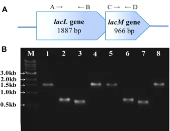
Some β-Gals from LAB are heterodimers consisting of a large subunit (72 kDa) encoded by lacL and a small sub- unit (35 kDa) encoded by lacM [5, 6]. β-Gal from Esche- richia coli is a homotetramer consisting of a 110 kDa subunit encoded by lacZ [7].

β-Gal gene from E. coli is used in molecular biological Lactobacillus zymae GU240 was previously isolated from Kimchi, a Korean fermented vegetable, as a strong GABA producer. The strain showed β-galactosidase (β-Gal) activity on MRS agar plates with X-gal.

When growth and β-Gal activities of GU240 were measured using MRS (glucose, 2%, w/v) and MRSL (lac- tose, 2%, w/v) broths, cells were found to grow slowly in MRSL, and the β-Gal activity (36 units at 4 h) was lower than that of cells grown in MRS (94 units at 16 h). The highest OD

600

value of the culture in MRS was 1.6 at 24 h at 37 ℃, whereas that of the culture in MRSL was 0.6 at 16 h. β-Gal activity of the culture in MRS reached the maximum (95.6 u/ml) at 16 h, decreased thereafter, and was not detected at 48 h. β-Gal activity for culture in MRSL reached its highest (36 u/ml) at 4 h and decreased gradually, but some activity (11.05 u/

ml) still remained at 72 h. The structural gene encoding β-Gal in L. zymae GU240 was cloned as a 3.1 kb frag- ment, and DNA sequencing confirmed the presence of complete lacLM genes. lacLM genes from L. zymae GU240 showed 98 −99% homologies in nucleotide sequences with other lacLM genes from L. brevis. Reverse transcription (RT)-PCR confirmed the operon structure of lacLM. The results indicated that L. zymae GU240 might be in the process of losing the ability to grow rapidly on lactose-containing medium, such as milk, due to adaptations to plant environments, including kimchi .

Keywords:

Lactobacillus zymae GU240, β-galactosidase, lacLM, Kimchi

*Corresponding author

Tel: +82-55-772-1904, Fax: +82-55-772-1909 E-mail: [email protected]

© 2020, The Korean Society for Microbiology and Biotechnology

(2)

researches as a reporter gene [8]. β-Gal genes from vari- ous sources are used for removal of lactose from milk in food industry and production of galactooligosaccharides [1, 9]. For heterologous gene expression among LAB, a gene involved in lactose transport is used in a food-grade expression vector as a selection marker [10, 11].

β-Gal gene from LAB isolated from fermented vegeta- ble foods are rarely studied because plants do not con- tain lactose in significant amount. In this report, we studied β-Gal activity of Lactobacillus zymae GU240, an isolate from Kimchi, producing GABA in large amount [12]. We also cloned the structural gene, and examined the structure of β-Gal gene. The strain and the β-Gal gene might be useful as a probiotic or a starter for foods containing lactose.

Materials and Methods

Bacterial strains and culture condition

L. zymae G9240 was grown on MRS broth (lactobacilli MRS, BD Difco, USA) and MRS agar (2%, w/v) without shaking at 37 ℃. When growth was tested on lactose as a carbon source, MRSL was used where all components were the same with MRS except glucose was replaced with lactose (Sigma-Aldrich, USA). E. coli DH5α was grown in Luria-Bertani broth (LB) at 37 ℃ with shaking.

When cultivating cells with a plasmid, ampicillin (50 μg/

ml) was included.

Determination of β-gal activity

The enzyme activity was measured according to Miller's method [13]. L. zymae GU240 was grown in MRS and MRSL broth at 37 ℃. Aliquots were taken and cells were harvested by centrifugation (8000 ×g, 10 min at 4 ℃).

After washing twice with sterile water, cell pellet was resus- pended with Z-buffer (pH 7.0, 1.61 g of Na

2

HPO

4

·H

2

O, 0.55 g of NaH

2

PO

4

· H

2

O, 0.075 g of KCl, 0.0246 g of MgSO

4

· 7H

2

O, 0.27 ml of β-mercaptoethanol, adjust the volume to 100 ml with sterile water). Cells were dis- rupted by sonication using an ultrasonicator (Bandelin Electronic, Germany). Cells were sonicated for 1 min fol- lowed by pause for 1min on ice (1 cycle), and total 5 cycles were done for each sample. Disrupted cells were centrifuged at 12,000 ×g for 20 min to collect supernatant.

β-gal activity was determined by using o-nitrophenyl-β- D-galactopyranoside (Sigma-Aldrich) as the substrate at

28 ℃. After yellow color appeard, 0.5 ml of 1 M sodium carbonate was added to stop the reaction. Absorbance was measured at 420 nm and 550 nm with a spectropho- tometer. β-Gal activities were calculated by this follow- ing method:

β-Gal Miller unit (U) = 1000 × (OD

420

– 1.75OD

550

)/

(T × V × OD

600

).

OD

420

and OD

550

were the values of the reaction mix- ture, and OD

600

was the cell density of the washed cell suspension. T was the reaction time (min), and V (ml) was the volume of culture used in the assay.

Cloning of lacLM genes

L. zymae GU240 was grown in MRS broth overnight at 37 ℃. Cells were harvested by centrifugation (12,000 ×g, 10 min, 4 ℃), and resuspended in 1 ml of lysis buffer (10 mM Tris-HCl, pH 8.0, 10 mM EDTA, 100 mM NaCl, 2% SDS) with 40 μl of proteinase K (Takara, Japan, 600 U/ml). After incubation for 30 min at 55 ℃, cell lysate was extracted with phenol and chloroform followed by ethanol precipitation. After 70% ethanol washing, DNA was dissolved in small volume of sterile water [14].

To amplify lacLM genes from L. zymae GU240, a primer pair was used: LMF (5'-CGGTGCGTCCCTACA- CATA-3') and LMR (5'-GACGTACTAATTTGCAATTT GCCATC-3'). Primers were designed based on the known lacLM sequences from National Center for Bio- technology Information (NCBI). PCR reaction mixture (50 μl) contained 2 μl of template DNA (20 ng/ul), 2 μl of each primer (10 μM), 5 μl of deoxynucleoside triphos- phates (0.25 mM), and 0.5 μl of Ex Taq DNA polymerase (Takara). The amplification conditions were as follows:

94 ℃ for 2 min; 30 cycles at 94℃ for 40 s, 53.7℃ for 40 s, 72 ℃ for 1 min; a final extension at 72℃ for 6 min. PCR product was examined by agarose gel electrophoresis using a 1% (w/v) agarose gel and stained with EcoDye™

DNA staining solution (Biofact, Korea).

Amplified fragment was ligated with pGEM T-Easy

vector (Promega, USA). E. coli DH5a competent cells

(Enzynomics, Korea) were used for transformation. Com-

petent cells were treated by heat-shock at 42 ℃ for 30 s

and left on ice for 2 min. Then 400 ul of SOC medium

(20 g tryptone, 5 g yeast extract, 4.8 g MgSO

4

, 3.603 g

dextrose, 0.5 g NaCl, 0.186 g KCl, per liter) was added

and cells were incubated for 1 h at 37 ℃ with shaking.

(3)

Aliquots (100 μl) were spreaded onto LB agar plates with ampicillin (100 μg/ml) and spreaded with 100 μl of 100 mM IPTG (isopropyl- β-D-thiogalactoside) and 20 μl of 50 mg/ml X-Gal (5-bromo-4-chloro-3-indoxyl- β-D- galactopyranoside). Plates were incubated overnight at 37°C. Transformants were screened to identify cells har- boring recombinant plasmid with lacLM genes. Plasmid DNA from E. coli DH5α was purified using DNA-spin™

plasmid DNA purification kit (iNtRON Biotechnology, Korea). Nucleotide sequencing was done for a recomn- inant, and the sequences were analyzed using BLAST.

RT-PCR experiment

RNA sample was prepared from L. zymae GU240 grown in MRS broth for 24 h by Trizol/bead method [15].

RNA sample was treated with DNase, RQ1 RNase-free (Promega), and RT-PCR was done using one-step RT- PCR premix kit (iNtRON Biotechnology). The reaction mixture consisted of 8 μl of premixture, 1 μl of forward primer, 1 μl of reverse primer, 2 μl of RNA (1 μg), and 8 μl of water. The reaction was started by 30 min incuba- tion at 45 ℃, followed by initial PCR denaturation at 94 ℃ for 5 min. PCR cycles consisted of denaturation at 94 ℃ for 1 min, annealing at 60℃ for 1 min, and exten- sion at 72 ℃ for 1 min. A total of 29 cycles were repeated, and the final extension was done at 72 ℃ for 5 min. lacL was amplified by using primer A (5'-AGTCATGT GCT- TAAACG GTCAGCG-3') and B (5'-ACCCACCCTGAT ACATTGGGTATTGG-3'), and lacM was amplified by using primer C (5'-AAGACGGC AAGGAATGGTTGTACC- 3') and D (5'-GCATTGTTTT GTGTGGTCCCTCGT-3').

Overexpression of lacLM in E. coli

lacLM gene was subcloned into pET26b(+) (Novagen, USA) after PCR product was digested with NcoI and XhoI. The recombinant (pETlacLM) was introduced into E. coli BL21(DE3) by electroporation. E. coli BL21(DE3) with pETlacLM was grown in LB until the OD

600

reached 0.6, then IPTG was added (1 mM), and the culture was grown for additional 12 h. Cells were recovered by centrifugation (12,000 × g, 10 min, 4℃), and resuspended with 4 ml of phosphate buffered saline (PBS, 10 mM PO

43−

, 137 mM NaCl, 2.7 mM KCl, pH 7.4). Cells were disrupted by sonication as describe above, and centrifuged.

Supernatant (soluble fraction) and pellet (insoluble fraction) were obtained. Insoluble fraction was dissolved with

3 ml of PBS. Soluble and insoluble fractions were analyzed by SDS-PAGE. 10% acrylamide gel was used and SDS-PAGE was done as described previously [12].

Results and Discussion

Growth of L. zymae GU240 on glucose and lactose L. zymae GU240 was grown in MRS (2% glucose, w/v) and MRSL (2% lactose, w/v) for 72 h at 37 ℃, and growth (OD

600

) and β-Gal activities were measured, respectively (Fig. 1). L. zymae GU240 grew rapidly after 4 h. In MRSL, the highest OD

600

was 0.6 at 16 h whereas the highest value was 1.6 at 24 h in MRS broth. L. zymae GU240 grew quicker in MRS than in MRSL, and the β- Gal activities of culture grown on MRS were also higher than those of culture on MRSL. One observation was that β-Gal activity of culture on MRS increased, and the highest activity of 95.6 U was observed at 16 h. But the activity decreased rapidly after 16 h, and not detected at 48 h and thereafter. Whereas β-Gal activity of culture in MRSL increased rapidly and reached the highest point (36 U) at 4 h, and then decreased until 8 h (21 U). The activity decreased gradually after 8 h, but remained rela- tively stable, and significant activity (11.7 U) was still observed at 72 h. The result indicated that β-Gal production, a result of β-Gal gene expression, is apparently not tightly repressed by glucose in MRS at least until 48 h. This is contrary to the known phenomenon of glucose repression of genes involved in utilization of alternative carbon sources such as lactose [16]. It is noteworthy that although L.

Fig. 1. Growth and β-Gal activities of L. zymae GU240 in

MRS and MRSL broth at 37 ℃. -●-, OD

600

in MRS; - ○-, β-Gal

activity in MRS; - ■-, OD

600

in MRSL; - □-, β-Gal activity in MRSL.

(4)

zymae GU240 grew slowly in MRSL compared to in MRS as shown by OD

600

values, β-Gal activity of culture in MRSL reached the highest point at 4 h, quite earlier than that of culture in MRS (16 h), and the activity remained stably until 72 h. These results indicated that β-Gal production in GU240 was induced by lactose, but not tightly repressed by glucose in MRS.

The results were contrary to those observed in Lacto- coccus lactis A2 [17] and L. lactis spp. lactis ATCC7962 [18]. In L. lactis A2 and 7962, maximum activity of β-Gal was observed in cells grown on lactose at 30°C, 210 U at 7 h and 325 U at 4 h, respectively. But cells grown on glucose showed very low activity, 2 −3 U for A2 and 3.6 U for 7962. The reason why L. zymae GU240 showed higher activities on glucose is not clear. The lack of an efficient lactose transport system in L. zymae GU240 might be one reason for poor growth of L. zymae GU240 on lactose. Since lactose is not a common sugar in plant environments, Lb. zymae GU240 might have a problem for efficient lactose utilization. Further studies on lac- tose transport of L. zymae GU240 are necessary to answer the question.

Cloning of lacLM genes from L. zymae GU240

A 3.1 kb fragment was amplified (Fig. 2), and a recom- binant plasmid, pGEM-TLM was obtained. Nucleotide sequence analysis confirmed that the fragment con- tained genes homologous to known lacLM genes. Nucle- otide sequences and translated amino acid sequences are shown in Fig. 3. The sequences were deposited into GenBank under the accession number MN188052.

Cloned lacLM gene is 2,836 bp in length and the nucleotide sequences are underlined. The ORF of LacL and LacM were located by using ExPASy tool (https://

web.expasy.org/translate/). The first codon of lacL gene is TTG, starting at 112

th

and ending at 1,998

th

, capable of encoding a protein of 628 amino acids. The first codon of lacM gene is ATG, starting at 1,982

nd

and ending at 2,947

th

, capable of encoding a protein of 321 amino acids.

One special feature of lacL and lacM genes is that both genes overlap partially, 17 nucleotides in length. The first codon of lacM (ATG) locates within the ORF of LacL, very close to the stop codon of lacL. But their read- ing frames are different. This kind of gene overlapping is also observed for other LAB [5, 19]. The pI and molecu- lar mass of LacL and LacM are 4.80/71,673.73 Da and

5.26/34,569.56 Da, respectively.

lacLM showed 98-99% identities in nucleotide sequence to those of homologous genes from L. brevis SRCM101106 (CP021674, 2,834/2,836), L. brevis ATCC367 (CP000416, 2,834/2,836), L. brevis BSO464 (CP005977, 2,821/2,836), L. brevis TMW1.2111 (CP019743, 2,798/2,836), L. brevis NPS-QW-145 (CP015398, 2,796/2,836), and L. brevis KB290 (AP012167, 2,792/2,836). The high homologies were expected because L. zymae is very similar to L. bre- vis except few differences [12].

The deduced amino acid sequences of LacL were shown above the nucleotide sequence and the deduced amino acid sequences of LacM were shown below the nucleotide sequences. The stop codons were marked as a short line after the amino acid sequences. Amino acid sequences were aligned with those of other homologous enzymes (Figs. 4 and 5).

RT-PCR experiment

RT-PCR was done for RNA sample to confirm the operon structure of lacLM. The expected amplicon size was 677 bp for lacL, and 577 bp for lacM. Both ampli- cons with the matching sizes were obtained (Fig. 6B, lanes 2 −3, and 6−7). Also, a transcript covering both LacL and LacM was detected (Fig. 6B, lanes 4 and 8), confirming the operon structure of lacLM genes.

Fig. 2. PCR amplification of lacLM genes. M, GeneRuler 1 kb

DNA Ladder (Thermo Fisher Scientific, SM0311); lane 2, PCR

product.

(5)

Fig. 3. Nucleotide sequence of lacLM. Nucleotide sequences of ORFs are underlined. Translated amino acids are shown above the

nucleotides (LacL) or below the nucleotides (LacM). The first codon of lacL gene is TTG, starting at 112

th

and the first codon of lacM

gene is ATG, starting at 1,982

nd

.

(6)

Fig. 4. Alignment of amino acid sequence of LacL with homologous enzymes from L. brevis strains. GU240 (L. zymae GU240,

MN188052), SRCM101106 (L. brevis SRCM101106, CP021674), ATCC367 (L. brevis ATCC367, CP000416), BSO464 (L. brevis BSO464,

CP005977), TMW 1.2111 (L. brevis TMW1.2111, CP019743), NPS-QW-145 (L. brevis NPS-QW-145, CP015398), and KB290 (L. brevis KB290,

AP012167). Amino acids different from other enzymes are marked as asterisks above the amino acids

(7)

Overexpression of lacLM in E. coli

SDS-PAGE results showed that LacM was overproduced in E. coli BL21(DE3) as shown in Fig. 7. A thick 35 kDa band was observed from insoluble fraction from E. coli

BL21(DE3) harboring pETlacLM (Fig. 7, lane 6), which

matched well with the expected size of LacM (34,569.56

Da). LacL production was not significant compared with

LacM, and the reason is not known. LacM was produced

Fig. 5. Alignment of amino acid sequence of LacM with homologous enzymes from Lactobacillus strains. GU240 (L. zymae GU240,

MN188052), SRCM101106 (L. brevis SRCM101106, CP021674), ATCC367 (L. brevis ATCC367, CP000416), BSO464 (L. brevis BSO464,

CP005977), TMW 1.2111 (L. brevis TMW1.2111, CP019743), NPS-QW-145 (L. brevis NPS-QW-145, CP015398), and KB290 (L. brevis KB290,

AP012167). Amino acids different from other enzymes are marked as asterisks above the amino acids

(8)

as inclusion body, and this is a common phenomenon when a foreign gene is overproduced in E. coli using an efficient expression system such as a pET vector and E.

coli BL21(DE3). Renaturation steps are necessary if overproduced β-Gal in E. coli is to be utilized.

L. zymae GU240 possesses intact lacLM genes but the strain grows poorly in lactose containing medium. The result indicates a possibility that L. zymae GU240 might

be in the process of adaptation to plant environments including Kimchi where lactose is not a major nutrient.

There is no need for a host cell to maintain an ability to grow rapidly in lactose as long as the environments contain no lactose since maintaining the ability is expen- sive for LAB which get less energy through metabolisms.

Although lactose utilization is not a major energy gener- ating pathway in L. zymae, still lacLM from L. zymae GU240 might be useful as reporter genes or a selection marker for hosts such as Lactococcus lactis and other LAB actively growing on milk and other dairy products.

Also the gene might be useful for the production of galactooligosaccharides, a prebiotic with industrial importance [20].

Acknowledgments

This work was supported by Korea Institute of Planning and Evalua- tion for Technology in Food, Agriculture, Forestry and Fisheries (IPET) through (Agricultural Microbiome R&D Program), funded by Ministry of Agriculture, Food and Rural Affairs (MAFRA) (918005043HD070). HG Le was supported by full time graduate student scholarship from Gyeong Sang Nat'l university. Z Yao, JA Kim, SJ Lee, Y Meng, and JY Park were supported by BK21 (+) program from MOE, Korea.

Conflict of Interest

The authors have no financial conflicts of interest to declare.

References

1. Juers DH, Matthews BW, Huber RE. 2012. LacZ β-galactosidase:

structure and function of an enzyme of historical and molecular biological importance. Protein Sci. 21: 1792-1807.

2. De Vos WM, Vaughan EE. 1994. Genetics of lactose utilization in lactic acid bacteria. FEMS Microbiol. Rev. 15: 217-237.

3. De Vos WM, Simons G. 1988. Molecular cloning of lactose genes in dairy lactic streptococci: the phospho-β-galactosidase and β- galactosidase genes and their expression products. Biochimie 70: 461-473.

4. Vaughan EE, Pridmore RD, Mollet B. 1998. Transcriptional regula- tion and evolution of lactose genes in the galactose-lactose operon of Lactococcus lactis NCDO2054. J. Bacteriol. 180: 4893- 4902.

5. Nguyen TH, Splechtna B, Yamabhai M, Haltrich D, Peterbauer C.

2007. Cloning and expression of the β-galactosidase genes from Lactobacillus reuteri in Escherichia coli. J. Biotechnol. 129: 581-591.

6. Obst M, Meding ER, Vogel RF, Hammes WP. 1995. Two genes encoding the β-galactosidase of Lactobacillus sake. Microbiology 141: 3059-3066.

Fig. 6. The operon structure of lacL and lacM. (A) Operon structure of lacL and lacM genes in L. zymae GU240. The arrows indicate the binding sites for each primer used for RT-PCR. (B) Agarose gel electrophoresis result. Lane M, iVDye 1kb DNA lad- der (Gendepot, USA); 1-4, RNA preps; 5-8, RNA preps treated with DNase I. Lanes 1 and 5, RT-PCR using universal primers (27F, 1492R) for 16S rRNA genes; 2 and 6, RT-PCR using primers A and B; 3 and 7, RT-PCR using primers C and D; 4 and 8, RT-PCR using primers A and D.

Fig. 7. Overexpression of lacLM in E. coli. M. DokDo-Marker

(EBM-1034, Elpisbiotech, Korea); lanes 1-3, culture supernatant

(1), soluble fraction (2), insoluble fraction (3) of E. coli BL21(DE3)

[pET-26b(+)]; lanes 4-6, culture supernatant (4), soluble fraction (5),

insoluble fraction (6) of E. coli BL21(DE3) [pETlacLM], no IPTG

added; lanes 7-9, culture supernatant (7), soluble fraction (8), insol-

uble fraction (9) of E. coli BL21(DE3) [pETlacLM], IPTG (1 mM)

added.

(9)

7. Adams RM, Yoast S, Mainzer SE, Moon K, Palombella AL, Estell DA. et al. 1994. Characterization of two cold-sensitive mutants of the β-galactosidase from Lactobacillus delbruckii subsp. bulgari- cus. J. Biol. Chem. 269: 5666-5672.

8. Franco D, de Boer PA, de Gier-de Vries C, Lamers WH, Mooman AF.

2001. Methods on in situ hybridization, immunohistochemistry and beta-galactosidase reporter gene detection. Eur. J. Morphol. 39: 3-25.

9. Oliveira C, Guimaraes PMR, Domingues L. 2011. Recombinant microbial systems for improved β-galactosidase production and biochemical applications. Biotechnol. Adv. 29: 600-609.

10. Platteeuw C, van Allen-Boerrigter I, van Schalkwijk S, de Vos EM.

1996. Food-grade cloning and expression system for Lactococcus lactis. Appl. Environ. Microbiol. 62: 1008-1013.

11. Landete JM. 2017. A review of food-grade vectors in lactic acid bacteria: from the laboratory to their application. Crit. Rev. Bio- technol. 37: 296-308.

12. Park JY, Jeong SJ, Kim JH. 2014. Characterization of a glutamate decarboxylase (GAD) gene from Lactobacillus zymae. Biotechnol.

Lett. 36: 1791-1799.

13. Miller JH. 1972. Experiments in molecular genetics, Cold Spring Harbor, NY: Cold Spring Harbor Laboratory Press.

14. Heo K, Cho KM, Lee CK, Kim GM, Shin JH, Kim JS, et al. 2013. Char- acterization of a fibrinolytic enzyme secreted by Bacillus amylo-

liquefaciens CB1 and its gene cloning. J. Microbiol. Biotechnol.

23: 974-983.

15. Sa HD, Park JY, Jeong SJ, Lee KW, Kim JH. 2015. Characterization of glutamate decarboxylase (GAD) from Lactobacillus sakei A156 isolated from Jeot-gal. J. Microbiol. Biotechnol. 25: 696-703.

16. Saier MH, Chauvaux S, Cook GM, Deutscher J, Paulsen IT, Reizer J, et al. 1996. Catabolite repression and inducer control in Gram- positive bacteria. Microbiology 142: 217-223.

17. Park RJ, Lee KW, Kim SJ, Park JY, Nam SJ, Park JY, et al. 2002. Isola- tion of Lactococcus lactis strain with β-galactosidase activity from Kimchi and cloning of lacZ gene from the isolated strain. J. Micro- biol. Biotechnol. 12: 157-161.

18. Kim TY, Lee JM, Chang HC, Chung DK, Lee JH, Kim JH, et al. 1999.

Effect of temperature and a carbon source on the expression of β-galactosidase gene of Lactococcus lactis spp. lactis ATCC7962.

J. Microbiol. Biotechnol. 9: 201-205.

19. David S, Stevens H, van Riel M, Sinons G, de Vos W. 1992. Leuco- nostoc lactis β-galactosidase is encoded by two overlapping genes. J. Bacteriol. 174: 4475-4481.

20. Chanalia P, Gandhi D, Attri P, Dhanda S. 2018. Purification and

characterization of β-galactosidase from probiotic Pediococcus

acidilactici and its use in milk lactose hydrolysis and galactooli-

gosaccharide synthesis. Bioorg. Chem. 77: 176-189.

수치

Fig.  1.  Growth  and  β-Gal activities of L. zymae GU240 in MRS and MRSL broth at 37 ℃
Fig. 2. PCR amplification of  lacLM genes. M, GeneRuler 1 kb DNA Ladder (Thermo Fisher Scientific, SM0311); lane 2, PCR product
Fig. 4. Alignment of amino acid sequence of LacL with homologous enzymes from  L. brevis strains
Fig. 6. The operon structure of  lacL  and  lacM.  (A) Operon structure of lacL and lacM genes in L

참조

관련 문서

Fabrication and Optical Properties of Polysiloxane Hybrimer Resin Using Oligohydrosiloxane 1 (OPH 1).. Fabrication and Optical Properties of Polysiloxane Hybrimer

캐나다정부간행물목록(Weekly checklist of Canadian government publications) 에 수록된 자료 중 Folder자료, 인구센서스, 전화번호 자료 등을 제외한

We determined the nucleotide sequences of the mitochondrial DNA (mtDNA) control region using cloning and sequencing, and obtained the complete sequence from the cattle bones

• Stormwater treatment.. 1) Domestic wastewater: Wastewater discharged from residences and from commercial, institutional, and similar facilities.. 2) Industrial

During culture, culture aliquots were withdrawn to monitor bacterial growth and to meas- ure β -galactosidase activity, and culture supernatants and

Full genome cloning and nucleotide sequence analysis of hepatitis C virus from sera of chronic hepatitis patients in Korea. Detection of hepatitis C virus RNA

회원국의 영토밖에서 다른 회원국의 , 영토내에서 회원국의 서비스 소비자에게

[r]