Introduction
Scutellaria baicalensis Georgi (Labiatae), called ‘Hwang-Gum’ in Korea and as ‘Huang- Qin’ in China, is one of the most widely used herbal medicines against bacterial infection of the respiratory and gastrointestinal tracts and various inflammatory diseases
1.2).
Prostaglandins (PGs) are key inflammatory mediators that are converted from arachidonic
acid by cyclooxygenase (COX). There are two isoforms of COX: cyclooxygenase-1 (COX-1) is constitutively expressed in nearly all tissues and provides PGs to maintain physiological func- tions such as cytoprotection of the stomach and regulation of renal blood flow
3,4). In contrast, cyclooxygenase-2 (COX-2) is inducible in the immune cells such as macrophages and synovi- ocytes in response to infection, injury or other stresses and COX-2 produces excessive amount of PGs that sensitize nociceptors and induce inflammatory states
5,6). Prostaglandin E2 (PGE2) is overproduced at sites of inflammation as a result of the activation of inducible COX-2
7,8).
The writhing test is a well-known animal test for the model of inflammatory and visceral pain.
received : 19 November 2007
received in revised from : 20 November 2007 accepted : 5 December 2007
Correspondence to : Hyung-Ho Lim
Department of Oriental Rehabilitation Medicine, Seoul Oriental Hospital Kyungwon University, 20-8 Songpa-Dong, Songpa- Gu, Seoul Korea
(Tel : +82-2-425-3456 / Fax : +82-2-425-3560, E-mail : omdlimhh@chol.com)
Analgesic and Anti-Inflammatory Effect of Scutellaria Baicalensis
Joong-Keun Lee, Yun-Kyung Song, Hyung-Ho Lim
Department of Oriental Rehabilitation Medicine, College of Oriental Medicine, Kyungwon University
Original Article
Backgrounds : Scutellaria baicalensis has been used as a medicinal plant to treat various disease conditions accompanying inflammatory response and oxidative stress.
Objectives : The aim of this study is to evaluate the effects of Scutellaria baicalensis against inflammatory, pain and edema Methods : In vitro, the effects of Scutellaria baicalensis against lipopolysaccharide-induced inflammation were investigated in mouse BV2 microglial cells. In vivo, the effects of Scutellaria baicalensis on acetic acid-induced writhing response, carrageenan-induced edema and the plantar test (nociceptive thermal stimulation) were investigated using rats and mice.
Results : The present results showed that pre-treatment with the aqueous extract of Scutellaria baicalensis suppressed the lipopolysaccharide-stimulated cyclooxygenase-2 expressions in mouse BV2 microglial cells. The aqueous extract of Scutellaria baicalensis inhibited acetic acid-induced abdominal pain in mice and also reduced thermal pain in rats. However, no significant inhibition on carrageenan-induced edema in rats.
Conclusions : The present study showed that Scutellaria baicalensis possesses anti-inflammatory and analgesic effects.
Key Words : Scutellaria baicalensis , analgesic, anti-inflammatory
It is useful test for studying anti-nociceptive effect. Carrageenan induced local inflammation is commonly used to evaluate analgesic effect.
Therefore, carrageenan induced local inflamm- ation (paw edema) is a useful model to assess the contribution of mediators involved in vascular changes associated with acute inflammation
9). Plantar test (Hargreaves method) is used to determine thermal pain threshold by exposing the animals to heat
10).
The effect of herbal medicine on inflammatory disease and pain is widely studied. For example, anti-inflamatory of Corydalis turtschaninovii
11-14)
, Clematis mandshurica
15,16)are generally evaluated. Scutellaira baicalensis is also studied about acute renal failure
17), hepatocytes cytotox- icity
18), ischemia-reperfusion injuries of hearts
19), neuroprotective of the brain ischemia
20)and so on. But, anti-inflammatory and analgesic actions of the Scutellaria baicalensis have not been clarified yet. In the present study, we investi- gated the effects of Scutellaria baicalensis against LPS-stimulated expressions of COX-1 and COX-2 in mouse BV2 microglial cells by using Western blot analysis in mouse BV2 microglial cells, in vitro. Moreover, the anti- nociceptive and anti-inflammatory effects of Scutellaria baicalensis using several experim- ental pain models, acetic acid-induced writhing response, carrageenan-induced for edema and response on thermal pain were also evaluated, in vivo.
Materials and Methods
1. Preparation of the aqueous extract of Scutellaria baicalensis
To obtain the aqueous extract of Scutellaria baicalensis, 100 g of Scutellaria baicalensis was
added to distilled water and extraction was performed by heating at 90°C for 2 h, concentr- ating with a rotary evaporator and lyophilization (Eyela, Tokyo, Japan). The resulting powder, weighing 20.58 g, was dissolved in saline solution and filtered through a 0.22 ㎛ syringe before use.
2. In vitro experiments 1) Reagents
The Scutellaira baicalensis was purchased from Oriental Medicine Hospital, Kyungwon University(Seoul, Korea). Rats and mice were purchased from Oriental Bio Co(Kyunggido, Korea).
2) Cell culture
Mouse BV2 macroglia cells were cultured in Dulbecco's modified Eagle's Medium (DMEM;
Gibco BRL, Grand Island, NY, USA) supplem- ented with 10 % heat-inactivated fetal bovine serum (FBS; Gibco BRL, Grand Island, NY, USA) at 37°C in 5 % CO2-95 % O2 in a humidified cell incubator. The cells were plated onto culture dishes at a density of 2 × 10⁴ cells/cm2 in culture dish, 24 h prior to drug treatments.
3) MTT cytotoxicity assay
Mouse BV2 macroglia cells were grown in a
final volume of 100 ㎕culture medium per well
in 96-well plates. In order to determine the
cytotoxicity of Scutellaria baicalensis, the cells
were treated with Scutellaria baicalensis at
concentrations of 1 ㎍/㎖, 10 ㎍/㎖, 100 ㎍/㎖,
1,000 ㎍/㎖ and 10,000 ㎍/㎖ for 24 h. Cultures
of the cells of the control group were left
untreated. After adding 10 ㎕ of the MTT labeling
reagent containing 5 ㎎/㎖3-(4,5-dimethyl thiazol
-2yl)-2,5-diphenyl tetrazolium bromide in phos-
phate-buffered saline to each well, the plates were incubated for 2 h. Solubilization solution 100 ㎕ containing 10 % sodium dodecyl sulfate in 0.01 M hydrochloric acid was added to each well and the cells were incubated for another 12 h. The absorbance was then measured with a microtiter plate reader (Bio-Tek, Winooski, VT, USA) at a test wavelength of 595 ㎚ with a reference wavelength of 690 ㎚. The optical density (O.D.) was calculated as the difference between the absorbance at the reference wave- length and that observed at the test wavelength.
Percent viability was calculated as (O.D. of drug-treated sample/control O.D.) × 100.
4) Western blot analysis
Mouse BV2 microglial cells were lysed in a ice-cold whole cell lysate buffer containing 50 mM HEPES (pH 7.5), 150 mM NaCl, 10 % glycerol, 1 % Triton X-100, 1.5 mM magnesium chloride hexahydrate, 1 mM ethyleneglycol-bis- ( β-aminoethyl ether)-N,N'-tetraacetic acid (EGTA), 1 mM phenylmethylsulfonyl fluoride (PMSF), 2
㎍/㎖ leupeptin, 1 ㎍/㎖ pepstatin, 1 mM sodium ortho vanadate and 100 mM sodium floride and the mixture were incubated 30 min at 4°C. Cell debris was removed by microcentrifugation, followed by quick freezing of the supernatant.
The protein concentration was measured using a Bio-Rad colorimetric protein assay kit (Bio-Rad, Hercules, CA, USA). Protein of 40 ㎍ was sepa- rated on SDS-polyacrylamide gels and transferred onto a nitrocellulose membrane (Schleicher &
Schuell GmbH, Dassel, Germany). Goat COX-1 antibody (1:1000; Santa Cruz Biotech, CA, USA) and Goat COX-2 antibody (1:1000; Santa Cruz Biotech, CA, USA) were used as a primary antibody. Horseradish peroxidase-conjugated anti-goat antibody (1:4000; Santa Cruz Biotech)
for COX-1 and COX-2 was used as a secondary antibody. Band detection was performed using the enhanced chemiluminescence (ECL) detection system (Amersham Pharmacia Biothech GmbH, Freiburg, Germany).
3. In vivo experiments 1) Animals and treatments
Male ICR mice weighing 28-30 g and male Sprague-Dawley rats weighing 150-160 g were used for the experiments. The experimental procedures were performed in accordance with the animal care guidelines of the National Institute of Health (NIH) and the Korean Academy of Medical Sciences. The animals were housed under laboratory conditions at a controlled temperature (20±2oC) and maintained under light-dark cycles, each consisting of 12 h of light and 12 h of darkness (lighting from 07:00 to 19:00 h) with food and water made available adlibitum.
2) Acetic acid-induced writhing response in mice
Male ICR mice were divided into five groups:
the control group, the acetic acid injection group,
the acetic acid injection and 50 ㎎/㎏ Scutellaria
baicalensis-treated group, the acetic acid injec-
tion and 100 ㎎/㎏ Scutellaria baicalensis-
treated group and the acetic acid injection and
200 ㎎/㎏ Scutellaria baicalensis-treated group
(n = 10 in each group). Scutellaria baicalensis
and control vehicle were orally administered 1 h
before the acetic acid injection. Then, the mice
received intraperitoneally with 0.15 ㎖ of 1.0 %
acetic acid as an irritant stimulus and placed in
an individual plastic cage (20 × 30 × 12 ㎝ high)
for observation. The number of writhes was
counted for 30 min after acetic acid injection.
3) Carrageenan-induced edema in rats
The volume of the paw edema in male Sprague- Dawley rats was measured in each animal using a plethysmometer (Ugo Basile, Italy) with a precision of two decimal places. To induce edema in the experimental animals, a single subplantar injection of carrageenan (1 %, 0.05 ㎖ Sigma Chemical Co., St. Louis, MO, USA) was given to each animal and the animals of the control group received injections of equivalent doses of normal saline
21)as a same method.
Animals of Scutellaria baicalensis-treated groups received orally with 1 ㎖ of the aqueous extract of Scutellaria baicalensis at the respective doses before 1 h of carrageenan injection and those of the control group and the carrageenan- induced edema group received equivalent amount of drinking water before 1 h of carrageenan injection. The paw volume was measured imme- diately before, 1 h, 2 h, 3 h, 4 h and 5 h after carrageenan injection. The animals were divided into five groups: the control group, the carrageenan- induced edema group, the carrageenan-induced edema and 100 ㎎/㎏ Scutellaria baicalensis- treated group, the carrageenan-induced edema and 200 ㎎/㎏ Scutellaria baicalensis-treated group and the carrageenan-induced edema and 400 ㎎/㎏ Scutellaria baicalensis-treated group (n = 10 in each group). The percentage of edema was calculated as follows:
Percentage of edema (%) = (Vt-Vn)/Vn × 100 Vt = The paw volume of each time after the
injection of carrageenan
Vn = The paw volume before the injection of carrageenan
4) Plantar test (Hargreaves's method)
To assess nociceptive responses to thermal stimuli, paw withdrawal latency of male Sprague-
Dawley rats was tested using the procedure previously described by Hargreaves et al
10). The center of a focused beam of radiant heat was applied to the plantar surface of the hind paw in rats and the withdrawal latency time was recorded. The infra-red intensity of the heat stimulus was 60 and stimulation was adjusted so that the baseline latency was 6 sec and 20 sec cut-off time was imposed to avoid tissue damage. Three minutes were allowed for the next test. The animals in the Scutellaria baicalensis-treated groups received orally with 1
㎖ of the aqueous extract of Scutellaria baicalensis at the respective doses before 1 h of test and those of the control group received equivalent amount of saline before 1 h of test.
The withdrawal latency time was measured immediately before, 1 h and 2 h after drug administration. The animals were divided into four groups: thermal stimulation-induced nocic- eption and drinking water-treated group, thermal stimulation-induced nociception and 50 ㎎/㎏
Scutellaria baicalensis-treated group, thermal stimulation-induced nociception and 100 ㎎/㎏
Scutellaria baicalensis-treated group and thermal stimulation-induced nociception and 200 ㎎/㎏
Scutellaria baicalensis-treated group (n = 6 in each group).
4. Data analysis
Results are presented as the mean ± standard error means (SEM). Statistical analysis was performed using one-way ANOVA followed by Duncan post-hoc test. Duncan's Test can be used to determine the significant differences between group means in an analysis of variance setting.
Duncan's test is based on the range statistic for a detailed discussion of different post hoc tests.
Difference was considered significant at p<0.05.
Results
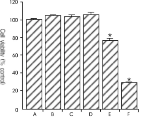
1. Cell viability of Scutellaria baicalensis on BV2 microglia cells
In order to assess the cytotoxic effect of the aqueous extract of Scutellaria baicalensis on BV2 microglia cells, the cells were cultured with the aqueous extract of Scutellaria baicalensis at final concentrations of 0.001, 0.01, 0.1, 1 and 10
㎎/㎖ for 24 h and MTT assays wasthen carried out. The cells cultured in Scutellaria baicalensis- free media were used as the control. The viability of cells incubated with Scutellaria baicalensis at concentrations of 0.001, 0.01, 0.1, 1 and 10 ㎎/㎖
for 24 h was 105.10 ± 0.94, 103.83 ± 1.70, 106.51 ± 1.71, 77.31 ± 2.06 and 30.07 ± 0.47 % of the control value, respectively.
The present results show that the Scutellaria baicalensis exerted no significant cytotoxicity until it was at a concentration of 0.1 ㎎/㎖.
However, a high concentration (1 ㎎/㎖ and 10
㎎/㎖) of Scutellaria baicalensis reduced cell viability. Then, we used the aqueous extract of Scutellaria baicalensis at concentration of 0.01 and 0.1 ㎎/㎖ for the next experiment (Fig. 1).
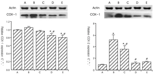
2. Effect of Scutellaria baicalensis on protein expression of COX-1 and COX-2 The level of COX-1 protein was 1.06 ± 0.02 following a treatment with 1 ㎍/㎖ LPS for 24 h.
The level of COX-1 protein was 0.97 ± 0.01, 0.86 ± 0.04 and 0.80 ± 0.05 in the cells pre- treated with the aqueous extract of Scutellaria baicalensis at 0.01 ㎎/㎖, 0.1 ㎎/㎖ and 500 μM acetylsalicylic acid (ASA), one hour before LPS treatment.
The level of COX-2 protein was markedly increased to 5.28 ± 0.81 following a treatment with 1 ㎍/㎖LPS for 24 h. The level of COX-2 protein was decreased to 3.64 ± 0.58, 1.27 ± 0.16 and 1.49 ± 0.08 in the cells pre-treated the aqueous extract of Scutellaria baicalensis at 0.01 ㎎/㎖, 0.1 ㎎/㎖ and 500 μM ASA one hour before LPS treatment.
The present results show that LPS enhanced COX-2 protein expression in mouse BV2 microglia cells and the aqueous extract of Scutellaria baicalensis suppress LPS-induced COX-2 protein expression. However, LPS and the aqueous extract of Scutellaria baicalensis exerted no significant effect on the expression of
*
*
120
100
80
60
40
20
0
A B C D E F
Fig. 1. Cell viagility of Scutellaria baicalensis on BV2 microglia cells. (A) Control group, (B) 0.001 /
Scutellaria baicalensis-treated group, (C) 0.01 / Scutellaria baicalensis-treated group, (D) 0.1 /
Scutellaria baicalensis-treated group, (E) 1 / Scutellaria baicalensis-treated group, (F) 10 /
Scutellaria baicalensis-treated group. * represents p < 0.05 compared to the control group.
COX-1 protein, except the aqueous extract of Scutellaria baicalensis at concentration of 0.1 ㎎ / ㎖ (Fig. 2).
3. Effect of Scutellaria baicalensis on acetic acid-induced writhing response in mice
The number of the writhing response in the control group was 0.25 ± 0.25. The number of writhing response in the acetic acid injection group was 39.88 ± 8.18. The number of writhing response in the acetic acid injection and Scutellaria baicalensis (50, 100 and 200 ㎎/
㎏)-treated group was 27.33 ± 1.61, 29.33 ± 6.10
*,
#*,
# AA B C D E
1.2
1.0
0.8
0.6
0.4
0.2
0.0 Actin
COX-1
B C D E
*
*,
## #
8
6
4
2
0
A B C D E
A B C D E
Actin
COX-1
Fig. 2. Results of Western blot analysis of the protein levels of COX-1 and COX-2. (A) Control, (B) LPS-treated group, (C) LPS- and 0.01 / Scutellaria baicalensis-treated group, (D) LPS- and 0.1 / Scutellaria baicalensis-treated group, (E) LPS- and 500 μM ASA-treated group. Actin was used as the internal control. * represents p < 0.05 compared to the control group. # represents p < 0.05 compared to the LPS-treated group.
*
*,
#*,
#*,
#A B C D E
50
40
30
20
10
0
Fig. 3. Effect of Scutellaria baicalensis on the number of writhing response. (A) control group, (B) 1 % acetic
acid-induced writhing response group, (C) 1 % acetic acid-induced writhing response and 50 /
Scutellaria baicalensis-treated group, (D) 1 % acetic acid-induced writhing response and 100 mg/kg
Scutellaria baicalensis-treated group, (E) 1 % acetic acid-induced writhing response and 200 mg/kg
Scutellaria baicalensis-treated group. * represents p < 0.05 compared to the control group. # represents
p<0.05 compared to the 1 % acetic acid-induced writhing response group.
and 25.63 ± 3.04.
The present results showed that acetic acid injection into the abdominal cavity induced writhing response. The acetic acid injection and Scutellaria baicalensis-treated group suppressed acetic acid-induced writhing response (Fig. 3).
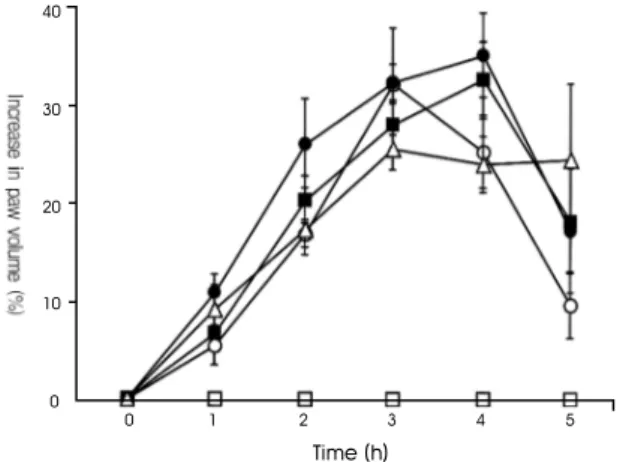
4. Effect of Scutellaria baicalensis on the volume of carrageenan-induced paw edema
After 1 h, paw volume in the control group was 0.00 ± 0.00 %. Paw volume in the 1 % carrageenan-induced edema group was increased to 5.96 ± 0.28 %. Paw volume of 1 % carrageenan- induced edema and Scutellaria baicalensis- treated groups at concentrations of 100, 200 and 400 ㎎/㎏ was 4.68 ± 1.79, 9.40 ± 1.52 and 7.87
± 2.17 %.
After 2 h, paw volume in the control group was 0.00 ± 0.00 %. Paw volume in the 1 % carrageenan-induced edema group was incre- ased to 17.57 ± 2.11 %. Paw volume in the 1 %
carrageenan-induced edema and Scutellaria baicalensis-treated groups at concentrations of 100, 200 and 400 ㎎/㎏ was 14.61 ± 1.23, 22.51
± 3.97 and 14.92 ± 2.26 %.
After 3 h, paw volume in the control group was 0.00 ± 0.00 %. Paw volume in the 1 % carrageenan-induced edema group was increased to 24.21 ± 2.21 %. Paw volume in the 1 % carrageenan-induced edema and Scutellaria baicalensis-treated groups at concentrations of 100, 200 and 400 ㎎/㎏ was 27.86 ± 1.77, 28.00
± 4.78 and 20.67 ± 2.52 %.
After 4 h, paw volume in the control group was 0.00 ± 0.00 %. Paw volume in the 1 % carrageenan-induced edema group was incre- ased to 26.20 ± 3.39 %. Paw volume in the 1 % carrageenan-induced edema and Scutellaria baicalensis-treated groups at concentrations of 100, 200 and 400 ㎎/㎏ was 21.80 ± 3.21, 30.38
± 3.74 and 20.67 ± 2.52 %.
After 5 h, paw volume in the control group was 0.00 ± 0.00 %. Paw volume in the 1 %
40
30
20
10
0
0 1 2 3 4 5
Time (h)
Fig. 4. Effect of Scutellaria baicalensis in response to doses and time on carrageenan-induced paw edema.
( ) control group, ( ) 1 % carrageenan-induced edema group, ( ) 1 % carrageenan-induced edema and 100 / Scutellaria baicalensis-treated group, ( ) 1 % carrageenan-induced edema and 200
/ Scutellaria baicalensis-treated group, ( ) 1 % carrageenan-induced edema and 400 /
Scutellaria baicalensis-treated group.
carrageenan-induced edema group was incre- ased to 15.71 ± 4.56 %. Paw volume in the 1 % carrageenan-induced edema and Scutellaria baicalensis-treated groups at concentrations of 100, 200 and 400 ㎎/㎏ was 8.24 ± 2.95, 14.83 ± 5.51 and 21.19 ± 6.63 %.
The present results showed that the paw volume in the control group was maintained at constant level and that paw volume in the carrageenan-induced edema group was increased as time-dependently during 4 h.
First of all, the concentrations of Scutellaria baicalensis-treated groups are 50, 100, 200 ㎎/
㎏. However, carrageenan-induced edema and Scutellaria baicalensis-treated groups exerted no significant inhibition on carrageenan-induced paw edema. And this time, the concentrations of Scutellaria baicalensis-treated groups changed 100, 200, 400 ㎎/㎏, but the result was also no significant inhibition (Fig. 4).
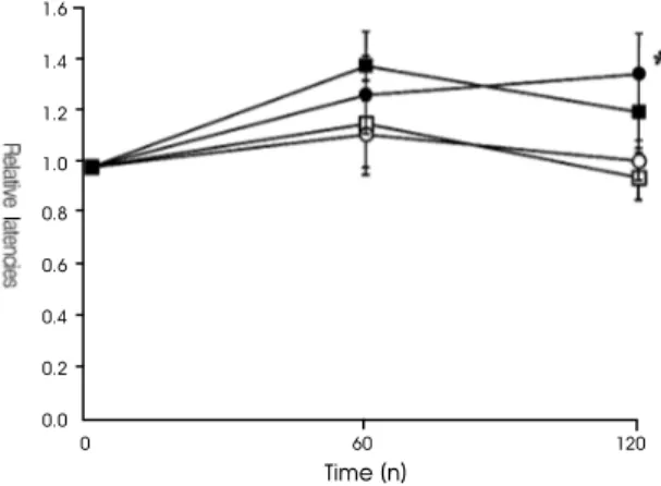
5. Effect of Scutellaria baicalensis on the plantar test (nociceptive thermal stim- ulation)
After 1 h, paw withdrawal threshold in the pre-treated value was considered as 1.00. The withdrawal latency in the thermal stimulation- induced nociception and drinking water-treated group was 1.17 ± 0.17. The withdrawal latency in the thermal stimulation-induced nociception and 50 ㎎/㎏ Scutellaria baicalensis-treated group was 1.39 ± 0.13. The withdrawal latency in the thermal stimulation-induced nociception and 100 ㎎/㎏ Scutellaria baicalensis-treated group was 1.13 ± 0.16. The withdrawal latency in the thermal stimulation-induced nociception and 200 ㎎/㎏ Scutellaria baicalensis-treated group was 1.28 ± 0.15. After 1 h, the withdrawal latency of thermal stimulation-induced nocic- eption was not significantly different in the animals treated with Scutellaria baicalensis.
After 2 h, the withdrawal latency in the thermal stimulation-induced nociception and drinking water-treated group was 0.96 ±0.09.
The withdrawal latency in the thermal stimulation- induced nociception and 50 ㎎/㎏ Scutellaria baicalensis-treated group was 1.22 ± 0.15. The
1.6 1.4 1.2 1.0 0.8 0.6 0.4 0.2
0.0
0 60 120
Time (n)